
The Biotechnology : Principles and Processes Class 12 chapter — Chapter 9 of the NCERT Class 12 Biology textbook, official edition on ncert.nic.in — runs across 16 PDF pages (printed pages 160–176). The official PDF is linked right below, and underneath it this page explains what the chapter contains: its sections, its figures, and the idea behind each one.
Everything on this page follows the NCERT edition page by page, so it works as a study companion in class or the night before a test.
| What the chapter holds | Count | Where it is used |
|---|---|---|
| Printed pages | 16 | |
| Sections in the chapter | 12 | |
| Figures with NCERT captions | 9 | |
| Exercise questions | 12 | answered in our NCERT Solutions |
| Official NCERT PDF | Download the chapter PDF | the chapter exactly as NCERT publishes it |

Official NCERT Class 12 Biology Chapter 9 PDF
Get the official NCERT Class 12 Biology Chapter 9 Biotechnology : Principles and Processes PDF from ncert.nic.in and study the exact current edition of the chapter, with the original figures and exercises as NCERT printed them. Downloading straight from the NCERT portal matters because the file is the complete, official chapter — nothing deleted, nothing reformatted by a third party.
To check the edition or find another chapter of the book, browse the NCERT textbook portal.
Reference: NCERT Class 12 Biology textbook, chapter 9, official edition on ncert.nic.in.
Chapter 9 at a Glance: Sections, Figures and Exercises
The table below lists what this chapter holds — numbered sections, figures and the exercises at the end — so you can see its size at a glance before you open the file.
Unit IX of the book opens with this chapter and closes with Chapter 10, Biotechnology and Its Applications. Inside Chapter 9 the order is consistent: principles first (Section 9.1), then tools (Section 9.2), then processes (Section 9.3), finished by the chapter summary and its exercises (p. 175).
What This Chapter Covers: Biotechnology : Principles and Processes Class 12
This section is a map of the chapter’s argument. Once you know why the sections sit in this order, each individual topic has a place to attach to.
Chapter 9 answers three questions in sequence:
- What is biotechnology? Section 9.1 opens with the European Federation of Biotechnology (EFB) definition and splits the subject into its two core techniques (p. 163).
- Which tools make recombinant DNA possible? Section 9.2 introduces restriction enzymes, polymerase enzymes, ligases, vectors and the host organism (p. 165).
- How are the tools used, step by step? Section 9.3 walks through the six processes from isolating DNA to a finished product (p. 171).
The anchor story is the construction of the first recombinant DNA molecule by Stanley Cohen and Herbert Boyer in 1972 — cutting an antibiotic resistance gene out of a Salmonella typhimurium plasmid, rejoining it with DNA ligase, and multiplying the result in E. coli (p. 164). That one experiment gives the chapter its shape.
The book also fixes three basic steps of genetically modifying an organism (p. 164):
- Identification — find the DNA that carries the desirable gene.
- Introduction — get that DNA into the host organism.
- Maintenance — keep the introduced DNA in the host and ensure it is passed to the host’s progeny.
| Section | What it answers | NCERT page |
|---|---|---|
| 9.1 Principles of Biotechnology | What biotechnology is, and the two core techniques behind it | p. 163 |
| 9.2 Tools of Recombinant DNA Technology | The tool kit: restriction enzymes, polymerase enzymes, ligases, vectors, host organism | p. 165 |
| 9.2.1 Restriction Enzymes | How enzymes cut DNA and why the cut ends can be rejoined | p. 165 |
| 9.2.2 Cloning Vectors | What a vector needs: ori, selectable marker, cloning sites | p. 168 |
| 9.2.3 Competent Host | How recombinant DNA is forced into cells that normally reject it | p. 170 |
| 9.3 Processes of Recombinant DNA Technology | The six steps from isolating DNA to a marketing-ready product | p. 171 |
| Summary and Exercises | The chapter in brief, plus the questions that test it | p. 175 |
This chapter stays strictly on principles and processes. The applications of these tools — what the products are actually used for — belong to Chapter 10, as the Unit IX opening states.
Key Concepts of Biotechnology : Principles and Processes Class 12
This is the teaching core of the page. Each subsection below matches one part of the chapter, states the idea in plain words, and carries the NCERT page number so you can follow along in the book.
9.1 Principles: Definition, the Two Core Techniques and the First Recombinant DNA
The chapter opens by defining its subject. The European Federation of Biotechnology (EFB) defines biotechnology as the integration of the natural sciences with organisms, cells, parts of cells or molecular analogues, to produce goods and deliver services (NCERT, p. 163). In plain words: biology plus technology, aimed at a product or a service.
Under that broad definition, making curd, bread or wine — all microbe-mediated — counts as biotechnology. The modern, restricted sense is narrower: processes that use genetically modified organisms on a large scale. Two core techniques made this modern form possible:
- Genetic engineering — altering the chemistry of genetic material (DNA and RNA), introducing it into host organisms, and thus changing the host’s phenotype (p. 163).
- Bioprocess engineering — keeping a sterile, contamination-free environment in chemical engineering processes so that only the desired microbe or eukaryotic cell grows, in large quantities, to manufacture products such as antibiotics, vaccines and enzymes (p. 163–164).
Why does an alien piece of DNA need help to multiply? If a foreign DNA somehow reaches a new organism, it usually cannot multiply in the progeny cells on its own.
It multiplies only when it becomes part of a chromosome that can replicate, or when it is linked to the specific sequence at which replication starts — the origin of replication (NCERT, p. 164). Linking a foreign DNA to this sequence and multiplying it is cloning: making multiple identical copies of a template DNA.
The first artificial recombinant DNA came in 1972.
Stanley Cohen and Herbert Boyer cut the gene for antibiotic resistance out of a plasmid of Salmonella typhimurium — a plasmid being the circular, autonomously replicating extra-chromosomal DNA of a bacterium — joined the cut gene to plasmid DNA with the enzyme DNA ligase, and let E. coli multiply the new circle using its own DNA polymerase (p. 164).
That new combination of circular, autonomously replicating DNA, created in vitro, is recombinant DNA, and the plasmid that carried it acted as a vector.
From this one experiment the chapter draws the three basic steps of genetically modifying an organism (p. 164): identification of the DNA with the desirable genes; introduction of that DNA into the host; and maintenance of the introduced DNA in the host, with transfer to its progeny.
9.2.1 Restriction Enzymes: Molecular Scissors and Sticky Ends
Restriction enzymes are the most-examined tool in this chapter, because the cut they make is what everything else builds on.
The history is short but classic. In 1963 two enzymes that restricted the growth of bacteriophage in E. coli were isolated: one added methyl groups to DNA, the other cut DNA. The cutter was named restriction endonuclease.
Five years later, Hind II became the first restriction endonuclease whose activity depended on a specific DNA sequence — a recognition sequence of six base pairs (NCERT, p. 165).
The naming convention is a code worth learning. The first letter comes from the genus of the prokaryote, the next two letters from its species, the following letter from the strain, and the Roman numeral is the order in which the enzyme was isolated from that strain. EcoRI, for instance, comes from Escherichia coli RY13 (p. 165).
Worked example — decoding Hind III, the same way the book decodes EcoRI:
- H — genus, Haemophilus
- in — species, influenzae
- d — the strain the enzyme was isolated from
- III — the third enzyme isolated from that strain
Restriction enzymes belong to a larger class of enzymes called nucleases, which come in two kinds (p. 166):
| Enzyme | Where it acts | What it does |
|---|---|---|
| Exonuclease | The ends of a DNA strand | Removes nucleotides from the ends of the DNA |
| Endonuclease | Inside the DNA | Makes cuts at specific positions within the DNA |
A restriction endonuclease is therefore a special type of endonuclease — one that cuts inside DNA, but only at its own recognition sequence.
Each restriction endonuclease recognises a palindromic nucleotide sequence. A DNA palindrome is not a word palindrome: the sequence must read the same on the two strands when the reading orientation is kept the same, that is, \(5′ \rightarrow 3’\) on both strands.
The book’s example is \(5′-\text{GAATTC}-3’\) paired with \(3′-\text{CTTAAG}-5’\); read either strand in the \(5′ \rightarrow 3’\) direction, you get GAATTC (NCERT, p. 166–167).
The enzyme cuts each strand a little away from the centre of the palindrome, between the same two bases on the opposite strands. That leaves single-stranded overhangs — sticky ends — which form hydrogen bonds with complementary sticky ends on any other DNA cut by the same enzyme. This stickiness is what makes DNA ligase able to seal the ends together (p. 167).
After cutting, the fragments must be separated. Gel electrophoresis forces the negatively charged DNA through an agarose matrix towards the anode; the agarose sieves molecules by size, so the smaller the fragment, the farther it moves (NCERT, p. 168).
The book’s own question — at which end of the gel was the sample loaded? — is answered by the same fact: at the negative end, near the wells, because DNA moves towards the positive electrode.
The bands are invisible in visible light; staining with ethidium bromide and exposing the gel to UV radiation makes them glow bright orange. The bands you want are cut out of the gel and the DNA extracted from the gel piece — a step called elution (p. 168).
9.2.2 Cloning Vectors: ori, Selectable Markers and Cloning Sites
A cloning vector is the vehicle that carries foreign DNA into a host. Plasmids and bacteriophages replicate inside bacteria independently of the chromosomal DNA, so any alien piece linked to them is copied with them; the copy number you recover equals the copy number of the vector (p. 168).
For cloning to work, a vector needs three features:
- Origin of replication (ori) — the sequence from which replication starts. Any DNA linked to it replicates inside the host, and this sequence also controls the copy number of the linked DNA: a high-copy-number origin lets you recover many copies (p. 169).
- Selectable marker — a gene that helps identify transformants and eliminate non-transformants. Genes for resistance to antibiotics such as ampicillin, chloramphenicol, tetracycline or kanamycin work well for E. coli because normal E. coli carries resistance to none of them (p. 169).
- Cloning sites — very few, preferably single, recognition sites for commonly used restriction enzymes. More than one site would generate several fragments and complicate cloning (p. 169).
Worked walkthrough — picking recombinants with pBR322. The E. coli vector pBR322 carries two antibiotic resistance genes, ampicillin (\(amp^R\)) and tetracycline (\(tet^R\)), plus single recognition sites for a set of restriction enzymes (p. 169). The double-plate method runs like this:
- Ligate the foreign DNA into the BamH I site, which lies inside the tetracycline resistance gene.
- The recombinant plasmid loses tetracycline resistance but keeps ampicillin resistance.
- Plate the transformants on ampicillin medium — every cell that took up a plasmid grows.
- Transfer the growing colonies to tetracycline medium.
- Compare: recombinants (foreign DNA in \(tet^R\)) fail to grow on tetracycline; non-recombinants with the intact plasmid grow on both media.
One resistance gene selects the transformants; the other, inactivated by the insert, identifies the recombinants.
Plating on two antibiotics is cumbersome, so a colour-based alternative was developed: insertional inactivation of the \(\beta\)-galactosidase gene. Foreign DNA is inserted into the coding sequence of this gene, which inactivates it (p. 170).
On a medium containing a chromogenic substrate, the difference shows as colour. A colony without an insert has a working \(\beta\)-galactosidase gene; the enzyme is made and converts the substrate into a coloured product, so the colony turns blue. A colony with the insert cannot make the enzyme, so no colour appears and the colony stays colourless.
The colourless colonies are the recombinants (NCERT, p. 170).
For plants and animals, we borrow tricks from pathogens. The tumour-inducing (Ti) plasmid of Agrobacterium tumefaciens — a pathogen of several dicot plants that delivers T-DNA and forces cells to form a tumour — has been disarmed, so it no longer causes disease but still delivers genes of interest into plants. Disarmed retroviruses do the same for animal cells (p. 170).
9.2.3 to 9.3.6: From a Competent Host to Downstream Processing
The last tool question is the doorway: how does recombinant DNA get through a cell membrane? Because DNA is a hydrophilic molecule, it cannot pass through cell membranes by itself (NCERT, p. 170).
To make bacteria take up plasmid DNA, cells are treated with a specific concentration of a divalent cation such as calcium, which raises the efficiency with which DNA enters through pores in the cell wall.
The cells are then incubated with the recombinant DNA on ice, given a brief \(42^\circ\text{C}\) heat shock, and put back on ice. Cells treated this way are said to be competent (p. 170).
Three other routes exist into cells that resist this treatment:
- Micro-injection — recombinant DNA is injected directly into the nucleus of an animal cell (p. 170).
- Biolistics (gene gun) — plant cells are bombarded with high-velocity gold or tungsten micro-particles coated with DNA (p. 170–171).
- Disarmed pathogen vectors — a pathogen that no longer causes disease is allowed to infect the cell and delivers the recombinant DNA (p. 171).
Recombinant DNA technology then runs as six processes in a fixed sequence (overview on p. 171):
- Isolation of the genetic material. Break cells open with lysozyme (bacteria), cellulase (plant cells) or chitinase (fungi). Remove RNA with ribonuclease, remove proteins with protease, and the purified DNA precipitates as fine threads after chilled ethanol is added — visible threads that can be spooled out (p. 171).
- Cutting of DNA at specific locations. Incubate the purified DNA with the restriction enzyme at optimal conditions and check the digestion on an agarose gel. The vector is cut with the same enzyme so the sticky ends match; the cut gene and cut vector are mixed, ligase is added, and recombinant DNA is formed (p. 172).
- Amplification of the gene of interest using PCR. PCR makes multiple copies of the gene in vitro. Each cycle has three steps: denaturation, primer annealing and extension of primers. Two sets of primers — small, chemically synthesised oligonucleotides complementary to regions of the DNA — flank the segment, and DNA polymerase extends them using the supplied nucleotides and the genomic DNA as template. Repeated cycling amplifies the segment to roughly a billion copies. The polymerase must be thermostable to stay active through the high-temperature denaturation, which is why it is isolated from the bacterium Thermus aquaticus (NCERT, p. 172).
- Insertion of recombinant DNA into the host and selection. Make the recipient cells competent, let them take up the surrounding DNA, then plate them on a medium containing the antibiotic. Only transformants carrying the resistance gene grow — the gene acts as a selectable marker (p. 173).
- Obtaining the foreign gene product. A protein-encoding gene expressed in a heterologous host gives a recombinant protein. Growing cells in continuous culture — draining used medium from one side while fresh medium enters from the other — keeps them in the log/exponential phase and yields a larger biomass. For large quantities, bioreactors process 100–1000 litres of culture and provide optimum temperature, pH, substrate, salts, vitamins and oxygen (p. 173–174).
- Downstream processing. After the biosynthetic stage, separate and purify the product, formulate it with suitable preservatives, run thorough clinical trials (as for drugs) and apply strict quality control before marketing (p. 174).
Figure Walkthrough: What Each Diagram in the Chapter Shows
The figures below carry the chapter’s visual argument. Each block gives the figure, tells you what to look at, and names the one idea it exists to show.
Figure 9.1: How EcoRI Forms Recombinant DNA
This diagram shows the cut-and-join logic the whole chapter depends on: how a single restriction enzyme creates DNA that can be spliced together.
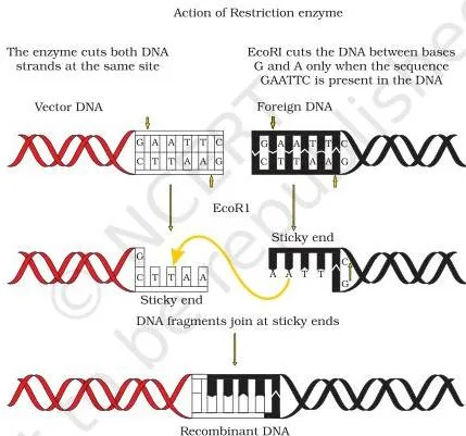
EcoRI recognises the GAATTC palindrome, cuts each strand between the same two bases, a little away from the centre of the palindrome, and leaves single-stranded overhangs — sticky ends (p. 166). Two molecules cut by the same enzyme carry complementary overhangs, which hydrogen-bond with each other, and DNA ligase seals the sugar-phosphate backbone.
The order an answer should reproduce is recognition \( \rightarrow \) cutting \( \rightarrow \) sticky ends \( \rightarrow \) joining.
Figure 9.2: The Recombinant DNA Technology Flow
This flowchart is the whole chapter in one picture — the route from cut DNA to multiplying host.
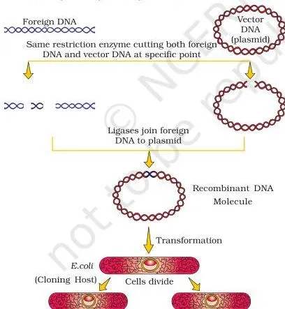
Vector DNA and source DNA are cut with the same restriction enzyme, so their sticky ends match; the gene of interest is ligated into the cut vector; the recombinant DNA enters a host and multiplies there (p. 167). Unless both are cut with the same enzyme, the recombinant molecule cannot form.
The five moves to remember: cut \( \rightarrow \) join \( \rightarrow \) ferry \( \rightarrow \) express \( \rightarrow \) purify.
Figure 9.3: Reading an Agarose Gel
The gel photograph answers a question the book puts directly to you: where was the sample loaded, and why?
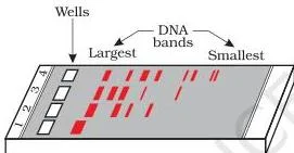
Lane 1 holds undigested DNA — one main band. Lanes 2 to 4 hold the same DNA digested, so you see several bands separated by size (p. 168).
The sample enters at the negative end, near the wells, because DNA carries a negative charge and moves towards the anode; the agarose sieves the fragments, so smaller ones travel farther.
None of this is visible in ordinary light — the bands glow orange only after ethidium bromide staining and UV exposure.
Figure 9.4: Reading the pBR322 Vector Map
A plasmid map is a diagram of addresses; this one labels everything a student must be able to locate on pBR322.
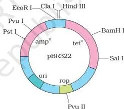
Read the circle as a street map. \(ori\) marks where replication starts and controls copy number; \(amp^R\) and \(tet^R\) are the selectable markers; each listed restriction site is a possible insertion address (p. 169). \(rop\) codes for proteins involved in the replication of the plasmid.
The link worth making in an answer: insert foreign DNA at the BamH I site inside \(tet^R\), and the plasmid’s tetracycline resistance disappears — the basis of the double-plate selection.
Figure 9.5: DNA Spooling After Precipitation
This is the visible proof that the isolation step worked — purified DNA you can see and collect.
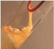
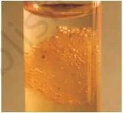
After ribonuclease has removed RNA, protease has removed proteins, and chilled ethanol has been added, the purified DNA precipitates out of suspension as fine threads (p. 171). These threads are wound onto a glass rod — spooling. Any answer about isolation of genetic material can end with this image: pure DNA is a visible, spoolable mass.
Figure 9.6: The Three Steps of a PCR Cycle
The PCR figure compresses one cycle into three steps — the minimum a student must be able to name in order.
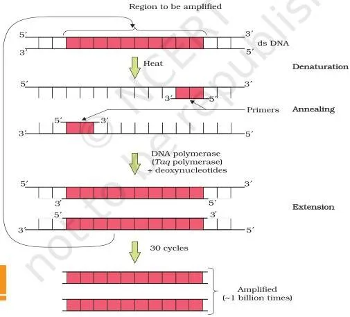
Each cycle doubles the target segment, and repeated cycling amplifies it to roughly a billion copies (p. 172). Two sets of primers flank the region; DNA polymerase extends them using the nucleotides provided and the genomic DNA as template. Because every cycle opens with a high-temperature denaturation, the polymerase must be thermostable — the book’s polymerase comes from Thermus aquaticus.
Figure 9.7: Stirred-Tank Bioreactors
The bioreactor diagram answers the production question: how do you grow culture on an industrial scale with everything under control?
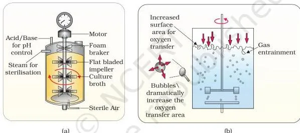
Bioreactors scale production from small laboratory flasks to 100–1000 litres (p. 173–174). The labels worth naming in an answer: agitator system, oxygen delivery system, foam control system, temperature control, pH control, and sampling ports for withdrawing small culture volumes periodically.
Type (b) adds the sparger that bubbles sterile air through the culture — the difference between the two drawings. The stirrer keeps mixing even and oxygen available throughout, which is why stirred-tank bioreactors outperform shake flasks at large scale, as exercise question 6 asks.
Key Terms and Definitions at a Glance
These are the chapter’s essential terms in exam-ready language. Each definition is short enough to reuse in a two-mark answer.
| Term | Plain definition | NCERT page |
|---|---|---|
| Biotechnology (EFB) | Combining the natural sciences with organisms, cells, parts of cells or molecular analogues to make products and services | p. 163 |
| Genetic engineering | Changing the chemistry of DNA and RNA, introducing it into a host, and thereby changing the host’s phenotype | p. 163 |
| Bioprocess engineering | Sterile, large-scale culture of only the desired microbe or eukaryotic cell to manufacture products | p. 163–164 |
| Recombinant DNA | A new circular, autonomously replicating DNA formed in vitro by joining DNA from different sources | p. 164 |
| Vector | A DNA vehicle, such as a plasmid, that carries an alien piece of DNA into a host organism | p. 164 |
| Restriction endonuclease | An enzyme that cuts DNA at its specific recognition sequence | p. 165 |
| Recognition sequence | A specific DNA sequence, usually six base pairs, that a restriction enzyme recognises before cutting | p. 165 |
| Palindrome (in DNA) | A base-pair sequence that reads the same on both strands in the \(5′ \rightarrow 3’\) direction | p. 166–167 |
| Sticky ends | Single-stranded overhangs left after a cut, which hydrogen-bond with complementary ends | p. 167 |
| Gel electrophoresis | A technique that separates DNA fragments by size in an agarose gel under an electric field | p. 168 |
| Origin of replication | The sequence where replication starts and which controls copy number of linked DNA | p. 169 |
| Selectable marker | A gene (usually antibiotic resistance) used to identify transformants and eliminate non-transformants | p. 169 |
| Insertional inactivation | Loss of a gene’s function because foreign DNA is inserted into its coding sequence | p. 170 |
| Competent host | A cell treated so DNA can enter it, e.g. with calcium and a heat shock | p. 170 |
| PCR | In vitro amplification of a DNA segment using two primers and a thermostable DNA polymerase | p. 172 |
| Recombinant protein | A protein-encoding gene expressed in a heterologous host | p. 173 |
| Downstream processing | Separation, purification, formulation, clinical trials and quality control after the biosynthetic stage | p. 174 |
Common Mistakes Students Make in This Chapter
These six errors show up regularly in revision and in answers. The table names each one, explains why it happens, and gives the correction.
| The mistake | Why it happens | Correct rule and how to check |
|---|---|---|
| Mixing up exonucleases and endonucleases | Both are nucleases, and the names sound alike | Exonucleases remove nucleotides from the ends of DNA; endonucleases cut at specific positions inside it (p. 166). Ask: where does the cut happen — at the free end, or in the middle of the strand? |
| Assuming an alien DNA piece multiplies in the host on its own | It feels as if DNA should simply copy itself | It must be linked to an origin of replication or become part of a chromosome that can replicate (p. 164). Ask: what lets this piece replicate — ori, or nothing? |
| Reading a DNA palindrome like a word palindrome | A palindrome usually means ‘same forwards and backwards’ on one line | In DNA the same \(5′ \rightarrow 3’\) sequence must appear on both strands, as GAATTC on one strand and CTTAAG on the other (p. 166–167). Write both strands and read each from the 5′ end. |
| Reversing the blue-white result | It is easy to think white means ‘no plasmid’ | No insert → \(\beta\)-galactosidase is active → blue colony. Insert → gene inactivated → colourless colony. White colonies are the recombinants (p. 170). Ask: which colonies contain foreign DNA — the colourless ones. |
| Thinking PCR produces the protein | PCR is associated with getting something useful from a gene | PCR amplifies DNA copies using two primers and a thermostable DNA polymerase; expression into protein happens separately in the host (p. 172). Check: PCR output is DNA, not protein. |
| Getting the gel loading side wrong | The wells sit at the top of the diagram and the anode is easy to confuse | Load at the negative end: DNA is negatively charged and moves towards the anode, and smaller fragments travel farther (p. 168). Check: the wells are at the end the DNA moves away from. |
What the Chapter’s Exercises Test: A Question Map
This chapter has no numerical problems — all twelve exercises are descriptive, reasoning or teacher-consultation tasks. The table maps each question to the concept it checks, so you can revise weaknesses instead of rereading everything. Textbook contents and the examinable syllabus are not always identical — check the current official CBSE syllabus before deciding what to prepare.
| Question | What it tests | What a good answer includes |
|---|---|---|
| Q1 | Recombinant proteins in medical practice | A research task: list 10 therapeutic recombinant proteins and say where each is used (internet sources allowed) |
| Q2 | Restriction enzyme action | A labelled chart showing the enzyme, its substrate DNA, the cut site and the cut product |
| Q3 | Molecular size: enzyme vs DNA | The reasoning that a gene sits on a long DNA molecule while an enzyme is a single protein; purified DNA is even visible as threads |
| Q4 | Molar concentration of human DNA in a cell | A teacher-consultation calculation — the chapter gives no shortcut for it |
| Q5 | Restriction endonucleases in eukaryotes | The justification that these enzymes are bacterial defences against bacteriophages, hence absent from eukaryotic cells |
| Q6 | Stirred-tank bioreactors vs shake flasks | The bioreactor features: optimum growth conditions, larger biomass, even mixing, and sampling ports for periodic withdrawal |
| Q7 | Palindromic DNA sequences | Building sequences by base-pair rules so the \(5′ \rightarrow 3’\) reading is identical on both strands |
| Q8 | Recombinant DNA and meiosis | Recall that crossing over during prophase I of meiosis produces recombinant DNA |
| Q9 | Reporter enzyme to monitor transformation | The insertional-inactivation logic: a reporter such as \(\beta\)-galactosidase changes colour only where foreign DNA has inserted and disrupted it |
| Q10 | ori, bioreactors, downstream processing | Short descriptive definitions of the three terms |
| Q11 | PCR, restriction enzymes and DNA, chitinase | Brief explanations of what each is and its role in recombinant DNA technology |
| Q12 | Plasmid vs chromosomal DNA, RNA vs DNA, exonuclease vs endonuclease | A comparison of each pair, discussed with your teacher |
One-Page Recap of Chapter 9
The whole chapter, compressed into what you should be able to recite in under a minute.
Two pillars carry modern biotechnology:
- Genetic engineering — change the chemistry of DNA and RNA, introduce them into a host, change the host’s phenotype.
- Bioprocess engineering — sterile conditions, large scale, only the desired microbe or eukaryotic cell.
Recombinant DNA technology in five moves — mnemonic: cut, join, ferry, express, purify:
- Cut — restriction endonucleases isolate the gene of interest and open the vector.
- Join — DNA ligase seals the gene into the vector.
- Ferry — a plasmid or viral vector carries the recombinant DNA into the host.
- Express — the foreign gene is expressed in the host to produce the protein.
- Purify — purify and formulate the product for marketing, using bioreactors for large-scale production.
The tool kit from Section 9.2: restriction enzymes, polymerase enzymes, ligases, vectors and the host organism (p. 165).
Related NCERT Resources
This listing is maintained for the 2026-27 academic session using the NCERT textbook information available to us. NCERT remains the authority for confirming the latest edition.
Move around the book and the site from here:
- The Class 12 Biology notes on this site cover the rest of the book, chapter by chapter.
- Biotechnology and Its Applications is Chapter 10, where the principles of this chapter are put to practical use, as the Unit IX opening states.
- For the microbes behind many biotechnological products, see the Microbes in Human Welfare notes.
- The Class 12 hub and the main CBSE notes index help you jump between subjects.
Sources and Data Verification
- This page describes the NCERT Class 12 Biology textbook, Chapter 9, ‘Biotechnology : Principles and Processes’, official edition on ncert.nic.in.
- Chapter page numbers and figure captions are taken from that edition; the chapter file is lebo109.pdf on the NCERT portal.
- The page is maintained for the current academic session using the NCERT information available to us.
- NCERT settles textbooks, editions and PDFs; CBSE settles curriculum, syllabus and examinations.
You can verify the current edition of any textbook on the NCERT textbook portal.
Frequently Asked Questions
Why are restriction enzymes called molecular scissors?
Because they cut DNA at precise, predictable points. A restriction endonuclease inspects the DNA, finds its own recognition sequence, and cuts both strands at specific positions in the sugar-phosphate backbones — the same way scissors cut at a chosen line (p. 164–166).
The name appears in the chapter when these enzymes are introduced as the ‘molecular scissors’ that made cutting DNA at specific locations possible.
What is the difference between an exonuclease and an endonuclease?
An exonuclease removes nucleotides from the ends of DNA, working inwards from the free ends. An endonuclease cuts at specific positions within the DNA. A restriction endonuclease is an endonuclease that cuts only at its own recognition sequence (p. 166).
Why does PCR need a thermostable DNA polymerase?
Because every PCR cycle opens with a high-temperature step that denatures the double-stranded DNA, and a normal enzyme would be destroyed at that temperature. The thermostable polymerase, isolated from the bacterium Thermus aquaticus, stays active through the denaturation so extension can continue cycle after cycle (p. 172).
How do blue and white colonies help identify recombinant plasmids?
The foreign DNA is inserted into the coding sequence of the \(\beta\)-galactosidase gene. With an insert, the gene is inactivated and no enzyme is made, so a chromogenic substrate stays unchanged and the colony is colourless (white). Without an insert, the enzyme is made and converts the substrate to a coloured product, so the colony turns blue.
The colourless colonies are the recombinants (p. 170).
Do eukaryotic cells have restriction endonucleases?
No — and that is exactly what exercise question 5 asks you to justify. Restriction endonucleases were discovered in bacteria, where they restrict the growth of bacteriophages; more than 900 of them have been isolated from over 230 strains of bacteria (p. 165). The chapter’s examples all come from prokaryotes, which is the reasoning the answer needs.
Where can I download the official NCERT Class 12 Biology Chapter 9 PDF?
From the download block at the top of this page: the NCERT Class 12 Biology Chapter 9 Biotechnology : Principles and Processes PDF is hosted on ncert.nic.in, so it is always the official current edition.
Reference: NCERT Class 12 Biology textbook, chapter 9, official edition on ncert.nic.in.
Explore Class 12 Biology Books
Class 12 Biology on LearnCBSE:
Related chapters: