
This is the molecular basis of inheritance class 12 chapter you are looking for: NCERT Biology Chapter 5, printed on pages 78 to 108 of the Class 12 textbook. The official chapter PDF is linked directly below, and the rest of this page tells you exactly what the chapter contains — every section, key idea, figure and exercise.
Download the Molecular Basis of Inheritance Class 12 PDF
The official NCERT Class 12 Biology Chapter 5 Molecular Basis of Inheritance PDF is hosted on ncert.nic.in, and opening it gives you the complete chapter exactly as printed — the section list on page 78, the full text, every figure, Table 5.1, and the exercises that close the chapter on page 108.
| What the chapter holds | Count | Where it is used |
|---|---|---|
| Printed pages | 31 | |
| Sections in the chapter | 24 | |
| Figures with NCERT captions | 19 | |
| Tables | 1 | |
| Exercise questions | 14 | answered in our NCERT Solutions |
| Official NCERT PDF | Download the chapter PDF | the chapter exactly as NCERT publishes it |
Chapter 5 at a Glance
The table below lists what this chapter holds — its sections, figures, worked examples and exercise questions — so you can see the size of the task before you start.
Three structural facts worth knowing before you open the file: the chapter is organised into ten numbered sections, from 5.1 The DNA to 5.10 DNA Fingerprinting; it opens with the section list on page 78 and closes with SUMMARY on pages 107–108, followed by EXERCISES; and its only data table, Table 5.1, the genetic code, sits on page 96.
What This Chapter Covers
The whole chapter tells one continuous story: what the genetic material is, how it is copied, how it is read into proteins, how that reading is regulated, and how the knowledge was applied to whole genomes and to individuals.
- 5.1 The DNA (pp. 79–83): the polynucleotide chain, the Watson–Crick double helix, and packaging into nucleosomes and chromatin.
- 5.2 The Search for Genetic Material (pp. 83–87): Griffith, Avery–MacLeod–McCarty, Hershey–Chase, and the properties a genetic material must have.
- 5.3 RNA World (pp. 87–88): why RNA is thought to have been the first genetic material.
- 5.4 Replication (pp. 87–91): the semiconservative scheme, the Meselson–Stahl proof, and the enzyme machinery.
- 5.5 Transcription (pp. 90–94): the transcription unit, template and coding strands, and RNA processing in eukaryotes.
- 5.6 Genetic Code (pp. 94–98): codons, the code table, mutations and tRNA.
- 5.7 Translation (pp. 97–99): protein synthesis on the ribosome.
- 5.8 Regulation of Gene Expression (pp. 98–101): the levels of regulation and the lac operon.
- 5.9 Human Genome Project (pp. 101–105): goals, methods and salient features.
- 5.10 DNA Fingerprinting (pp. 104–107): polymorphism, VNTRs and the technique.
Learn the sections in this order. Each one builds an idea the next section needs.
Key Concepts in Molecular Basis of Inheritance
The subsections below follow the book’s own sequence, with NCERT page numbers so you can follow along in your copy. Read them in order — structure first, then the evidence, then each process.
The DNA Double Helix: Structure and Packaging
DNA is a very long polymer of nucleotides, and its structure explains both how information is stored and how it is copied. Four ideas matter most here: the chemistry of a nucleotide, the 5′ and 3′ ends, the base-pairing rules, and how 2.2 metres of DNA fits into a nucleus.
A nucleotide has three parts — a nitrogenous base, a pentose sugar and a phosphate group (NCERT, p. 79). The bases are of two kinds: purines (adenine, A, and guanine, G) and pyrimidines (cytosine, C, thymine, T, and uracil, U). DNA uses deoxyribose and thymine; RNA uses ribose and uracil.
The linkages have fixed names, and examiners expect them. A base linked to the 1′ carbon of the sugar through an N-glycosidic linkage forms a nucleoside. A phosphate on the 5′ carbon, through a phosphoester linkage, makes a nucleotide. Successive nucleotides join by 3′–5′ phosphodiester linkages (NCERT, p. 79).
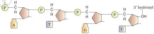
Figure 5.1 shows the product: a chain with a sugar-phosphate backbone and the bases projecting out of it. One end carries a free 5′ phosphate; the other carries a free 3′ OH. Those two ends are the key to every direction-based question in the chapter.
In 1953 Watson and Crick proposed the double helix, building on X-ray diffraction data from Maurice Wilkins and Rosalind Franklin and on Erwin Chargaff’s observation that, in double-stranded DNA, A = T and G = C (NCERT, p. 80). This Chargaff rule means the two strands are complementary — know one strand and you can write the other.
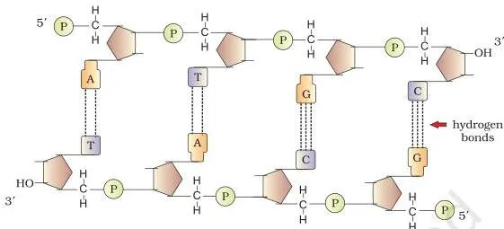
Figure 5.2 shows the pairing. The strands are antiparallel: if one runs 5′ to 3′, the other runs 3′ to 5′. A pairs with T through two hydrogen bonds, G with C through three (NCERT, p. 82). Because a purine always faces a pyrimidine, the distance between the two backbones stays almost uniform.
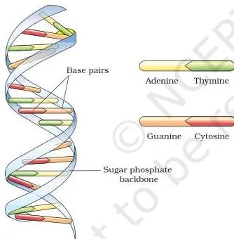
Figure 5.3 gives the numbers you will be asked for. The helix coils right-handed with a pitch of 3.4 nm, roughly 10 base pairs per turn, and therefore about 0.34 nm per base pair. The stacked base pairs add stability to the structure (NCERT, p. 82).
The structural message of the model became the central dogma: genetic information flows \( \text{DNA} \rightarrow \text{RNA} \rightarrow \text{protein} \). Some viruses reverse the flow, going from RNA to DNA (NCERT, p. 82).
Worked example — base percentages with fresh numbers. A double-stranded DNA sample contains 22% guanine. Find the percentage of every base.
- Step 1: Chargaff’s rule gives G = C, so cytosine is also 22%.
- Step 2: G + C = 44%, so A + T must be the remaining 56%.
- Step 3: A = T, so each is half of 56%.
\[ G = C = 22\%;\quad A + T = 100\% – 44\% = 56\%;\quad A = T = 28\% \]
Final answer: A = 28%, T = 28%, G = 22%, C = 22%. Check by adding: 28 + 28 + 22 + 22 = 100%.
Packaging solves the size problem. A typical mammalian cell carries \( 6.6 \times 10^9 \) bp; multiplying by 0.34 nm per base pair gives about 2.2 metres of DNA (NCERT, p. 82):
\[ 6.6 \times 10^9\ \text{bp} \times 0.34 \times 10^{-9}\ \text{m/bp} \approx 2.2\ \text{m} \]
Prokaryotes keep their DNA in a nucleoid, held in large loops by proteins. Eukaryotes wrap DNA around histones — basic proteins rich in lysine and arginine. Eight histones make a histone octamer, and DNA wound around it forms a nucleosome of about 200 bp (NCERT, p. 83).
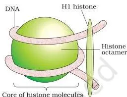
Figure 5.4a shows the nucleosome: negatively charged DNA wrapped around the positively charged octamer. Nucleosomes are the repeating unit of chromatin, the stained thread-like material of the nucleus.
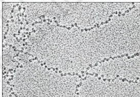
The electron micrograph in Figure 5.4b explains the common name for chromatin: a beads-on-string arrangement of nucleosomes. Loosely packed, lightly staining euchromatin is transcriptionally active; dense, darkly staining heterochromatin is inactive (NCERT, p. 83). Further coiling condenses the fibre into chromosomes at metaphase.
Self-check from the book (p. 82): if E. coli DNA measures 1.36 mm, how many base pairs does it contain?
\[ \frac{1.36 \times 10^{-3}\ \text{m}}{0.34 \times 10^{-9}\ \text{m/bp}} = 4 \times 10^6\ \text{bp} \]
The chapter gives E. coli about \( 4.6 \times 10^6 \) bp, so the calculation lands in the right range — a good check that you have handled units correctly.
How DNA Was Proven to Be the Genetic Material
Before DNA was accepted, three experiments closed the case, and each answered a different question: is there a transforming substance, what is its chemical nature, and which molecule actually enters the cell?
Griffith (1928) worked with Streptococcus pneumoniae (NCERT, p. 84). The S strain has a polysaccharide coat, forms smooth colonies and kills mice; the R strain lacks the coat and does not kill. Heat-killed S bacteria are harmless — but a mixture of heat-killed S and live R kills the mice, and live S bacteria are recovered from their bodies.
Griffith’s conclusion was that some transforming principle had passed from the dead S bacteria into the live R bacteria. The biochemical nature of that principle was still unknown.
Avery, MacLeod and McCarty (1933–44) purified the components of heat-killed S cells and tested each one (NCERT, p. 85). Only DNA transformed R into S. Protein-digesting enzymes (proteases) and RNA-digesting enzymes (RNases) did not stop transformation; DNase did. The transforming substance was DNA.
Hershey and Chase (1952) proved it beyond doubt using bacteriophages — viruses that infect bacteria (NCERT, pp. 85–86). They grew phages with radioactive phosphorus, which labels DNA, and separately with radioactive sulfur, which labels protein.
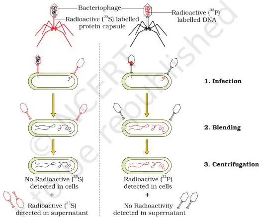
Work through Figure 5.5 step by step. The radioactive phages attach to E. coli and inject their genetic material. A blender strips the viral coats off the bacterial surface, and a centrifuge separates bacteria from viruses.
Only the bacteria infected by phosphorus-labelled phages became radioactive — the DNA had entered them. Sulfur-labelled protein stayed behind in the coats. DNA, not protein, is what enters the cell.
Why DNA and not RNA? A genetic material must replicate, be chemically and structurally stable, allow slow change for evolution, and express itself as Mendelian characters (NCERT, pp. 86–87). RNA fails the stability test: the 2′-OH group on every nucleotide makes it labile and easily degraded.
DNA’s thymine, in place of uracil, adds further stability, and its double strand with repair mechanisms protects it more. RNA mutates faster, which is why RNA viruses evolve rapidly.
The RNA World: Why RNA Came First
If DNA is the better genetic material, why does RNA exist at all? The chapter’s answer (NCERT, pp. 87–88): essential life processes — metabolism, translation, splicing — show evidence of having evolved around RNA, and RNA can act as a catalyst.
RNA was the first genetic material. It did both jobs, storing information and catalysing reactions, but a catalyst is reactive and therefore unstable. DNA evolved from RNA through chemical modifications that made it more stable, and being double-stranded, with a complementary strand and repair, protected it further. The book returns to this argument in the chapter on chemical evolution.
DNA Replication: Semiconservative Scheme and Its Proof
Replication copies the whole genome once per cell cycle. The double helix itself suggested the copying mechanism.
It has not escaped our notice that the specific pairing we have postulated immediately suggests a possible copying mechanism for the genetic material.
That remark by Watson and Crick, quoted in the textbook on page 88, became the semiconservative scheme: the two parental strands separate, each acts as a template, and every daughter DNA molecule contains one parental and one newly synthesised strand.
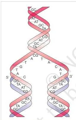
Figure 5.6 is the model in one picture: the parental helix opens, each strand builds its complement, and two identical double helices result.
Meselson and Stahl (1958) proved the scheme in E. coli (NCERT, pp. 88–89). They grew bacteria in medium containing \( ^{15}\text{NH}_4\text{Cl} \), so the DNA became heavy with \( ^{15}\text{N} \), then shifted the cells to normal \( ^{14}\text{N} \) medium and centrifuged DNA samples in a cesium chloride density gradient, where heavier molecules sediment lower.
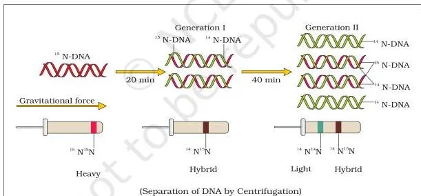
Read Figure 5.7 band by band. Generation zero: all DNA is heavy, so one band sits low in the gradient. One generation after the transfer (20 minutes — E. coli divides every 20 minutes), all DNA is hybrid, one band at intermediate density. This single result eliminates the conservative model, which would have produced heavy and light bands.
After two generations (40 minutes), hybrid and light DNA appear in equal amounts: two bands, because half the molecules still carry one heavy strand and half are entirely new.
The book’s self-check (p. 89): after 80 minutes — four generations — the proportion of hybrid DNA falls to \[ \frac{2}{16} = \frac{1}{8}\ \text{hybrid, and}\ \frac{7}{8}\ \text{light} \]
because each doubling halves the share of molecules that still contain a heavy strand.
The machinery (NCERT, pp. 90–91): the main enzyme is DNA-dependent DNA polymerase, which polymerises only in the 5′ to 3′ direction. Replication runs in a small opened region called the replication fork. On the 3′–5′ template, synthesis is continuous; on the 5′–3′ template it is discontinuous, the fragments later joined by DNA ligase.
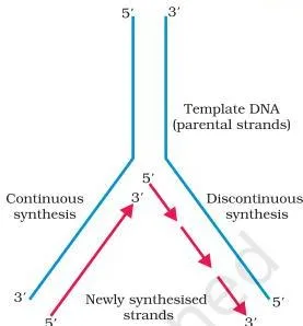
Figure 5.8 shows that asymmetry: a continuous arrow on one side, short fragments waiting for ligase on the other.
DNA polymerase cannot initiate a chain on its own, so replication begins at a definite origin of replication — the reason a cloning vector must provide one. Deoxynucleoside triphosphates supply the energy, and E. coli maintains an average pace of about 2000 base pairs per second (NCERT, p. 90).
Transcription: Reading One Strand into RNA
Transcription copies genetic information from one strand of DNA into RNA. Unlike replication, it copies only one strand of a limited segment of DNA.
Why not both strands? First, the two strands are complementary, not identical, so both would code for two different proteins. Second, the two RNA products would pair into a double-stranded RNA that could not be translated (NCERT, p. 90).
A transcription unit is defined by three regions: a promoter, a structural gene and a terminator (NCERT, pp. 90–91). The strand with 3′–5′ polarity is the template strand, which is actually transcribed.
The other strand, 5′–3′, is the coding strand: its sequence matches the RNA, with U in place of T. All reference points in the unit are named from the coding strand — the promoter lies upstream, towards its 5′ end, and the terminator downstream (NCERT, p. 92).
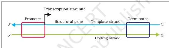
Figure 5.9 is the map to reproduce from memory: promoter and terminator flanking the structural gene, with both strands labelled by polarity.
Worked example — template strand and mRNA, fresh numbers. Use this coding strand: \( \text{5′-AAG TCC GTA CGA-3′} \). Write the template strand and the mRNA.
Step 1: The template strand is complementary and antiparallel, so it is written 3′ to 5′:
\[ \text{Template: 3′-TTC AGG CAT GCT-5′} \]
Step 2: The mRNA has the coding strand’s sequence with U in place of T:
\[ \text{mRNA: 5′-AAG UCC GUA CGA-3′} \]
Final answer: template 3′-TTC AGG CAT GCT-5′; mRNA 5′-AAG UCC GUA CGA-3′. If you are given the template instead, build the mRNA by complementing it.
Genes are not always clean segments of DNA. A cistron is a DNA segment coding for a polypeptide; structural genes are monocistronic mostly in eukaryotes and polycistronic mostly in bacteria. In eukaryotes genes are split: exons are the sequences that appear in mature RNA, and introns are removed (NCERT, p. 92).
In bacteria, one RNA polymerase makes all three RNA types — mRNA, tRNA and rRNA. The enzyme only elongates; it needs a sigma factor to initiate and a rho factor to terminate (NCERT, pp. 92–93). Because bacteria have no nuclear membrane, translation can begin before transcription is finished — the two are said to be coupled.
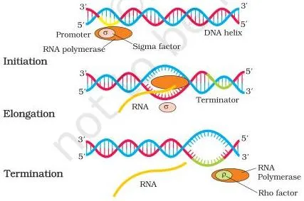
Figure 5.10 labels the helpers: sigma loads the polymerase at the promoter, rho ends transcription at the terminator.
Eukaryotes add two complications (NCERT, p. 94). Three nuclear RNA polymerases divide the work: I makes rRNAs, II makes the mRNA precursor hnRNA, and III makes tRNA, 5S rRNA and snRNAs.
The primary transcript also needs processing — capping at the 5′ end, tailing with 200–300 adenylate residues at the 3′ end, and splicing to remove introns and join exons.
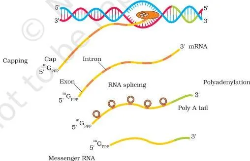
Figure 5.11 is the processing pipeline in the nucleus: cap, splice, tail, then export the finished mRNA.
Replication versus transcription is a comparison examiners reuse, and the chapter gives you every fact for it. The table summarises the differences.
| Feature | Replication | Transcription |
|---|---|---|
| Template | Both strands of the whole DNA molecule | Only the 3′–5′ template strand of one segment |
| Enzyme | DNA-dependent DNA polymerase | DNA-dependent RNA polymerase |
| Base pairing | A–T, G–C | A–U, G–C |
| Product | Two identical double-stranded DNA molecules | RNA (mRNA, tRNA or rRNA) |
| Direction | Polymerisation only 5′ to 3′ | Polymerisation only 5′ to 3′ |
| Location (eukaryotes) | Nucleus, at S-phase of the cell cycle | Nucleus; mRNA then moves to the cytoplasm |
Both polymerases share the same rule: extension happens only in the 5′ to 3′ direction (NCERT, pp. 90–91).
The Genetic Code and Translation: From mRNA to Protein
The genetic code is the dictionary that translates a nucleotide language into an amino acid language, and tRNA is the adapter molecule that reads it.
Four bases cannot code for 20 amino acids one at a time. George Gamow argued that the code must be a combination of bases, and three nucleotides per codon works: \( 4^3 = 64 \) possible codons (NCERT, p. 95). The code was deciphered using Khorana’s defined RNA synthesis, Nirenberg’s cell-free protein-synthesising system and Ochoa’s enzyme; the finished table is Table 5.1 on page 96.
The code has six salient features (NCERT, pp. 96–97). It is a triplet code: 61 codons specify amino acids and UAA, UAG, UGA are stop codons. It is degenerate — some amino acids have more than one codon. It is read contiguously with no punctuation, and it is nearly universal, from bacteria to humans.
Finally, AUG has dual roles: initiator codon and the codon for methionine.
Memory hooks: the stop codons read like farewells — “You Are Away (UAA), You Are Gone (UAG), U Go Away (UGA)”. For the bases, pyrimidines are just CUT — cytosine, uracil, thymine — and purines are the pair left over: A and G.
Mutations tie the code to disease (NCERT, p. 97). A point mutation in the beta-globin gene changes glutamate to valine, causing sickle cell anaemia. Inserting or deleting one or two bases shifts the reading frame from that point on; inserting or deleting three bases leaves the frame intact. The book’s word example — RAM HAS RED CAP — demonstrates this on page 97.
tRNA is the adapter Crick predicted (NCERT, pp. 97–98). One end carries an anticodon loop with bases complementary to the codon; the other is an amino acid acceptor end. Each amino acid has its own tRNA, there is a special initiator tRNA for AUG, and there are no tRNAs for stop codons.
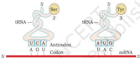
Figure 5.12 shows the cloverleaf: the anticodon loop sits opposite the acceptor end. In three dimensions the molecule folds into a compact inverted L.
Translation assembles the polypeptide on the ribosome (NCERT, pp. 98–99). First, charging of tRNA — aminoacylation — links each amino acid to its tRNA, using ATP. The ribosome, made of structural RNAs and about 80 proteins, then moves along the mRNA, matching each codon with its anticodon. In bacteria the peptide bond is actually catalysed by an RNA, the 23S rRNA — a ribozyme.
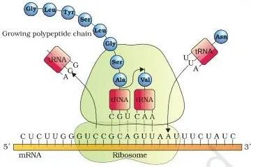
Figure 5.13 shows the ribosome in action: mRNA threaded through the two subunits, tRNAs arriving one by one, and the polypeptide growing out as the ribosome advances. A release factor meeting a stop codon ends the process.
The mRNA section between the start and stop codons is the translational unit, flanked by untranslated regions (UTRs) at both ends, which the chapter says are needed for efficient translation (NCERT, p. 99).
Gene Regulation and the Lac Operon
Cells do not make every protein all the time. The lac operon is the classic example of a genetic switch controlled by the substrate itself.
In eukaryotes, gene expression can be regulated at four levels: transcriptional, processing, mRNA transport, and translational (NCERT, p. 98). In prokaryotes the dominant control is the rate of transcription initiation, set by activator and repressor proteins acting near the promoter. The operator is the sequence where the repressor binds, and each operon has its own operator and repressor (NCERT, pp. 99–100).
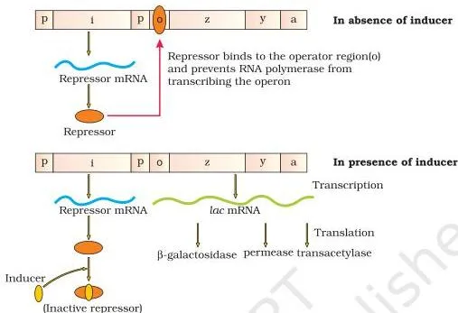
The lac operon (NCERT, pp. 100–101) is a polycistronic arrangement: the i gene (i for inhibitor) codes for the repressor and is always expressed; z codes for beta-galactosidase, y for permease, and a for transacetylase. Lactose is the substrate of these enzymes and the inducer of the operon.

Figure 5.14 draws the same operon twice. In the upper state the repressor sits on the operator and RNA polymerase is blocked. In the lower state lactose has inactivated the repressor, the operator is free, and transcription of z, y and a proceeds. This is negative regulation — the default is off.
Why the switch turns itself off — the reasoning question this chapter keeps returning to:
- The i gene makes the repressor all the time.
- The repressor binds the operator, so RNA polymerase cannot transcribe the structural genes.
- When lactose enters the cell, it or allolactose binds the repressor and inactivates it.
- The operator is free, so RNA polymerase transcribes z, y and a, and lactose is metabolised.
- Once the lactose is used up, no inducer remains.
- The repressor becomes active again, binds the operator, and the operon switches off.
Glucose and galactose cannot act as inducers (NCERT, p. 101). The whole loop is regulation of enzyme synthesis by its own substrate.
The Human Genome Project and DNA Fingerprinting
Once DNA sequence is understood as the source of genetic information, two applied ideas follow: reading the whole human genome, and comparing genomes between individuals.
HGP is a mega project by any measure (NCERT, p. 102). The genome holds about \( 3 \times 10^9 \) bp; at an early estimated cost of about US $3 per base pair that is roughly 9 billion dollars; and the typed sequence would fill some 3300 books of 1000 pages. Launched in 1990 and completed in 2003, the project drove the growth of bioinformatics.
Its six goals (NCERT, p. 102): identify all genes; determine the sequences of the three billion base pairs; store the information in databases; improve analysis tools; transfer technology to industry; and address ethical, legal and social issues (ELSI).
Two methods were used (NCERT, p. 103). One looked for expressed genes using expressed sequence tags (ESTs). The other sequenced the whole genome, coding and non-coding, and assigned functions later — sequence annotation.
Fragments were cloned in BAC and YAC vectors, sequenced by the Sanger method, and assembled by overlapping regions with computer programs. Chromosome 1, the last of the 24 to be finished, was completed in May 2006.
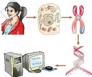
Figure 5.15 condenses the workflow into one picture: long DNA broken into fragments, cloned, sequenced, and assembled computationally.
Salient features of the genome to quote with precision (NCERT, p. 104): 3164.7 million bp; an average gene of 3000 bases, though the largest, dystrophin, is 2.4 million bases; about 30,000 genes; and 99.9 per cent of bases identical in all humans.
Also worth remembering: functions are unknown for over half the genes; less than 2 per cent codes for proteins; chromosome 1 carries the most genes (2968) and the Y the fewest (231); and there are about 1.4 million SNP locations (NCERT, p. 104). The book uses “approximately \( 3 \times 10^9 \) bp” for the project and “3164.7 million bp” for the completed genome — use each figure where the book does.
DNA fingerprinting is the fast way to compare individuals without sequencing everything (NCERT, pp. 105–107). It relies on polymorphism — an inheritable variant at a locus occurring with a frequency greater than 0.01.
Regions of repetitive DNA separate from bulk DNA in density gradients as satellite DNA peaks and show high polymorphism. Alec Jeffreys used a probe for VNTRs — variable number of tandem repeats.
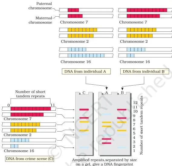
In Figure 5.16, each band is a VNTR fragment of a particular size, coloured to trace its chromosome of origin. The banding pattern from the crime scene matches individual B, not A.
The six steps in order (NCERT, p. 106): isolate DNA, digest it with restriction endonucleases, separate the fragments by electrophoresis, blot them onto a membrane, hybridise with a labelled VNTR probe, and detect the result by autoradiography.
Because the bands are inherited, the technique serves forensics and paternity testing; with PCR, studied in Chapter 9, even the DNA from a single cell is enough (NCERT, p. 106).
How to Read the Chapter’s Key Diagrams
The diagrams above are the ones the chapter’s reasoning depends on. Learn to read these five rather than memorise them.
- Figure 5.2 — strand polarity (p. 82): follow the arrows. The strands run in opposite directions, and every purine faces a pyrimidine, which is why the backbone spacing stays even.
- Figure 5.7 — density bands (p. 89): in a CsCl gradient, position means density — the heavier the DNA, the lower the band. One hybrid band after one generation, then hybrid and light in equal amounts after two, is the complete proof of semiconservative replication.
- Figure 5.10 — transcription machinery (p. 93): the promoter and terminator are the start and stop signs flanking the structural gene; sigma loads the polymerase, rho pulls it off.
- Figure 5.14 — the operon switch (p. 101): read the two states side by side. The only question is whether the repressor occupies the operator or the inducer has removed it.
- Figure 5.16 — the fingerprint (p. 107): each band is a VNTR fragment; the pattern is inherited, and the crime scene lane matches individual B, not A.
Definitions to Remember
Quick look-up glossary: each definition is one line, and the page given is where the term first appears in the chapter.
| Term | Meaning | NCERT page |
|---|---|---|
| Nucleotide | A unit of a nucleic acid: nitrogenous base + pentose sugar + phosphate group | 79 |
| Nucleoside | A nitrogenous base linked to the 1′ carbon of a pentose sugar | 79 |
| Chargaff’s rule | In double-stranded DNA, the ratios A:T and G:C are constant and equal one | 80 |
| Histone | Positively charged basic protein, rich in lysine and arginine | 83 |
| Nucleosome | About 200 bp of DNA wrapped around a histone octamer | 83 |
| Euchromatin | Loosely packed, lightly staining, transcriptionally active chromatin | 83 |
| Heterochromatin | Densely packed, darkly staining, inactive chromatin | 83 |
| Transforming principle | The substance in Griffith’s experiment that converted live R strain bacteria into S strain | 84 |
| Bacteriophage | A virus that infects bacteria | 85 |
| Semiconservative replication | Each daughter DNA holds one parental strand and one newly synthesised strand | 88 |
| Replication fork | The small opening of the DNA helix where replication occurs | 90 |
| DNA ligase | Enzyme that joins the discontinuously synthesised fragments | 90 |
| Origin of replication | The definite region in DNA where replication begins | 90 |
| Template strand | The 3′–5′ strand of a transcription unit, the one transcribed into RNA | 91 |
| Coding strand | The 5′–3′ strand with the same sequence as the RNA, U in place of T | 91 |
| Promoter | DNA sequence upstream of the structural gene where RNA polymerase binds | 92 |
| Terminator | DNA sequence downstream of the structural gene that ends transcription | 92 |
| Exon | Coding sequence that remains in mature RNA | 92 |
| Intron | Intervening sequence removed from RNA during splicing | 92 |
| Splicing | Removal of introns and joining of exons in a defined order | 94 |
| Degenerate code | A code in which an amino acid is specified by more than one codon | 96 |
| Stop codon | UAA, UAG or UGA; codes for no amino acid and ends translation | 96 |
| Anticodon | The tRNA triplet complementary to the mRNA codon | 98 |
| Charging of tRNA | Linking an amino acid to its specific tRNA, using ATP | 98 |
| Ribozyme | An RNA that acts as an enzyme, such as 23S rRNA in bacteria | 98 |
| Operon | A cluster of bacterial genes transcribed together under one promoter | 100 |
| Inducer | Lactose or allolactose, which inactivates the lac repressor | 101 |
| Polymorphism | An inheritable variant at a locus with a frequency greater than 0.01 | 105 |
| Satellite DNA | Small density-gradient peaks of repetitive DNA separated from bulk DNA | 105 |
| VNTR | Variable number of tandem repeats — the probe Jeffreys used in fingerprinting | 106 |
Common Mistakes Students Make in This Chapter
The ten errors below are the characteristic ones for this chapter. Check your answer against the third column before moving on.
| Mistake | Correct rule | How to check your answer |
|---|---|---|
| Writing the complementary strand in the same direction as the given strand | The strands are antiparallel: 5′-ATGC… pairs with 3′-TACG… (NCERT, p. 80) | Write the partner 3′ to 5′ beneath the given strand, then read it back 5′ to 3′ if asked (Exercise 3, p. 109) |
| Confusing template and coding strand when writing the mRNA | mRNA matches the coding strand with U for T; it is complementary to the template strand (NCERT, pp. 90–91) | If the question gives the coding strand 5′ to 3′, simply swap every T for U (Exercise 4) |
| Wrong base-percentage arithmetic | In double-stranded DNA, A = T and G = C; one given percentage fixes its partner, and the remaining pair splits the rest equally | Add all four percentages — they must total 100 (Exercise 2, p. 80) |
| Assuming both strands are transcribed | Only the template strand is copied; copying both would give two different proteins and a non-translatable double-stranded RNA (NCERT, p. 90) | Ask which strand the RNA sequence matches — it should match the coding strand |
| Swapping the Hershey–Chase labels | Phosphorus labels DNA, sulfur labels protein — DNA has phosphorus, protein has sulfur (NCERT, pp. 85–86) | Remember: P is in phosphate (DNA), S is in sulfur (protein) (Exercise 7) |
| Thinking DNA polymerase can start a new chain by itself | It cannot initiate; it only extends 5′ to 3′ and needs an origin of replication to begin (NCERT, pp. 90–91) | Recall why a recombinant vector must carry an origin of replication |
| Mixing up capping, tailing and splicing | Capping modifies the 5′ end, tailing adds adenylates at the 3′ end, splicing removes introns and joins exons (NCERT, p. 94) | Cap at the top, tail at the bottom, splice in the middle |
| Frameshift direction confusion | Inserting or deleting one or two bases shifts every downstream codon; inserting or deleting three keeps the frame (NCERT, p. 97) | Test with the book’s RAM HAS RED CAP example — insert 1, then 2, then 3 letters |
| Lac operon timing error | The operon shuts down because lactose, the inducer, is consumed; the repressor then binds the operator again. Glucose and galactose do not induce (NCERT, pp. 100–101) | Trace the order: inducer gone → repressor active → operator occupied → transcription stopped (Exercise 10) |
| Reversing the sickle-cell amino acid change | Glutamate changes to valine — not valine to glutamate (NCERT, p. 97) | The beta-globin point mutation swaps a single amino acid: Glu → Val |
Exam Notes: How to Study This Chapter
The exercise list at the end of the chapter is the best map of what matters — fourteen questions, each testing a named section. Use the table below to target your weak areas.
| Question | What it tests | Where it comes from |
|---|---|---|
| 1 | Nitrogenous bases versus nucleosides | 5.1.1, p. 79 |
| 2 | Base percentage by Chargaff’s rule | p. 80 |
| 3 | Complementary strand and its direction | p. 80 |
| 4 | Writing the mRNA from the coding strand | 5.5.1, p. 91 |
| 5 | The helix property that suggested semiconservative replication | 5.4, p. 88 |
| 6 | Nucleic acid polymerases by template and product | 5.2.2 and 5.4.2, pp. 86–90 |
| 7 | Hershey–Chase radioactive labels | 5.2.1, p. 86 |
| 8 | Differentiations: repetitive vs satellite DNA, mRNA vs tRNA, template vs coding strand | 5.10, 5.5.3, 5.5.1 |
| 9 | Two essential roles of the ribosome during translation | 5.7, p. 98 |
| 10 | Why the lac operon shuts down after lactose is added | 5.8.1, pp. 100–101 |
| 11 | Functions of promoter, tRNA and exons | 5.5.1, 5.6.2, 5.5.2 |
| 12 | Why the Human Genome Project is a mega project | 5.9, p. 102 |
| 13 | DNA fingerprinting and its applications | 5.10, p. 105 |
| 14 | Brief descriptions: transcription, polymorphism, translation, bioinformatics | 5.5, 5.10, 5.7, 5.9 |
The chapter’s own order is the best revision order: structure, then the evidence for DNA, then replication, transcription, the genetic code, translation, regulation, and finally the genome and fingerprinting. Each step is a prerequisite for the next.
Two italicised self-check prompts inside the text make quick revision tests: the E. coli base-pair calculation on page 82 and the 80-minute Meselson–Stahl question on page 89.
One honest warning: textbook contents and the examinable syllabus are not always identical, so check the current official CBSE syllabus for what is examinable this session.
If you need the condensed version, the Class 12 Biology notes sit alongside this chapter, and the notes on the previous chapter, Principles of Inheritance and Variation, cover the mutations and Mendelian patterns this chapter refers back to.
Chapter 5 in One Page: Quick Revision Summary
One section before the exam? Read this. It condenses the chapter’s own SUMMARY (NCERT, pp. 107–108) into the points you must be able to reproduce.
- DNA stores genetic information; RNA mostly transfers and expresses it.
- DNA is the more stable genetic material, but RNA evolved first and DNA was derived from it.
- The double helix is held together by hydrogen bonding: A–T with two bonds, G–C with three, making the strands complementary.
- DNA replicates semiconservatively, guided by complementary base pairing.
- Transcription copies one strand of a segment of DNA, not the whole molecule.
- In eukaryotes genes are split; splicing removes introns and joins exons to make functional RNA.
- The code is read in triplets by tRNA adapter molecules.
- Translation happens on ribosomes, where one rRNA acts as a ribozyme for peptide bond formation.
- Gene expression is regulated most often at transcription; bacteria group genes into operons, and the lac operon is regulated by its own substrate, lactose.
- The Human Genome Project sequenced the human genome and opened bioinformatics.
- DNA fingerprinting detects variation using polymorphism in repetitive DNA — the basis of forensics and paternity testing.
Related Resources
This listing is maintained for the 2026-27 academic session using the NCERT textbook information available to us. NCERT remains the authority for confirming the latest edition.
Keep moving through the book from here. The NCERT Class 12 Biology book page lists every chapter in this textbook, and the Class 12 hub collects all subjects.
This chapter opens by referring back to the previous chapter, Principles of Inheritance and Variation, and returns to it for mutations on page 97. The next chapter, Evolution, is the chapter on chemical evolution that the RNA World section points to on pages 87–88. PCR, mentioned on page 106, is taken up in a later chapter.
For revision versions, start at the CBSE notes home, go to Class 12, and open the Biology notes. The Evolution notes continue the RNA World story from this chapter.
Sources and Data Verification
- This page describes the NCERT Biology textbook for Class 12, Chapter 5, Molecular Basis of Inheritance, as published on ncert.nic.in.
- It covers this NCERT book only — not other Class 12 biology textbooks, guides or separate editions.
- The listing is maintained for the current academic session using the NCERT information available to us.
- NCERT settles textbooks, editions and PDFs; CBSE settles the curriculum, syllabus and examinations.
Reference: NCERT Class 12 Biology textbook, chapter 5, official edition on ncert.nic.in.
FAQs on Molecular Basis of Inheritance
Why is DNA a better genetic material than RNA?
Because it is more stable. The 2′-OH group on every RNA nucleotide makes RNA reactive, labile and easily degradable, while DNA is chemically less reactive and structurally more stable; thymine in place of uracil adds further stability, and a double strand with repair protects DNA (NCERT, pp. 86–87).
Both can replicate and mutate, and RNA is actually better at expressing characters directly — but for long-term storage of information, DNA wins.
Why are both strands of DNA not copied during transcription?
Two reasons, both on NCERT page 90. First, the two strands are complementary, not identical, so transcribing both would produce two different RNAs and therefore two different proteins from one DNA segment. Second, the two RNA molecules would pair with each other into a double-stranded RNA that could not be translated.
What is the difference between the template strand and the coding strand?
The template strand runs 3′ to 5′ and is the one actually transcribed; the coding strand runs 5′ to 3′ and has the same sequence as the RNA, with U in place of T (NCERT, pp. 90–91). All reference points in a transcription unit are named using the coding strand, and the promoter sits upstream of it.
Why is DNA replication called semiconservative?
Because each daughter DNA molecule keeps one parental strand and carries one newly synthesised strand (NCERT, p. 88). Meselson and Stahl proved it in E. coli: after one generation in a \( ^{14}\text{N} \) medium, all DNA was of hybrid density, which is only possible if every daughter molecule contains one old heavy strand and one new light strand (NCERT, p. 89).
Why does the lac operon shut down some time after lactose is added to the medium?
Because the inducer gets used up. Lactose is both the substrate and the inducer: while it is present it inactivates the repressor, so transcription proceeds. Once the lactose is consumed, no inducer remains, the repressor becomes active again, binds the operator, and blocks transcription (NCERT, pp. 100–101; this is also Exercise 10 at the end of the chapter).
What are VNTRs and why are they used in DNA fingerprinting?
VNTRs — variable number of tandem repeats — are short DNA sequences repeated in tandem whose copy number varies from chromosome to chromosome and from person to person (NCERT, pp. 106–107). They are inherited and highly polymorphic, so the band pattern they produce on a gel is effectively a personal barcode — the basis of the DNA fingerprint.
Explore Class 12 Biology Books
- Previous: Principles of Inheritance and Variation
- Next: Evolution
Class 12 Biology on LearnCBSE:
Related chapters: