
Looking for the sexual reproduction in flowering plants class 12 chapter — this page has it: the official NCERT Class 12 Biology Chapter 1, which opens the Reproduction unit of the NCERT Biology book. The chapter runs 25 pages, and the official PDF from ncert.nic.in is in the download section right below.
Keep scrolling and you will find what each part of the chapter teaches, the diagrams you must know, common mistakes, and how the closing exercises test you.
Sexual Reproduction in Flowering Plants Class 12 PDF Download
The file below is the chapter exactly as printed in the NCERT Biology textbook for Class 12 — the same PDF that ncert.nic.in hosts officially — so the page numbers, diagrams and exercises match your book.
Download the official NCERT Class 12 Biology Chapter 1 Sexual Reproduction in Flowering Plants PDF if you need the chapter on your phone or to print. If you would rather browse the whole book, the complete Class 12 Biology textbook page on ncert.nic.in lists every chapter of the same edition.
| What the chapter holds | Count | Where it is used |
|---|---|---|
| Printed pages | 25 | |
| Sections in the chapter | 11 | |
| Figures with NCERT captions | 21 | |
| Exercise questions | 18 | answered in our NCERT Solutions |
| Official NCERT PDF | Download the chapter PDF | the chapter exactly as NCERT publishes it |
Chapter at a Glance: What the NCERT Chapter Contains
The table below lists what the chapter holds — its sections, figures and exercise questions — as printed in the NCERT edition, so what you see always matches your book.
“Sections” here means the chapter’s main numbered headings, 1.1 Flower through 1.5 Apomixis and polyembryony, not every sub-heading inside them.
The gallery below gathers the chapter’s figures with their original captions — a quick way to revise every diagram without flipping through the PDF.
What This Chapter Covers: From the Flower to the Seed
The chapter opens with a simple claim: flowers exist for sexual reproduction, and all flowering plants show it (NCERT p. 3). Everything that follows — anther, pollen, pistil, ovule, pollination, fertilisation, seed, fruit — is one continuous journey toward the two end products, the seed and the fruit.
The chapter belongs to Unit VI, Reproduction, whose three chapters cover flowering plants, human reproduction and reproductive health (NCERT p. 1).
| Section | What it does | NCERT page |
|---|---|---|
| 1.1 Flower — a fascinating organ of angiosperms | Recalls floral parts and asks where the male and female gametophytes develop | 3 |
| 1.2 Pre-fertilisation: structures and events | Stamen to pollen grain; pistil to embryo sac; pollination and its agents | 4 |
| 1.3 Double fertilisation | The two fusions in the embryo sac, unique to flowering plants | 18 |
| 1.4 Post-fertilisation: structures and events | Endosperm, embryo, seed and fruit | 18 |
| 1.5 Apomixis and polyembryony | Seeds that form without fertilisation | 22 |
| Summary | The chapter’s own one-page recap | 23 |
| Exercises | The closing question set | 24 |
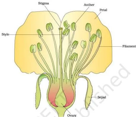
The L.S. (longitudinal section) of a flower in Figure 1.1 is the chapter’s way of making you recall the floral parts you met in Class 11 — the whorls of sepals, petals, stamens and the carpel.
From it the chapter poses the question that structures the whole chapter: in which two parts do the two most important units of sexual reproduction develop? The answer is the anther, which holds the male gametophyte (the pollen grain), and the ovule, which holds the female gametophyte (the embryo sac). Exercise 1 asks exactly this.
The Male Side: Stamen, Microsporangium and Pollen Grain
The stamen is the male reproductive organ of the flower, and the pollen grain is its male gametophyte. This whole section is the chain of events that turns anther tissue into a pollen grain ready to be shed.
From filament to pollen sac
A typical stamen has two parts: the long, slender filament and the terminal, generally bilobed anther. Each lobe carries two theca, so the anther is called dithecous (NCERT p. 5).
In cross-section the anther is four-sided, with four microsporangia at its corners, two in each lobe. As the anther matures, these microsporangia develop into pollen sacs, which run the length of the anther and become packed with pollen grains (NCERT p. 5).
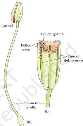
Figure 1.2(a) labels the two parts of the stamen — the filament stalk and the bilobed anther. Part (b) shows how each anther lobe holds two theca, which is why the text calls the anther both bilobed and dithecous: two lobes, and two theca within each.
Microsporangium wall and the tapetum
A microsporangium is ringed by four wall layers — from outside in, the epidermis, endothecium, middle layers and tapetum (NCERT p. 6). The outer three protect the contents and help the anther dehisce to release pollen.
The tapetum is the innermost layer. It nourishes the developing pollen grains, and its cells have dense cytoplasm, usually with more than one nucleus. The chapter asks you to think about how tapetal cells could become binucleate — keep that question next to the free-nuclear divisions of the embryo sac on p. 10: nuclear division without a wall forming immediately.
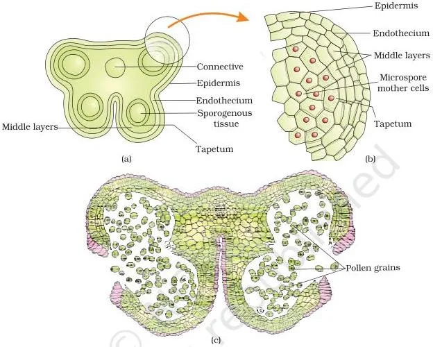
Figure 1.3(a) shows the young anther’s four microsporangia in a tetragonal arrangement. Part (b) enlarges one microsporangium so you can see the four wall layers around the central sporogenous tissue; part (c) shows a mature, dehisced anther releasing pollen.
Memory hook for the wall layers, outside to inside: Epidermis, Endothecium, Middle layers, Tapetum — “Every Elephant Must Train” (E-E-M-T).
Microsporogenesis: meiosis makes the tetrad
While the anther is young, the centre of each microsporangium holds compactly arranged cells called the sporogenous tissue. Each cell is a potential pollen mother cell (PMC). When the anther develops, each PMC undergoes meiosis and forms a cluster of four cells — the microspore tetrad.
This process is microsporogenesis (NCERT p. 6). As the anther dries, the tetrads dissociate and each microspore becomes a pollen grain. Because meiosis halves the chromosome number, the answer to the chapter’s italic question — the ploidy of the tetrad cells — is haploid.
The pollen grain: wall and cells
The pollen grain represents the male gametophyte. It is generally spherical, about 25–50 micrometres across, with a two-layered wall (NCERT p. 7):
- Exine — the hard outer layer of sporopollenin, which NCERT calls one of the most resistant organic materials known. It survives high temperature, strong acids and alkalis, no enzyme that digests it is known, and it is why pollen grains are found as fossils. The exine is absent at the germ pores — the apertures through which the pollen tube will emerge.
- Intine — the thin, continuous inner layer made of cellulose and pectin.
Why must the exine be hard? Protection. A shed pollen grain is a free, air-borne cell with no plant body around it, so the resistant wall shields it until it lands on a stigma. The germ pores give the pollen tube an exit.
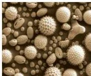
Figure 1.4 explains why the chapter pauses on pollen architecture: even a few species show an astonishing range of size, shape, colour and surface design.
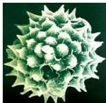
A mature pollen grain contains two cells: the large vegetative cell, with abundant food reserve and a big, irregularly shaped nucleus, and the small, spindle-shaped generative cell, which floats in the vegetative cell’s cytoplasm.
In over 60 per cent of angiosperms, pollen is shed at this two-celled stage. In the remaining species, the generative cell has already divided mitotically into two male gametes, so the pollen is shed at the three-celled stage (NCERT p. 7).

Figure 1.5(b) is worth reading carefully: the young microspore has vacuoles and an asymmetric spindle; after mitosis it holds the large vegetative cell and the small generative cell nested inside it.
Viability — how long shed pollen stays alive — varies wildly. Rice and wheat pollen loses viability within about 30 minutes; members of Rosaceae, Leguminosae and Solanaceae stay viable for months; and pollen of many species can be stored for years in liquid nitrogen at –196°C. Such stored pollen forms pollen banks for crop breeding programmes (NCERT p. 7).
One health note from the same page: pollen of some species, such as Parthenium (carrot grass), causes allergy and respiratory problems.
The Female Side: Pistil, Ovule and the Embryo Sac
The pistil is the female reproductive organ of the flower, and its ovule produces the embryo sac, the female gametophyte. This section is the mirror image of the previous one: a chain of events inside the ovary that ends in a gamete-bearing structure.
The pistil and the ovule
The pistil has three parts — the stigma, a landing platform for pollen; the style, the elongated slender part beneath it; and the ovary, the basal bulged part (NCERT p. 8). A gynoecium may hold a single pistil (monocarpellary) or several (multicarpellary); several may be fused (syncarpous, as in Papaver) or free (apocarpous, as in Michelia).
Inside the ovary, the ovules arise from the placenta.
A typical ovule (NCERT p. 9) is attached to the placenta by a stalk, the funicle. The body of the ovule fuses with the funicle at the hilum. One or two protective integuments encircle the ovule except at the tip, where a small opening — the micropyle — is left. Opposite the micropyle is the chalaza, the basal part of the ovule.
Inside the integuments lies the nucellus, a mass of food-rich cells, and within the nucellus sits the embryo sac — the female gametophyte, usually one per ovule, formed from a megaspore. The number of ovules per ovary ranges from one (wheat, paddy, mango) to many (papaya, watermelon, orchids) (NCERT p. 8–9).
Megasporogenesis and monosporic development
Megasporogenesis is the formation of megaspores from the megaspore mother cell (MMC). The MMC is a large cell, with dense cytoplasm and a prominent nucleus, that differentiates in the micropylar region of the nucellus. It undergoes meiosis, producing four megaspores (NCERT p. 10).
In most flowering plants, three of the four megaspores degenerate and only one is functional. Embryo sac formation from a single functional megaspore is monosporic development.
The two sporogenesis pathways are easy to mix up, so keep the comparison above your work:
| Microsporogenesis | Megasporogenesis | |
|---|---|---|
| Where | Microsporangium of the anther | Nucellus of the ovule |
| Starting cell | Pollen mother cell (PMC), diploid | Megaspore mother cell (MMC), diploid |
| Division | Meiosis | Meiosis |
| Direct product | Microspore tetrad — four microspores, all functional | Four megaspores — three degenerate, one functional |
| End product | Pollen grain (male gametophyte) | Embryo sac (female gametophyte) |
Notice the one difference that decides everything: in the anther, all four products of meiosis survive as pollen; in the ovule, only one megaspore goes on.
From one megaspore to the 8-nucleate embryo sac
The functional megaspore’s nucleus divides mitotically, and the daughter nuclei move to opposite poles — the 2-nucleate stage. Two more mitotic divisions give the 4-nucleate and 8-nucleate stages. These divisions are free-nuclear: nuclei divide without cell walls forming between them. Only after the 8-nucleate stage are cell walls laid down (NCERT p. 10).
The mature embryo sac has a fixed geography:
- At the micropylar end — the egg apparatus: two synergids and one egg cell. The synergids carry cellular thickenings called filiform apparatus at their micropylar tip, which guide the pollen tube into the synergid.
- At the chalazal end — three antipodal cells.
- In the middle — the large central cell, holding two polar nuclei.
Count the cells: 2 synergids + 1 egg + 3 antipodals + 1 central cell = 7 cells. Count the nuclei: 6 inside those cells + 2 polar nuclei = 8 nuclei. Hence “8-nucleate but 7-celled.”
Memory hook for the arrangement, from the micropylar end: 2 synergids, 1 egg, 2 polar nuclei, 3 antipodals — “S-E-C-A: Synergids, Egg, Central cell, Antipodals.”
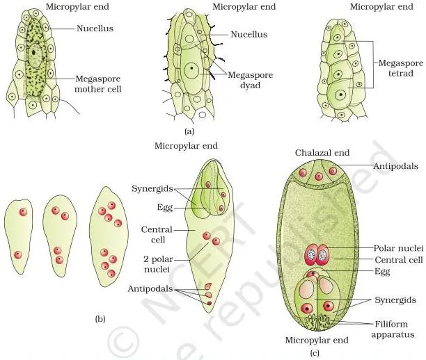
Figure 1.8 is the chapter’s centrepiece diagram. Read it left to right: the megaspore mother cell, then the dyad and tetrad of megaspores, then the 2-, 4- and 8-nucleate stages, and finally the mature embryo sac with every cell in place.
Worked counting: one MMC and a ten-ovule ovary
Chain it from the start. One MMC gives:
\[1\text{ MMC} \xrightarrow{\text{meiosis}} 4\text{ megaspores} \rightarrow 1\text{ functional},\; 3\text{ degenerate}\]
\[1\text{ functional megaspore} \xrightarrow{3\text{ free-nuclear mitoses}} 2 \rightarrow 4 \rightarrow 8\text{ nuclei} \rightarrow 7\text{ cells}\]
Now scale up, since an ovary can hold many ovules. Take an ovary with ten ovules, with one MMC in each:
- Ten MMCs undergo meiosis → 40 megaspores, of which 30 degenerate and 10 survive.
- Ten functional megaspores give 10 embryo sacs.
- Each embryo sac is 8-nucleate and 7-celled, so the ten ovules together hold 80 nuclei and 70 cells.
\[10 \times 8 = 80\text{ nuclei}\qquad 10 \times 7 = 70\text{ cells}\]
Pollination: Autogamy, Geitonogamy, Xenogamy and the Agents That Carry Pollen
Both gametes of flowering plants are non-motile — the pollen grain and the embryo sac cannot move. So the pollen must be carried to the stigma; that transport is pollination, the transfer of pollen grains from the anther to the stigma of a pistil (NCERT p. 11).
The three kinds, and their genetics
Pollination is classified by where the pollen comes from:
| Type | Pollen moves from | To | Genetic meaning |
|---|---|---|---|
| Autogamy | Anther of a flower | Stigma of the same flower | True self-pollination |
| Geitonogamy | Anther of a flower | Stigma of another flower on the same plant | Functionally a cross (an agent is needed) but genetically self — the pollen comes from the same plant |
| Xenogamy | Anther of a flower | Stigma of a flower on a different plant | The only type that brings genetically different pollen to the stigma |
Why does geitonogamy need an agent if it is genetically self? Because the two flowers are separate — pollen cannot fall from one anther to the other stigma on its own. The plant still needs wind, water or an animal to move it, which is why the chapter calls it functionally cross-pollination (NCERT p. 12).
Autogamy inside a normal, open flower is rather rare; it needs synchrony between pollen release and stigma receptivity, with anthers and stigma close together. Some plants — Viola, Oxalis, Commelina — carry two flower types: chasmogamous flowers open and expose anthers and stigma, while cleistogamous flowers never open at all.
In a cleistogamous flower, anthers and stigma stay close and self-pollinate in the bud. Because no cross-pollen can ever enter, cleistogamous flowers are invariably autogamous and give assured seed-set even when no pollinator visits (NCERT p. 11).
Agents: wind, water and animals
Plants use two abiotic agents — wind and water — and one biotic: animals. The majority of flowering plants are pollinated by animals (NCERT p. 12–13).
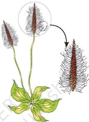
Wind-pollinated flowers trade show for quantity. Their pollen must be light and non-sticky, stamens are well exposed, stigmas are large and often feathery, and each ovary usually holds a single ovule. Grasses are the classic example — the corn cob you eat is the mass of stigmas and styles waving in the air to trap pollen (NCERT p. 12–13).
Because wind pollination is a chance event, these flowers produce enormous amounts of pollen.
Water pollination is rare — limited to about 30 genera, mostly monocotyledons: Vallisneria and Hydrilla in fresh water, and marine seagrasses such as Zostera. In Vallisneria, the female flower reaches the surface on a long stalk while pollen is released onto the water and carried to it by currents, as Figure 1.11 shows.
In most water-pollinated species, the pollen carries a mucilaginous covering that protects it from wetting.
A very common trap: not all aquatic plants are water-pollinated. Water hyacinth and water lily raise their flowers above the surface and are pollinated by insects or wind, like land plants (NCERT p. 13).
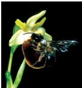
Animal pollination dominates. Bees are the dominant insect pollinators, with butterflies, flies, beetles, wasps, moths, sunbirds, hummingbirds and bats also taking part, along with some reptiles and primates in certain species.
Animal-pollinated flowers are generally large, colourful, fragrant and rich in nectar, and they reward visitors with nectar and sticky pollen — sticky, so it coats the visitor’s body and then rubs onto the next stigma (NCERT p. 13–14). Flies and beetles are attracted by foul odours.
In the obligate pair of Yucca and its moth, the moth lays its eggs in the ovary and the flower is pollinated in return — neither can complete its life cycle without the other. Visitors that take nectar or pollen without touching anthers and stigma are pollen/nectar robbers (NCERT p. 14–15).
Outbreeding devices
Most flowering plants are hermaphrodite, so self-pollination is a constant risk — and continued selfing leads to inbreeding depression. The chapter lists four devices that discourage self-pollination (NCERT p. 15):
- Non-synchronised timing — pollen is released before the stigma becomes receptive, or the stigma matures much before the pollen.
- Different positions — anther and stigma placed so that pollen cannot fall on the same flower’s stigma. Both of these prevent autogamy.
- Self-incompatibility — a genetic mechanism that blocks self-pollen by inhibiting germination or pollen tube growth in the pistil.
- Unisexual flowers — monoecious plants (castor, maize) carry male and female flowers on one plant: this prevents autogamy but not geitonogamy. Dioecious plants (papaya) have male and female flowers on different plants, preventing both.
Pollen-pistil interaction and artificial hybridisation
Landing on the stigma is not enough — the pollen must be the right type. The pistil recognises the pollen, accepts it if compatible and promotes the events that follow, or rejects it if incompatible. This continuous dialogue, from pollen deposition until the pollen tube enters the ovule, is pollen-pistil interaction (NCERT p. 15–17).
When pollen is accepted, it germinates through a germ pore, the tube grows through the style, reaches the ovary, enters the ovule through the micropyle, and finally enters one of the synergids through the filiform apparatus (NCERT p. 17).
Breeders who want to cross chosen parents use emasculation and bagging. In a bisexual flower, the anthers are removed from the bud with forceps before they dehisce — that is emasculation. The flower is then covered with a bag, usually of butter paper, so no unwanted pollen reaches the stigma — that is bagging.
When the stigma is receptive, pollen from the chosen male parent is dusted on and the flower is bagged again. If the female parent has unisexual flowers, emasculation is unnecessary — the buds are simply bagged before opening (NCERT p. 17).
Double Fertilisation: The Event Unique to Flowering Plants
One pollen tube delivers two male gametes into one synergid — and the embryo sac uses both. No other plant group does this; it is unique to flowering plants (NCERT p. 18).
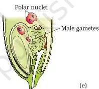
Figure 1.12 is the entire story in five frames: (a) germination on the stigma; (b) pollen tubes through the style; (c) the tube’s path through the pistil; (d) entry into a synergid through the filiform apparatus; (e) discharge of the two male gametes, one moving to the egg and the other to the central cell.
The two fusions
Once inside the synergid, the two male gametes do two different jobs (NCERT p. 18):
- Syngamy — one male gamete fuses with the egg cell nucleus, forming the diploid zygote (\(2n\)). The zygote develops into the embryo.
- Triple fusion — the other male gamete fuses with the two polar nuclei in the central cell, forming the triploid primary endosperm nucleus (\(3n\)). It is called triple fusion because three haploid nuclei fuse. The central cell becomes the primary endosperm cell and develops into endosperm.
Because these two fusions — syngamy and triple fusion — happen in the same embryo sac, the phenomenon is double fertilisation (NCERT p. 18).
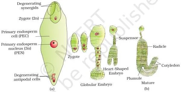
Figure 1.13(a) shows the fertilised embryo sac — the zygote and the primary endosperm nucleus sitting side by side. Part (b) previews the next section: the zygote developing through proembryo, globular and heart-shaped stages into a mature dicot embryo.
The complete ploidy table
Tracking ploidy is where marks slip away, so here is the whole chain in one place. Meiosis halves the chromosome number; fertilisation restores it (NCERT p. 6, 10, 18):
| Structure | Ploidy | How it is obtained |
|---|---|---|
| Nucellus | \(2n\) | Sporophytic tissue of the ovule (NCERT p. 10 question) |
| Megaspore mother cell | \(2n\) | Differentiates in the nucellus; will undergo meiosis |
| Megaspore | \(n\) | Product of meiosis of the MMC |
| Embryo sac (all its cells) | \(n\) | Builds from the functional haploid megaspore |
| Egg cell | \(n\) | One of the three cells of the egg apparatus |
| Polar nuclei | \(n\) each (two of them) | Lie together in the central cell |
| Microspore | \(n\) | Product of meiosis of the PMC (tetrad is haploid) |
| Male gamete | \(n\) | From the generative cell by mitosis |
| Zygote | \(2n\) | \(n\) egg + \(n\) male gamete (syngamy) |
| Endosperm (PEN) | \(3n\) | \(n\) male gamete + \(n + n\) polar nuclei (triple fusion) |
\[ \text{syngamy: } n + n = 2n \qquad \text{triple fusion: } n + n + n = 3n \]
Post-Fertilisation: Endosperm, Embryo, Seed and Fruit
After double fertilisation, the ovule matures into a seed and the ovary into a fruit — but the endosperm and the embryo must develop first, in that order. All of this is the “post-fertilisation” half of the chapter (NCERT p. 18).
Endosperm develops first
The primary endosperm cell divides repeatedly and forms triploid endosperm tissue, filled with reserve food. Endosperm development precedes embryo development — the embryo will need that food.
In the commonest pattern, the PEN first divides into free nuclei (free-nuclear endosperm) and only later are cell walls laid down (cellular endosperm). Tender coconut water is free-nuclear endosperm — thousands of nuclei — and the white kernel around it is cellular endosperm (NCERT p. 18–19).
Whether endosperm survives depends on the plant. It is completely consumed by the embryo in pea, groundnut and beans, but persists in castor and coconut (NCERT p. 19).
Embryo: from zygote to mature structure
The zygote sits at the micropylar end of the embryo sac, and most zygotes divide only after some endosperm has formed — an adaptation that guarantees nutrition for the developing embryo. That is the answer to Exercise 12 (NCERT p. 19).
The early stages of development, or embryogeny, run proembryo → globular → heart-shaped → mature embryo, and are similar in monocots and dicots (NCERT p. 19).
A typical dicot embryo (Figure 1.14a) is an embryonal axis with two cotyledons. Above the cotyledons the axis is the epicotyl, ending in the plumule (stem tip); below them it is the hypocotyl, ending in the radicle (root tip) covered by a root cap.
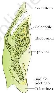
A grass embryo (Figure 1.14b) has just one cotyledon, the scutellum, placed to one side of the axis. Its radicle and root cap are enclosed in a sheath, the coleorrhiza; the epicotyl carries the shoot apex and leaf primordia inside a hollow sheath, the coleoptile (NCERT p. 19–20).
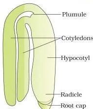
Use Figure 1.14 to make the terms concrete: in the dicot, epicotyl ends in the plumule and hypocotyl ends in the radicle; in the grass embryo, the coleoptile wraps the shoot tip and the coleorrhiza wraps the root tip. Exercise 13(b) asks exactly these two pairs.
Seed
The seed is a fertilised ovule — the final product of sexual reproduction in angiosperms (NCERT p. 20). The integuments harden into the tough seed coat, and the micropyle survives as a pore through which oxygen and water enter during germination. Mature seeds fall into two classes:
- Non-albuminous (ex-albuminous) — no residual endosperm; the embryo consumed it. Examples: pea, groundnut.
- Albuminous — endosperm persists in the mature seed. Examples: wheat, maize, barley, castor.
One more tissue can linger: the perisperm, a persistent nucellus present in black pepper and beet. It is nucellus, not ovary wall — that distinction is Exercise 13(d).
As the seed matures, its water content falls to about 10–15 per cent, the embryo’s metabolic activity slows, and it may enter dormancy until moisture, oxygen and temperature are right (NCERT p. 20).
Viability varies enormously — months for some seeds, years for many, and the record holders: a Lupinus arcticus seed from Arctic tundra germinated after an estimated 10,000 years, and a 2,000-year-old date palm (Phoenix dactylifera) seed was discovered during excavation at King Herod’s palace (NCERT p. 20–21).
Dormancy and dehydration are exactly what allow seeds to be stored as food and for next season’s crop.
Fruit: true, false and seedless
Ovary and ovules mature together: the ovary wall becomes the pericarp, and the fruit may be fleshy (guava, orange, mango) or dry (groundnut, mustard) (NCERT p. 20).
Most fruits develop only from the ovary — these are true fruits. But in apple, strawberry and cashew, the thalamus also contributes to fruit formation, and such fruits are false fruits — the fleshy part of an apple you eat is mostly thalamus.
A few species form fruits without fertilisation at all: parthenocarpic fruits, of which banana is the example. Parthenocarpy can also be induced by applying growth hormones, and such fruits are seedless (NCERT p. 20–21).
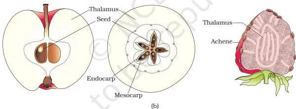
Figure 1.15 pairs the two ideas you must keep apart: the seed with its hardened coats, and the false fruits whose fleshy part comes from the thalamus, not the ovary wall.
Apomixis and Polyembryony: Seeds Without Fertilisation
Not every seed needs fertilisation. Some flowering plants — certain species of Asteraceae and grasses — make seeds without it, and that quirk is commercially important (NCERT p. 22).
Two ways apomictic seeds form
- A diploid egg is formed without reduction division and develops into an embryo without fertilisation.
- In many Citrus and Mango varieties, nucellar cells around the embryo sac start dividing, protrude into the embryo sac, and develop into embryos themselves.
Either way, the seed forms without fertilisation — so apomixis is a form of asexual reproduction that mimics sexual reproduction. When nucellar embryos form, one ovule can end up with many embryos: that is polyembryony, the occurrence of more than one embryo in a seed.
The chapter’s suggestion: squeeze orange seeds and observe the several embryos of different sizes inside each. Because those embryos arise from the mother plant’s own nucellar cells, they are genetically identical to the mother — the chapter’s italic question asks you to recognise them as clones.
Why hybrid seed companies care
Hybrid crops grow vigorously, but their seeds segregate: the next generation does not keep the hybrid’s characters, so farmers must buy fresh hybrid seed every year — and hybrid seed is costly. If the hybrid is apomictic, there is no segregation, so farmers can keep using saved seed year after year instead of buying it.
That is why laboratories worldwide are working to transfer apomictic genes into hybrid varieties (NCERT p. 22).
Key Terms and Definitions in This Chapter
These are the terms NCERT itself defines in this chapter, gathered in one place for a final-scan revision. Page references are given once per group so you can check any term in the book.
Male side: stamen to pollen grain (NCERT pp. 5–7)
| Term | Meaning |
|---|---|
| Stamen | The flower’s male reproductive organ: filament + anther. |
| Filament | The long, slender stalk of the stamen. |
| Anther | Terminal, generally bilobed part of the stamen. |
| Bilobed | Having two lobes, each carrying two theca. |
| Dithecous | Having two theca per anther lobe. |
| Microsporangium | Pollen-producing chamber of the anther; four in a typical anther. |
| Pollen sac | A microsporangium that has matured and become packed with pollen grains. |
| Tapetum | Innermost anther wall layer; nourishes developing pollen grains. |
| Sporogenous tissue | Central cell mass in a young microsporangium that gives rise to pollen mother cells. |
| Microsporogenesis | Formation of microspores from a PMC by meiosis. |
| Microspore tetrad | The cluster of four haploid microspores from one PMC. |
| Pollen grain | The male gametophyte released from the anther. |
| Exine | Hard outer pollen wall made of sporopollenin. |
| Sporopollenin | The highly resistant organic substance of the exine. |
| Germ pore | Aperture in the exine where sporopollenin is absent; the pollen tube emerges through it. |
| Intine | Thin inner pollen wall of cellulose and pectin. |
| Vegetative cell | The larger pollen cell, with food reserves and a large irregular nucleus. |
| Generative cell | The smaller pollen cell; divides into the two male gametes. |
Female side: pistil to embryo sac (NCERT pp. 8–10)
| Term | Meaning |
|---|---|
| Pistil | The flower’s female reproductive organ: stigma + style + ovary. |
| Stigma | The pistil’s top, where pollen lands. |
| Style | The slender part of the pistil below the stigma. |
| Ovary | The basal, bulged part of the pistil holding the ovules. |
| Ovule | The megasporangium; becomes the seed after fertilisation. |
| Funicle | Stalk attaching the ovule to the placenta. |
| Hilum | Junction where the ovule body meets the funicle. |
| Integument | Protective envelope around the nucellus; hardens into the seed coat. |
| Micropyle | Small opening at the ovule tip left by the integuments. |
| Chalaza | Basal part of the ovule, opposite the micropyle. |
| Nucellus | Food-rich tissue inside the integuments, housing the embryo sac. |
| Megasporogenesis | Formation of megaspores from the MMC by meiosis. |
| Megaspore mother cell | The large nucellus cell that undergoes meiosis. |
| Embryo sac | The female gametophyte; 7-celled, 8-nucleate. |
| Synergid | One of the two cells flanking the egg cell at the micropylar end. |
| Filiform apparatus | Thickenings at the synergids’ micropylar tips that guide the pollen tube. |
| Antipodal | One of the three cells at the chalazal end of the embryo sac. |
| Central cell | The large embryo sac cell holding the two polar nuclei. |
| Polar nuclei | The two haploid nuclei in the central cell; one male gamete fuses with them. |
| Monosporic | Embryo sac development from a single functional megaspore. |
Pollination and breeding (NCERT pp. 11–17)
| Term | Meaning |
|---|---|
| Pollination | Transfer of pollen grains from anther to stigma. |
| Autogamy | Pollination within the same flower. |
| Geitonogamy | Pollen transfer to another flower of the same plant. |
| Xenogamy | Pollen transfer to a flower of a different plant. |
| Chasmogamous | Flowers that open and expose anthers and stigma. |
| Cleistogamous | Flowers that never open; invariably autogamous. |
| Self-incompatibility | Genetic mechanism preventing self-pollen from fertilising ovules. |
| Outbreeding devices | Adaptations that discourage self-pollination. |
| Emasculation | Removal of anthers from a bisexual flower bud before dehiscence. |
| Bagging | Covering flowers with a bag to keep unwanted pollen away. |
| Pollen-pistil interaction | All events from pollen landing on the stigma to pollen tube entry into the ovule. |
Fertilisation and after (NCERT pp. 18–22)
| Term | Meaning |
|---|---|
| Double fertilisation | Syngamy + triple fusion in one embryo sac; unique to flowering plants. |
| Syngamy | Fusion of a male gamete with the egg → zygote. |
| Triple fusion | Fusion of a male gamete with two polar nuclei → PEN. |
| Primary endosperm nucleus | The triploid product of triple fusion. |
| Endosperm | Food-storing triploid tissue that nourishes the embryo. |
| Embryogeny | Early embryo development. |
| Proembryo | First stage of embryo development after the zygote divides. |
| Epicotyl | Embryonal axis above the cotyledons, ending in the plumule. |
| Hypocotyl | Embryonal axis below the cotyledons, ending in the radicle. |
| Plumule | The stem tip. |
| Radicle | The root tip. |
| Scutellum | The single cotyledon of a grass embryo, placed laterally. |
| Coleoptile | Hollow sheath enclosing the epicotyl’s shoot apex in grasses. |
| Coleorrhiza | Sheath enclosing the radicle and root cap in grasses. |
| Albuminous | Seeds retaining endosperm at maturity (wheat, maize, castor). |
| Non-albuminous | Mature seeds with no residual endosperm (pea, groundnut). |
| Perisperm | Residual, persistent nucellus in some seeds (black pepper, beet). |
| Dormancy | Inactive state of a mature embryo or seed. |
| True fruit | Fruit formed only from the ovary. |
| False fruit | Fruit to which the thalamus also contributes. |
| Pericarp | Fruit wall, developed from the ovary wall. |
| Parthenocarpy | Fruit development without fertilisation. |
| Apomixis | Asexual reproduction that mimics sexual reproduction; seeds form without fertilisation. |
| Polyembryony | Occurrence of more than one embryo in a seed. |
Common Mistakes Students Make in This Chapter
Most errors in this chapter come from counting cells, tracking ploidy, and the self-versus-cross distinction. Each row names the mistake, the correct rule, and a way to catch yourself.
| Mistake | Correct rule | How to check your answer |
|---|---|---|
| “The embryo sac is 8-celled” | It is 7-celled and 8-nucleate: the two polar nuclei share one central cell (NCERT p. 10). | Count cells: 3 egg apparatus + 3 antipodals + 1 central = 7. Count nuclei: 3 + 3 + 2 = 8. |
| Microsporangium and pollen sac treated as different organs | One structure at two stages — microsporangia develop further and become pollen sacs (NCERT p. 5). | Ask which came first: the young anther has microsporangia, the mature anther has pollen sacs. |
| “All aquatic plants are water-pollinated” | Water pollination is rare, limited to about 30 genera; water hyacinth and water lily are insect- or wind-pollinated (NCERT p. 13). | Ask whether the flower emerges above the surface — if it does, treat it like a land plant. |
| “Geitonogamy is cross-pollination” | Functionally cross because an agent carries the pollen, but genetically self — the pollen comes from the same plant (NCERT p. 12). | Check the pollen’s source plant, not the flower: same plant = self. |
| Ploidy slips — “endosperm is 2n”, “zygote is 3n” | Zygote \(2n\) (n + n); endosperm \(3n\) (n + n + n). Nucellus and MMC are \(2n\); megaspore and egg are \(n\) (NCERT pp. 10, 18). | Ask “was meiosis involved?” and “how many nuclei fused?” before writing ploidy. |
| “Cleistogamous flowers can be cross-pollinated” | They never open, so cross-pollen cannot reach the stigma — they are invariably autogamous (NCERT p. 11). | A closed flower has no route for an external pollen agent. |
| Perisperm and pericarp confused | Perisperm = persistent nucellus (black pepper, beet); pericarp = ovary wall, the fruit wall (NCERT p. 20). | Ask which tissue it comes from: nucellus or ovary wall? |
| “The zygote is simply resting” | The zygote delays division until some endosperm exists to nourish the embryo (NCERT p. 19). | Link it to the rule: endosperm always precedes embryo. |
One question NCERT poses but does not answer directly: how tapetal cells become binucleate (NCERT p. 6). Think about the free-nuclear divisions you meet later in the embryo sac (p. 10) — nuclear division without an immediate cell wall — and you can reason it out.
Exam Notes: What the NCERT Exercises Ask You to Know
The chapter’s closing exercises are its exam map: each question targets a single idea named in the table below. The “differentiate” and “explain” questions need both sides or all steps stated; the diagram questions need neat, correctly positioned labels.
| Q | Concept tested | What a complete answer includes |
|---|---|---|
| 1 | Sites of gametophyte development | Anther (male), ovule (female) |
| 2 | Micro- vs megasporogenesis | Both by meiosis; microspore tetrad vs megaspores; pollen grain vs embryo sac |
| 3 | Developmental sequence | Sporogenous tissue → pollen mother cell → microspore tetrad → pollen grain → male gametes |
| 4 | Ovule structure | Neat labelled diagram: funicle, hilum, integuments, micropyle, chalaza, nucellus |
| 5 | Monosporic development | One of four megaspores functional; three degenerate |
| 6 | Embryo sac structure | Diagram showing 7 cells and 8 nuclei, with names and positions |
| 7 | Chasmogamous vs cleistogamous | Cross-pollination impossible in cleistogamous flowers: they never open |
| 8 | Outbreeding devices | Any two: timing, position, self-incompatibility, unisexuality |
| 9 | Self-incompatibility | Genetic mechanism; self-pollen inhibited at germination or tube growth |
| 10 | Bagging | Bagging flowers to keep contamination out; why a breeder needs it |
| 11 | Triple fusion | In the central cell; male gamete + two polar nuclei → PEN |
| 12 | Zygote dormancy | Divides only after endosperm forms — assured nutrition |
| 13 | Four differentiations | Epicotyl/hypocotyl; coleoptile/coleorrhiza; integument/testa; perisperm/pericarp |
| 14 | False fruit | Apple: thalamus contributes; ovary wall → pericarp |
| 15 | Emasculation | Anther removal before dehiscence; used on bisexual female parents |
| 16 | Parthenocarpy | Growth hormones give seedless fruits; banana is the chapter’s natural example |
| 17 | Tapetum’s role | Nourishes the developing pollen grains, supplying what the pollen wall needs |
| 18 | Apomixis and its importance | Seeds without fertilisation; apomictic hybrids keep characters, so farmers reuse seed |
Two exercises carry printed diagrams in the NCERT book — Question 6 (embryo sac) and Question 18 — on NCERT page 25; open the PDF above to see them.
The book’s italic in-text questions are also part of the exam map: the ploidy of tetrad cells (p. 6), why the exine is hard and what germ pores do (p. 7), whether cleistogamy is advantageous (p. 11), and why wind- and water-pollinated flowers are not colourful and produce no nectar (p. 13) are all little explanations you should be able to give.
One standard caution: textbook contents and the examinable syllabus are not always identical — check the current official CBSE syllabus. For revision help on this chapter and the rest of the unit, see the Class 12 Biology notes hub.
Figure Walkthrough: Drawing the Ovule and Labelling the Embryo Sac


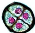
Exercises 4 and 6 both demand neat, labelled diagrams, and these are the two diagrams on which students lose marks — usually because labels are missing or misplaced, not because the drawing is bad.
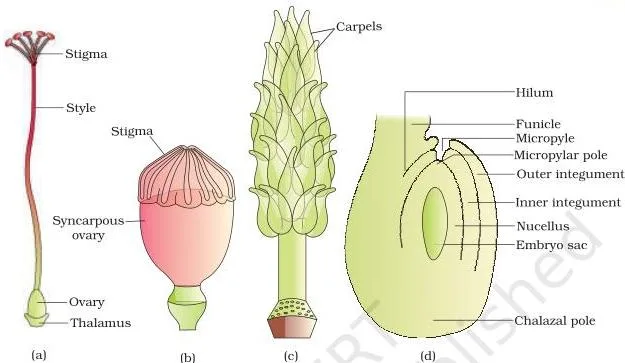
Drawing the anatropous ovule (Exercise 4)
Draw in this order so no part gets forgotten:
- Draw the stalk, the funicle, at the base.
- Mark the hilum where the ovule’s body meets the funicle.
- Draw the two integuments as envelopes that cover the ovule but leave a gap at the tip.
- Label the gap as the micropyle; the opposite, closed end is the chalaza.
- Inside the integuments, draw the nucellus as the central mass.
- Within the nucellus, sketch the embryo sac; in an anatropous ovule the body is bent back along the funicle, as Figure 1.7(d) shows.
Labelling the mature embryo sac (Exercise 6)
Using Figure 1.8, shown in the female-side section above, place the labels in this order:
- Micropylar end: the egg apparatus — two synergids flanking one egg cell, with filiform apparatus at the synergids’ tips.
- Centre: the large central cell with two polar nuclei.
- Chalazal end: three antipodal cells.
Repeat the counting trick as you label: 2 + 1 + 1 + 3 = 7 cells; 6 nuclei in cells + 2 polar nuclei = 8 nuclei. And one warning that appears in exam diagrams again and again: the egg cell sits at the micropylar end, never the chalazal end.
The Chapter in One Read: The Sequence from Anther to Seed
If you have two minutes, hold this single chain in your head — it is the chapter’s own summary (NCERT p. 23) rebuilt as one line:
- Flower → androecium (stamens) and gynoecium (pistils).
- Bilobed, dithecous anther → four microsporangia, which become pollen sacs.
- Microsporogenesis: PMC → meiosis → microspore tetrad → pollen grains.
- Pollen grain = male gametophyte: exine + intine, vegetative + generative cells.
- Pistil: stigma + style + ovary → ovule with integuments, micropyle, nucellus.
- Megasporogenesis: MMC → meiosis → 4 megaspores, 1 functional.
- Monosporic embryo sac: 7-celled, 8-nucleate.
- Pollination: autogamy / geitonogamy / xenogamy; wind, water, animals.
- Pollen-pistil interaction → pollen tube → synergid.
- Double fertilisation: syngamy (zygote \(2n\)) + triple fusion (PEN \(3n\)).
- Endosperm first, embryo second: proembryo → globular → heart-shaped → mature.
- Ovule → seed; ovary → fruit (true, false, parthenocarpic).
- Exceptions: apomixis (seeds without fertilisation) and polyembryony.
Hands-On Activities Suggested in the Chapter
NCERT scatters five low-cost observations through this chapter. Doing even one of them makes the diagrams stick.
- Watch flowers of cucumber, mango, peepal, papaya and others, note which animals touch anthers and stigma, and spot pollen/nectar robbers — visitors that take the reward without pollinating (p. 14–15).
- Dust pollen of pea, chickpea, Crotalaria, balsam or Vinca onto a drop of about 10% sugar solution and look for pollen tubes under the low-power lens after 15–30 minutes (p. 17).
- Split open seeds of castor, pea, bean, groundnut and a coconut, and look for endosperm in each (p. 19).
- Soak wheat, maize, pea, chickpea or groundnut seeds overnight, then dissect and identify the embryo parts (p. 19–20).
- Squeeze orange seeds and count the embryos of different sizes inside each seed — polyembryony you can see for yourself (p. 22).
Related Resources: The Reproduction Unit and More
This listing is maintained for the 2026-27 academic session using the NCERT textbook information available to us. NCERT remains the authority for confirming the latest edition.
This chapter is the first of the three-chapter Reproduction unit in the Class 12 NCERT Biology book. The sibling chapters continue the story — Human Reproduction (Chapter 2) and Reproductive Health (Chapter 3) — and the unit opener profiles the botanist Panchanan Maheshwari (NCERT p. 1–2), whose work on embryology and tissue culture underlies much of what this chapter describes.
- Class 12 Biology book page — browse every chapter of the NCERT book.
- Class 12 hub — all Class 12 NCERT books on this site.
- Human Reproduction revision notes — the chapter that follows this one.
- CBSE notes home — revision notes for every class and subject.
- Class 12 revision notes — the whole year’s subjects in one place.
Sources and Data Verification
Four things to know about where this page’s facts come from:
- All figures, section titles, page numbers, definitions and exercise descriptions on this page come from the NCERT Class 12 Biology textbook, Chapter 1 (Sexual Reproduction in Flowering Plants), official edition on ncert.nic.in.
- This page covers that NCERT textbook chapter only — not other Class 12 biology books or state-board variants.
- The page is maintained for the current academic session using the NCERT information available to us.
- NCERT settles textbooks, editions and PDFs; CBSE settles curriculum, syllabus and examinations. A textbook chapter is not proof that every part of it is examinable — check the current official syllabus.
Reference: NCERT Class 12 Biology textbook, chapter 1, official edition on ncert.nic.in.
Frequently Asked Questions About This Chapter
Why is the embryo sac described as 7-celled but 8-nucleate?
Because two of the eight nuclei share one cell. Six nuclei sit inside six cells (two synergids, one egg, three antipodals), while the remaining two polar nuclei lie together in the large central cell. The count is therefore 7 cells but 8 nuclei (NCERT p. 10).
What is the difference between apomixis and parthenocarpy?
Both skip fertilisation, but the products differ. Apomixis produces seeds without fertilisation (NCERT p. 22), while parthenocarpy produces fruits without fertilisation — and such fruits are seedless, banana being the example (NCERT p. 20–21).
Why is geitonogamy called cross-pollination even though it is genetically self-pollination?
Because a pollinating agent must carry the pollen between two separate flowers, it is functionally a cross-pollination event. But both flowers belong to the same plant, so the pollen carries the same genetic material as the stigma it lands on — genetically it is self-pollination, like autogamy (NCERT p. 12).
Is the endosperm of flowering plants diploid or triploid, and why?
Triploid. The endosperm comes from the primary endosperm nucleus, produced by triple fusion: one male gamete fuses with two polar nuclei in the central cell (NCERT p. 18). The ploidy is \(n + n + n = 3n\).
Why is apple called a false fruit?
Because the edible part of the apple is formed not only from the ovary but also from the thalamus, which contributes to fruit formation. Fruits that develop only from the ovary are true fruits; most of the apple’s fleshy tissue is thalamus, so it counts as a false fruit (NCERT p. 20).
How do apomictic hybrids help farmers avoid buying new seeds every year?
Hybrid seeds segregate in the next generation, so the progeny do not keep the hybrid’s characters and farmers must buy fresh seed each year. Apomictic hybrids form seeds without fertilisation, so no segregation occurs and farmers can sow saved seed year after year (NCERT p. 22).
Explore Class 12 Biology Books
Class 12 Biology on LearnCBSE:
Related chapters:
- Reproductive Health
- Principles of Inheritance and Variation
- Molecular Basis of Inheritance