
This is the carbohydrates class 12 chapter: Section 10.1 of Unit 10 (Biomolecules) in the NCERT Chemistry Part II textbook. The official NCERT PDF is right below, and this page teaches what the chapter contains — glucose, disaccharides, polysaccharides, then proteins and nucleic acids. This description follows the NCERT reprint for the 2026-27 session.
Download the carbohydrates class 12 chapter PDF (Official NCERT)
For the official textbook file, Download the NCERT carbohydrates class 12 chapter PDF (Biomolecules, Unit 10) straight from ncert.nic.in — the same file teachers use in class, covering Section 10.1 Carbohydrates and the five sections that follow it.
| What the chapter holds | Count | Where it is used |
|---|---|---|
| Printed pages | 22 | |
| Sections in the chapter | 19 | |
| Figures with NCERT captions | 19 | |
| Tables | 5 | |
| Exercise questions | 25 | answered in our NCERT Solutions |
| Official NCERT PDF | Download the chapter PDF | the chapter exactly as NCERT publishes it |
Chapter at a glance: what is inside this file
The file opens with Unit 10 Biomolecules, and Section 10.1 Carbohydrates is the first and largest section of the unit. The headings below show what the unit contains from start to finish, and the table at the end of this section gives the file’s scale at a glance.
- Section 10.1 Carbohydrates — definition, classification, glucose and fructose, disaccharides and polysaccharides
- Section 10.2 Proteins — amino acids, peptide linkage, structure and denaturation
- Section 10.3 Enzymes — biocatalysts and how they act
- Section 10.4 Vitamins — the two classes and their deficiency diseases
- Section 10.5 Nucleic Acids — composition, structure and functions of DNA and RNA
- Section 10.6 Hormones — intercellular messengers and their chemical classes
- The unit closes with a Summary and an exercise set, with in-text questions after the carbohydrate, protein and nucleic acid sections
What the carbohydrates class 12 chapter covers
Unit 10 surveys the biomolecules that keep a living system running. The book opens with a striking framing: a living system is composed of non-living atoms and molecules, and the study of what goes on chemically inside it is biochemistry. The six sections of the unit each take one family of biomolecules.
- 10.1 Carbohydrates — sugars and their polymers; the focus of this page, with glucose as its centrepiece
- 10.2 Proteins — polymers of amino acids, from sequence to shape
- 10.3 Enzymes — the biocatalysts that run reactions under mild conditions
- 10.4 Vitamins — small dietary requirements with named deficiency diseases
- 10.5 Nucleic Acids — DNA and RNA, the molecules of heredity
- 10.6 Hormones — chemical messengers carried by the blood
Textbook contents and the examinable syllabus are not always identical — check the current official CBSE syllabus before deciding what to revise.
Carbohydrates: the definition and the formula trap
Carbohydrates are a very large group of naturally occurring organic compounds produced mainly by plants, and most of them fit the general formula \( \mathrm{C_x(H_2O)_y} \). Because glucose, \( \mathrm{C_6H_{12}O_6} \), fits as \( \mathrm{C_6(H_2O)_6} \), the old name hydrates of carbon stuck — that is where the word carbohydrate comes from.
Cane sugar, glucose and starch are everyday examples.
The formula is a trap, not a definition. Acetic acid, \( \mathrm{CH_3COOH} \), fits the formula as \( \mathrm{C_2(H_2O)_2} \), yet it is not a carbohydrate. Rhamnose, \( \mathrm{C_6H_{12}O_5} \), is a carbohydrate and does not fit the formula at all.
The working definition this chapter uses is chemical (Section 10.1): a carbohydrate is an optically active polyhydroxy aldehyde or ketone, or a compound that produces such units on hydrolysis. Sweet-tasting carbohydrates are called sugars — sucrose is the sugar of the home, lactose the sugar of milk — and carbohydrates are also called saccharides, from the Greek sakcharon, meaning sugar.
Classification of carbohydrates: mono, oligo, poly and the reducing test
The chapter classifies carbohydrates twice, because hydrolysis behaviour and reducing behaviour answer two different questions. Learn both schemes and the words that belong to each.
By hydrolysis: carbohydrates fall into three groups (Section 10.1.1).
- Monosaccharides cannot be hydrolysed further to a simpler unit of polyhydroxy aldehyde or ketone. About 20 are known in nature; examples are glucose, fructose and ribose.
- Oligosaccharides yield two to ten monosaccharide units on hydrolysis; the common ones are disaccharides. Sucrose gives glucose and fructose; maltose gives two glucose units only.
- Polysaccharides release a large number of monosaccharide units on hydrolysis. They are not sweet, so they are called non-sugars; examples are starch, cellulose, glycogen and gums.
By reducing power: carbohydrates that reduce Fehling’s solution and Tollens’ reagent are reducing sugars. Every monosaccharide, whether aldose or ketose, is a reducing sugar.
Within monosaccharides, the name itself carries the functional group and the carbon count: an aldehyde gives an aldose, a ketone gives a ketose, and the carbon count adds a tail, as in the scheme below.
| Carbon atoms | General term | Aldehyde | Ketone |
|---|---|---|---|
| 3 | Tetrose? No — triose | Aldotriose | Ketotriose |
| 4 | Tetrose | Aldotetrose | Ketotetrose |
| 5 | Pentose | Aldopentose | Ketopentose |
| 6 | Hexose | Aldohexose | Ketohexose |
| 7 | Heptose | Aldoheptose | Ketoheptose |
So glucose is an aldohexose: an aldehyde group on a six-carbon chain. Fructose, with a ketone at C2, is a ketohexose.
Glucose: how its structure was proved
A molecular formula alone does not reveal a structure, so the chapter walks through the experiments that settled glucose’s, evidence by evidence. The preparation comes first, then the proof chain.
Glucose occurs free in sweet fruits and honey, and ripe grapes contain large amounts. In the laboratory, boiling sucrose with dilute HCl or \( \mathrm{H_2SO_4} \) in alcoholic solution gives glucose and fructose in equal amounts (Section 10.1.2.1):
\[ \mathrm{C_{12}H_{22}O_{11} + H_2O \xrightarrow{\mathrm{H^+}} C_6H_{12}O_6 + C_6H_{12}O_6} \]
Commercially, glucose is made by hydrolysing starch with dilute sulphuric acid at about 393 K under 2–3 atm pressure:
\[ \mathrm{(C_6H_{10}O_5)_n + nH_2O \xrightarrow[393\ \mathrm{K},\ 2\text{–}3\ \text{atm}]{\mathrm{H^+}} nC_6H_{12}O_6} \]
Glucose is an aldohexose, also called dextrose, the monomer of starch and cellulose, and probably the most abundant organic compound on earth. The structure the chapter assigns rests on five reagents, each of which removes one possibility and leaves one certainty.
| Reagent | Product | What it proves |
|---|---|---|
| HI, prolonged heating | n-Hexane | All six carbons are linked in a straight chain |
| Hydroxylamine, \( \mathrm{NH_2OH} \) | An oxime | A carbonyl group is present |
| Hydrogen cyanide, \( \mathrm{HCN} \) | A cyanohydrin | A carbonyl group is present |
| Bromine water (mild oxidant) | Gluconic acid | The carbonyl group is an aldehyde, not a ketone |
| Acetic anhydride | Glucose pentaacetate | Five \( -\mathrm{OH} \) groups, attached to different carbons |
| Nitric acid, \( \mathrm{HNO_3} \) | Saccharic acid (a dicarboxylic acid) | A primary alcoholic \( -\mathrm{OH} \) group is present |
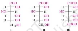
What the evidence leaves open is the arrangement of the OH groups in space. Fischer assigned that arrangement, and the figure shows the three structures the chapter draws — glucose (I), gluconic acid (II) and saccharic acid (III) — in the projection used throughout the chapter. This reagent chain is exactly what exercise 10.9 asks for, with HI, bromine water and nitric acid.
What D(+) glucose means: configuration and rotation are different
Glucose is named D(+)-glucose, and students routinely collapse the two labels into one. They describe two entirely different facts about the molecule (Section 10.1.2.1).
- D and L describe configuration — the spatial arrangement of atoms — fixed by comparing the compound to a reference isomer of glyceraldehyde.
- (+) and (−) describe rotation — the measured optical activity. Dextrorotatory means the molecule rotates plane-polarised light to the right; laevorotatory means to the left.
To assign D or L, compare the lowest asymmetric carbon with glyceraldehyde. Write the structure with the most oxidised carbon (the \( -\mathrm{CHO} \) group) at the top; if the \( -\mathrm{OH} \) on that lowest asymmetric carbon lies on the right, the compound is D; on the left, it is L. The other asymmetric carbons are ignored.
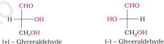
The figure shows the two enantiomers of glyceraldehyde at the heart of the scheme: the isomer with the \( -\mathrm{OH} \) on the right sets the D family, the one on the left the L family. Glucose, whose lowest asymmetric carbon has the \( -\mathrm{OH} \) on the right, is D, and the (+) records that it is dextrorotatory.
Fructose is D(−): same D family, but laevorotatory. The chapter is explicit that D and L have no relation to optical activity, and no relation to the lower-case letters d and l — never deduce rotation from D or L.
Cyclic structure of glucose: the open chain is not the whole story
The open chain structure explains most of glucose’s reactions, but three observations refuse to fit. The chapter uses them to force the next step in the model (Section 10.1.2.1).
- Despite having an aldehyde group, glucose gives no Schiff’s test and forms no hydrogen sulphite addition product with \( \mathrm{NaHSO_3} \).
- The pentaacetate of glucose does not react with hydroxylamine, which means there is no free \( -\mathrm{CHO} \) group left.
- Glucose crystallises in two forms: the \( \alpha \)-form (melting point 419 K) from a concentrated solution at 303 K, and the \( \beta \)-form (melting point 423 K) from a hot saturated solution at 371 K.
The resolution is that one of the \( -\mathrm{OH} \) groups adds across the aldehyde to form a cyclic hemiacetal. For glucose it is the C5 \( -\mathrm{OH} \) that forms the ring, and the ring has six members.
The former aldehyde carbon becomes C1, now called the anomeric carbon, and the \( \alpha \) and \( \beta \) forms differ only in the orientation of the \( -\mathrm{OH} \) at C1. Such isomers are anomers, not enantiomers.
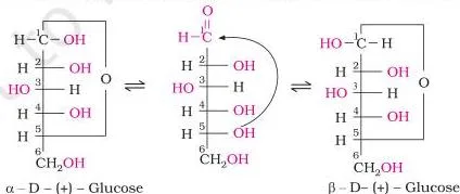
Read the figure for one thing: the single carbon where the two rings differ. In \( \alpha \)-D-glucose the C1 \( -\mathrm{OH} \) sits on one side of the ring; in \( \beta \)-D-glucose it sits on the other; everywhere else the drawings match. That is the anomeric carbon at work.
Six-membered sugar rings are pyranose structures, named by analogy with pyran (a six-membered ring of five carbons and one oxygen), and they are drawn as Haworth structures — the flat hexagon style you see in the figure. This material is the entire answer to exercise 10.10, which asks for the reactions of D-glucose that the open chain structure cannot explain.
Fructose: the ketohexose that forms a five-membered ring
Fructose shares glucose’s formula but not its functional group, and its ring is smaller. It is the sugar that rides along with glucose in sucrose (Section 10.1.2.2).
Fructose is \( \mathrm{C_6H_{12}O_6} \) with a ketone at C2 — a ketohexose. It belongs to the D-series and is laevorotatory, so it is written D(−)-fructose. It occurs in fruits, honey and vegetables, and in pure form it is used as a sweetener.
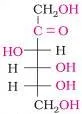
Cyclisation works exactly as for glucose, except the ring closes from the C5 \( -\mathrm{OH} \) to the ketone, and the result is a five-membered ring.

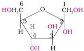
Five-membered sugar rings are furanose structures, named after furan, so the two forms are \( \alpha \)-D-fructofuranose and \( \beta \)-D-fructofuranose. Hold the comparison that trips students: glucose closes to a six-membered pyranose ring, fructose to a five-membered furanose ring, and fructose’s anomeric carbon is C2.
Disaccharides: sucrose, maltose and lactose compared
Disaccharides are two monosaccharide units joined by one oxygen bridge, and the single structural detail hidden in that bridge decides whether a disaccharide reduces Fehling’s solution (Section 10.1.3).
A glycosidic linkage is the oxide linkage formed between two monosaccharide units through an oxygen atom, with the loss of a water molecule. If the reducing groups — the aldehyde and ketone groups — are locked into that bond, the sugar is non-reducing; if one stays free, it is reducing.
| Sucrose | Maltose | Lactose | |
|---|---|---|---|
| Units | \( \alpha \)-D-glucose + \( \beta \)-D-fructose | Two \( \alpha \)-D-glucose units | \( \beta \)-D-galactose + \( \beta \)-D-glucose |
| Linkage | C1 of glucose to C2 of fructose | C1 of one glucose to C4 of the other | C1 of galactose to C4 of glucose |
| Reducing? | No — both reducing groups tied up | Yes — free aldehyde possible at C1 of the second unit | Yes — free aldehyde possible at C1 of glucose |
| Source | The common sugar of the home | — | Milk (milk sugar) |
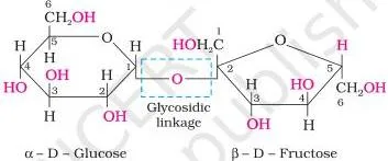
In the sucrose figure, find the oxygen that bridges the two rings: it joins C1 of the glucose unit to C2 of the fructose unit. Because both the aldehyde group of glucose and the ketone group of fructose take part in that bond, neither is free to reduce Tollens’ reagent — that is why sucrose alone among the three is non-reducing.
The rotation story completes it. Sucrose is dextrorotatory, but hydrolysis releases dextrorotatory glucose and laevorotatory fructose. The laevorotation of fructose (\( -92.4^\circ \)) outweighs the dextrorotation of glucose (\( +52.5^\circ \)), so the mixture is laevorotatory: the sign of rotation flips from (+) to (−), and the product is named invert sugar. Keep the two numbers together — they are the whole explanation.
Polysaccharides: starch, cellulose and glycogen
Polysaccharides are the most commonly encountered carbohydrates in nature, and they do one of two jobs: store food or provide structure (Section 10.1.4). All three polymers here are built from glucose, and the twist that separates them is which glucose isomer is used and how the chains branch.
- Starch is the main storage polysaccharide of plants, a polymer of \( \alpha \)-glucose in two components. Amylose, about 15–20%, is a water-soluble unbranched chain of 200–1000 \( \alpha \)-D-glucose units joined by C1–C4 glycosidic linkage. Amylopectin, about 80–85%, is water-insoluble and branched: C1–C4 chains with C1–C6 branch points.
- Cellulose is a straight-chain polysaccharide of only \( \beta \)-D-glucose units joined C1 to C4. It is the most abundant organic substance in the plant kingdom and the predominant constituent of plant cell walls.
- Glycogen is the carbohydrate store of the animal body, also called animal starch because its structure is similar to amylopectin but more highly branched. It is present in liver, muscles and brain, and enzymes break it down to glucose when the body needs it; it is also found in yeast and fungi.
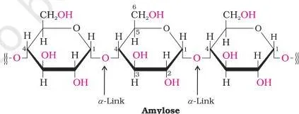
In the amylopectin figure, look for the branch points: the main chain is C1–C4 links, and where a side chain leaves, the link is C1–C6. That single change is what the exercise set tests when it asks about glycogen and starch.
| Polymer | Monomer | Linkage and shape | Role |
|---|---|---|---|
| Starch | \( \alpha \)-D-glucose | C1–C4 chains; amylopectin branched at C1–C6 | Main storage polysaccharide of plants |
| Cellulose | \( \beta \)-D-glucose | C1–C4 straight chains only | Main constituent of plant cell walls |
| Glycogen | \( \alpha \)-D-glucose | Like amylopectin, but more highly branched | Storage in animals: liver, muscles, brain |
Keep the one-line distinction: starch is the \( \alpha \)-glucose polymer, cellulose is the \( \beta \)-glucose polymer. Exercises 10.6 and 10.8 ask exactly this — how glycogen differs from starch, and the basic structural difference between starch and cellulose.
Why carbohydrates matter: energy, storage and structure
Carbohydrates are essential for life in both plants and animals, and they form a major portion of our food (Section 10.1.5).
- Honey has long been used as an instant source of energy by the Vaids in the ayurvedic system of medicine.
- Storage: carbohydrates are stored as starch in plants and glycogen in animals.
- Structure: cellulose makes up the cell wall of plants and bacteria — wood for furniture, cotton fibre for clothing.
- Raw materials: carbohydrates feed industries like textiles, paper, lacquers and breweries.
- Two aldopentoses, D-ribose and 2-deoxy-D-ribose, appear later in nucleic acids (Section 10.5.1).
- In the biosystem, carbohydrates are found combined with many proteins and lipids.
Proteins and enzymes: sections 10.2 and 10.3 in brief
The second block of the unit builds proteins from amino acids and then asks how the body runs these reactions — with enzymes. Same file, same exercises, so keep the terms exact.
Amino acids: monomers, zwitter ions, and the ten essentials
Proteins are the most abundant biomolecules of the living system; the word comes from the Greek proteios, meaning of prime importance. All proteins are polymers of \( \alpha \)-amino acids — compounds that carry both an amino group and a carboxyl group, with the general skeleton \( \mathrm{R\text{-}CH(NH_2)COOH} \) (Section 10.2.1).
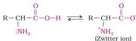
The figure makes the point of the section literally: amino acids are solids you can hold — colourless, crystalline, water-soluble and high-melting. They behave like salts rather than simple amines or acids because the carboxyl group can lose a proton while the amino group accepts one, giving a neutral dipolar ion called the zwitter ion.
In this form amino acids show amphoteric behaviour, reacting with both acids and bases — the mechanism behind exercise 10.16.
Essential amino acids cannot be synthesised in the body and must come from the diet; the ten marked with an asterisk in the chapter’s Table 10.2 are valine, leucine, isoleucine, arginine, lysine, threonine, methionine, phenylalanine, tryptophan and histidine. Except glycine, naturally occurring \( \alpha \)-amino acids are optically active; most have the L-configuration, written with the \( -\mathrm{NH_2} \) group on the left.
Peptide linkage and the four levels of protein structure
A peptide linkage is an amide bond: the \( -\mathrm{COOH} \) group of one amino acid combines with the \( -\mathrm{NH_2} \) group of another, a water molecule is eliminated, and the bond \( -\mathrm{CO\text{-}NH{-}} \) forms.
Two amino acids give a dipeptide, three a tripeptide, and more than ten a polypeptide; a protein is generally a polypeptide with more than a hundred amino acid residues and a molecular mass above 10,000 u. Insulin is the stated exception — 51 amino acids, still called a protein because it has a well defined protein conformation (Section 10.2.3).
Proteins come in two molecular shapes: fibrous (parallel chains held by hydrogen and disulphide bonds, insoluble — keratin in hair, wool and silk, myosin in muscle) and globular (chains coiled into a sphere, usually soluble — insulin and albumins). Structure is described at four levels:
- Primary — the sequence of amino acids in the polypeptide chain; changing the sequence makes a different protein.
- Secondary — regular folding of the backbone by hydrogen bonding between \( -\mathrm{C=O} \) and \( -\mathrm{NH{-}} \) groups of the peptide bond; two types, the \( \alpha \)-helix and the \( \beta \)-pleated sheet.
- Tertiary — overall folding of the chain into a shape, giving fibrous or globular forms; stabilised by hydrogen bonds, disulphide linkages, van der Waals and electrostatic forces.
- Quaternary — the spatial arrangement of two or more polypeptide chains, called subunits.
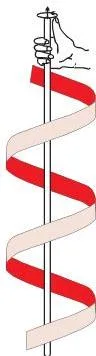
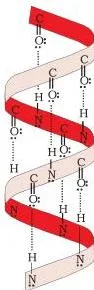
The helix figure shows the backbone twisted into a right-handed screw, with the \( -\mathrm{NH} \) group of each amino acid residue hydrogen-bonded to the \( \gt\mathrm{C=O} \) of an adjacent turn — that hydrogen bonding is the stabilising force exercise 10.14 asks about. The other secondary structure, the \( \beta \)-pleated sheet, is decoded in the figure walkthrough below.
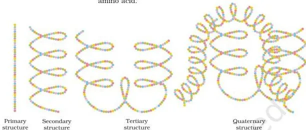
In the diagrammatic figure, each coloured ball is one amino acid. Watch the same chain gain a helix, fold into a globular shape, then assemble with other chains into the quaternary unit — the four levels of exercise 10.12 in one drawing.
Denaturation of proteins
A protein in its biological form, with a unique three-dimensional structure and biological activity, is a native protein. When temperature or pH changes, the hydrogen bonds are disturbed: globules unfold, helices uncoil, and the protein loses its biological activity. This is denaturation. Secondary and tertiary structures are destroyed; the primary structure remains intact.
The coagulation of egg white on boiling and the curdling of milk (lactic acid from bacteria) are the everyday examples (Section 10.2.4).
Enzymes
Enzymes are biocatalysts — they drive the body’s reactions under very mild conditions and are almost all globular proteins. They are specific for a particular reaction and a particular substrate, and are generally named after the compound they work on, as the ending -ase signals (Section 10.3). The chapter’s example:
\[ \mathrm{C_{12}H_{22}O_{11} \xrightarrow{\text{Maltase}} 2C_6H_{12}O_6} \]
Some enzymes are named after the reaction type — oxidoreductases catalyse the oxidation of one substrate with simultaneous reduction of another. The activation energy numbers make the mechanism point: acid hydrolysis of sucrose needs about 6.22 kJ mol⁻¹, while hydrolysis by the enzyme sucrase needs only about 2.15 kJ mol⁻¹.
Enzymes lower the activation energy, which is why they work in such small quantities.
Vitamins, nucleic acids and hormones: sections 10.4 to 10.6 in brief
The last three sections of the unit move from chemistry to biology, but the exam answers still live in the chemical definitions here. Keep each definition exact.
Vitamins
Vitamins are organic compounds required in the diet in small amounts to perform specific biological functions for normal growth and health; their deficiency causes specific diseases. Most cannot be synthesised in the body, though gut bacteria can produce some.
The name “vitamine” was coined from vital + amine; the final e was dropped when later work showed most vitamins lack amino groups. Excess vitamins are harmful, so the chapter warns against vitamin pills without a doctor’s advice (Section 10.4).
- Fat-soluble: vitamins A, D, E and K — stored in the liver and adipose (fat-storing) tissues.
- Water-soluble: the B group and vitamin C — readily excreted in urine, so they must be supplied regularly; the exception is vitamin B₁₂.
Mnemonic for the two classes: the fat-soluble four spell ADEK (a hook: “All Day Eat Kale”) and they stay in the body; the water-soluble set is the B group plus C — the ones that go down the drain in urine and must be replaced.
| Vitamin | Deficiency disease | Key sources |
|---|---|---|
| A | Night blindness; xerophthalmia (hardening of the cornea) | Fish liver oil, carrots, butter, milk |
| B₁ (Thiamine) | Beriberi (loss of appetite, retarded growth) | Yeast, milk, green vegetables, cereals |
| B₂ (Riboflavin) | Cheilosis (fissuring at the corners of the mouth), digestive disorders | Milk, egg white, liver, kidney |
| B₆ (Pyridoxine) | Convulsions | Yeast, milk, egg yolk, cereals, grams |
| B₁₂ | Pernicious anaemia (RBC deficient in haemoglobin) | Meat, fish, egg, curd |
| C (Ascorbic acid) | Scurvy (bleeding gums) | Citrus fruits, amla, green leafy vegetables |
| D | Rickets (bone deformities in children); osteomalacia (soft bones in adults) | Sunlight, fish, egg yolk |
| E | Increased fragility of RBCs, muscular weakness | Vegetable oils such as wheat germ oil |
| K | Increased blood clotting time | Green leafy vegetables |
Nucleic acids
Nucleic acids are long-chain polymers of nucleotides, so they are also called polynucleotides. Complete hydrolysis of DNA or RNA gives a pentose sugar, phosphoric acid and nitrogen-containing heterocyclic bases. In DNA the sugar is \( \beta \)-D-2-deoxyribose; in RNA it is \( \beta \)-D-ribose (Section 10.5.1).
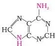
The base figure shows the heterocyclic bases. DNA and RNA share three — adenine, guanine and cytosine — and the fourth differs: thymine in DNA, uracil in RNA.
A base attached to the 1′ position of the sugar is a nucleoside; when phosphoric acid is linked at the 5′ position, the result is a nucleotide. Sugar carbons are numbered 1′, 2′, 3′ and so on precisely to distinguish them from the base atoms. Nucleotides are joined by a phosphodiester linkage between the 5′ and 3′ carbons of the pentose sugar (Section 10.5.2).
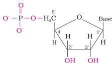
In the nucleoside/nucleotide figure, the difference is a single phosphate: (a) has only sugar plus base, (b) has the phosphate on board at the 5′ position. That one group is the entire answer to exercise 10.22.
Structurally, DNA is a double helix — two nucleic acid chains wound about each other and held by hydrogen bonds between base pairs: adenine pairs with thymine, cytosine with guanine. The two strands are complementary, not identical. RNA is single-stranded, with single-stranded helices in its secondary structure that sometimes fold back on themselves.
The three types of RNA are messenger RNA, ribosomal RNA and transfer RNA (m-RNA, r-RNA, t-RNA).
Functionally, DNA is the chemical basis of heredity — the reserve of genetic information that self-duplicates during cell division, with identical strands passed to daughter cells. The message for protein synthesis sits in DNA, while the RNA molecules carry out the synthesis.
DNA fingerprinting exploits the unique base sequence of each individual, which is the same in every cell and cannot be altered by any known treatment; it is used to identify criminals, determine paternity, identify bodies after accidents and study racial groups.
Hormones
Hormones are intercellular messengers produced by endocrine glands and poured directly into the blood, which carries them to the site of action (Section 10.6). Chemically they are steroids (estrogens and androgens), polypeptides (insulin and endorphins) or amino acid derivatives (epinephrine and norepinephrine).
- Insulin is released in response to a rapid rise in blood glucose and lowers it; glucagon tends to increase blood glucose. The two together keep blood glucose within a narrow limit.
- Thyroxine is an iodinated derivative of the amino acid tyrosine. Low thyroxine causes hypothyroidism (lethargy, obesity); low dietary iodine leads to hypothyroidism and goitre — the reason commercial table salt is iodised.
- Glucocorticoids control carbohydrate metabolism, modulate inflammatory reactions and take part in stress responses; mineralocorticoids control water and salt excretion by the kidney. Failure of the adrenal cortex can cause Addison’s disease.
- Testosterone drives male secondary sex characters (deep voice, facial hair, physical constitution); estradiol drives female secondary characters and controls the menstrual cycle; progesterone prepares the uterus for implantation of the fertilised egg.
Key reactions at a glance: what each reagent proves
Most of the chapter’s organic chemistry is one evidence table. This is the whole chain on one screen, with the glucose reagent column matching exercise 10.9.
| Reaction | Product | What it proves |
|---|---|---|
| Sucrose + dilute acid | Glucose + fructose in equal amounts | Sucrose is a disaccharide of glucose and fructose |
| Starch + dilute \( \mathrm{H_2SO_4} \), 393 K, 2–3 atm | Glucose | Starch is a glucose polymer; commercial preparation of glucose |
| Glucose + HI, prolonged heating | n-Hexane | All six carbons form a straight chain |
| Glucose + hydroxylamine / HCN | Oxime / cyanohydrin | A carbonyl group is present |
| Glucose + bromine water | Gluconic acid | The carbonyl group is an aldehyde group |
| Glucose + acetic anhydride | Glucose pentaacetate | Five \( -\mathrm{OH} \) groups on different carbons |
| Glucose + nitric acid | Saccharic acid | A primary alcoholic \( -\mathrm{OH} \) group is present |
| Sucrose + water (hydrolysis) | Laevorotatory mixture — invert sugar | Fructose’s rotation (\( -92.4^\circ \)) outweighs glucose’s (\( +52.5^\circ \)) |
| Maltose + maltase | Two glucose units | Maltose is two glucose units; enzymes are named after their substrate |
Figure walkthrough: the densest diagrams decoded
Three figures carry most of the visual load in the second half of the unit. Here is what to look for in each.
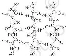
Beta-pleated sheet: the peptide chains are stretched out to nearly maximum extension and laid side by side, held together by intermolecular hydrogen bonds. The name comes from its resemblance to the pleated folds of drapery.
Contrast it with the \( \alpha \)-helix: both are secondary structure, but the helix is one chain twisting, while the sheet is parallel chains side by side.
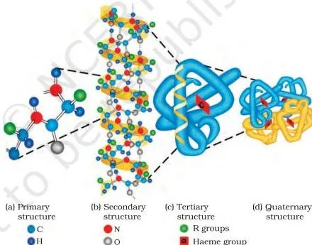
Haemoglobin: read the panels left to right to watch the same molecule climb the four levels — the amino acid sequence, the folding into helices, the overall globular fold, then the quaternary arrangement of subunits. This is the payoff figure of Section 10.2.3.
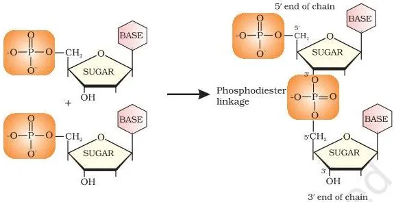
Dinucleotide: two nucleotides joined by a phosphodiester linkage between the 5′ carbon of one pentose sugar and the 3′ carbon of the next. The repeating phosphate–sugar spine is what makes nucleic acids polynucleotides.
Haworth projections, one line each, referring back to figures already read: in the glucose cyclic figure, the ring is a flat hexagon, the anomeric carbon is C1 — the former aldehyde carbon, now bonded to the ring oxygen — and it is the only carbon whose \( -\mathrm{OH} \) orientation changes between \( \alpha \) and \( \beta \); in the fructose Haworth pair, the ring is a pentagon because furanose is five-membered, and the anomeric carbon is C2.
Key terms defined: the vocabulary of the chapter
The end-of-unit exercises are largely definition-driven. Here is every term they lean on, in the order the chapter presents them.
| Term | Definition in this chapter |
|---|---|
| Monosaccharide | A carbohydrate that cannot be hydrolysed further to a simpler unit of polyhydroxy aldehyde or ketone; e.g., glucose, fructose, ribose. |
| Reducing sugar | A carbohydrate that reduces Fehling’s solution and Tollens’ reagent; every monosaccharide, aldose or ketose, is reducing. |
| Glycosidic linkage | The oxide linkage between two monosaccharide units through an oxygen atom, formed with the loss of a water molecule. |
| Anomer | One of two cyclic forms of a sugar that differ only in the configuration of the \( -\mathrm{OH} \) at the anomeric carbon. |
| Anomeric carbon | The aldehyde (or ketone) carbon before cyclisation, which becomes asymmetric on ring formation — C1 in glucose, C2 in fructose. |
| Pyranose | A six-membered cyclic sugar ring, named in analogy with pyran. |
| Furanose | A five-membered cyclic sugar ring, named in analogy with furan. |
| Invert sugar | The equimolar glucose–fructose mixture from sucrose hydrolysis, so named because the rotation flips from (+) to (−). |
| Zwitter ion | A dipolar ion, neutral overall but carrying both positive and negative charges, formed when the carboxyl group loses a proton and the amino group accepts one. |
| Essential amino acid | An amino acid the body cannot synthesise and so must obtain through the diet; ten are marked in the chapter’s Table 10.2. |
| Peptide linkage | The amide bond \( -\mathrm{CO\text{-}NH{-}} \) formed between the carboxyl group of one amino acid and the amino group of another, with loss of water. |
| Denaturation of proteins | Loss of the three-dimensional structure and biological activity of a protein when temperature or pH change disturbs hydrogen bonds; secondary and tertiary structure are destroyed, primary survives. |
| Enzyme | A biocatalyst, almost always a globular protein, specific for a particular reaction and a particular substrate. |
| Vitamin | An organic compound required in the diet in small amounts for specific biological functions; deficiency causes specific diseases. |
| Nucleoside | A base attached to the 1′ position of a pentose sugar. |
| Nucleotide | A nucleoside linked to phosphoric acid at the 5′ position of the sugar. |
| Phosphodiester linkage | The bond joining nucleotides through the 5′ and 3′ carbon atoms of the pentose sugar. |
| Hormone | A molecule produced by endocrine glands and carried by the blood, acting as an intercellular messenger. |
Common mistakes in this chapter and how to avoid them
The errors below are the ones that cost marks in this unit. Each row names the mistake before you can make it.
| Mistake | Correct rule | How to check your answer |
|---|---|---|
| Judging by the formula \( \mathrm{C_x(H_2O)_y} \) | The definition is functional: optically active polyhydroxy aldehyde or ketone, or a source of such units on hydrolysis | Ask: does it behave as a polyhydroxy carbonyl? Acetic acid fits the formula but is not a carbohydrate; rhamnose does not fit but is one. |
| D means dextrorotatory | D and L are configuration labels fixed by glyceraldehyde at the lowest asymmetric carbon; (+) and (−) are measured rotation | Glucose is D(+) and fructose is D(−) — same D family, opposite rotations, so D cannot mean +. |
| Writing only the open chain structure for glucose | The open chain cannot explain no Schiff’s test, no \( \mathrm{NaHSO_3} \) product, a pentaacetate that gives no oxime, and two crystalline forms | Any question mentioning Schiff’s test or two melting points is pointing at the cyclic hemiacetal structure. |
| Calling \( \alpha \) and \( \beta \) glucose enantiomers | They are anomers — they differ only in the \( -\mathrm{OH} \) orientation at C1, the anomeric carbon | Enantiomers are mirror images at every asymmetric carbon; here only C1 changes. |
| Assuming every disaccharide is reducing | A disaccharide is reducing only if a free aldehyde or ketone can regenerate; sucrose has both locked in the glycosidic linkage | Ask: can hydrolysis free a reducing group? Maltose and lactose yes, sucrose no. |
| Saying starch is the \( \beta \)-glucose polymer | Starch is \( \alpha \)-glucose (amylose + amylopectin); cellulose is the \( \beta \)-glucose straight-chain polymer | Check the monomer: alpha for starch, beta for cellulose — that is the whole answer to exercise 10.8. |
| Swapping nucleoside and nucleotide | A nucleotide is a nucleoside plus a phosphate at the 5′ position | Look for the phosphate: no phosphate, it is a nucleoside. |
| Forgetting water-soluble vitamins are excreted | The B group and C are excreted in urine and must be taken regularly — the exception is vitamin B₁₂ | Classify first: A, D, E, K are fat-soluble; B group and C are water-soluble. |
How to revise this chapter: exercises, sections and a sensible order
The unit’s exercise set follows the sections in order, so mapping it tells you which section you have and have not understood. No question numbers are promised here — the point is the pattern.
- The first block of exercises tests Section 10.1: monosaccharides, reducing sugars, classification, glycosidic linkage, glycogen, the hydrolysis products of sucrose and lactose, the starch–cellulose difference, and the glucose reagent reactions — exercise 10.9, for instance, asks for HI, bromine water and nitric acid.
- The middle block tests Sections 10.2 and 10.3: essential and non-essential amino acids, peptide linkage, protein structure levels, the bonding in the \( \alpha \)-helix, globular versus fibrous proteins, denaturation, amphoteric behaviour and enzymes.
- The final block tests Sections 10.4 to 10.6: vitamin classification and deficiency diseases, nucleic acid functions, nucleoside versus nucleotide, complementary strands in DNA, DNA versus RNA, and the RNA types.
In-text questions appear at the end of the carbohydrate, protein and nucleic acid sections; attempt them as you read, because they target exactly the facts that trip students later — glucose solubility, the hydrolysis products of lactose, and the missing aldehyde in the pentaacetate.
A workable order: definition and classification first, then the glucose evidence chain, then cyclic structure and anomers, then the disaccharide and polysaccharide comparisons, then proteins and nucleic acids. Before the exam, confirm the examinable portion against the current official syllabus.
Revision summary: the chapter in ten lines
The unit closes with its own Summary; here is that ground, regrouped by biomolecule.
Carbohydrates
- Carbohydrates are optically active polyhydroxy aldehydes or ketones, or compounds that give such units on hydrolysis.
- They are grouped into monosaccharides, disaccharides and polysaccharides, and monoscaccharide units are held together by glycosidic linkages.
- Glucose, the most important source of energy for mammals, is obtained by the digestion of starch.
Proteins and enzymes
- Proteins are polymers of about twenty \( \alpha \)-amino acids linked by peptide bonds; ten amino acids are essential because the body cannot synthesise them.
- Proteins perform structural and dynamic functions; a change of pH or temperature disturbs their secondary or tertiary structure, causing denaturation while the primary structure stays intact.
- Enzymes are biocatalysts that speed up biosystem reactions; they are specific, selective, and chemically mostly proteins.
Vitamins
- Vitamins are accessory food factors, classified as fat-soluble (A, D, E, K) and water-soluble (B group and C); their deficiency leads to many diseases.
Nucleic acids
- Nucleic acids are polymers of nucleotides, each nucleotide being a base, a pentose sugar and a phosphate.
- DNA carries 2-deoxyribose and thymine, is double-stranded, and is the chemical basis of heredity; RNA carries ribose and uracil, is single-stranded, and its three types — mRNA, rRNA and tRNA — carry out protein synthesis.
Related chapters and official sources
This chapter sits inside the larger NCERT Class 12 Chemistry book page, which lists the units of both Chemistry Part I and Part II; the Class 12 hub holds the other subjects of the class.
The previous chapter of this book, Amines, covers the nitrogen compounds whose chemistry carries into the amino acids here, and the CBSE notes index organises revision material by class and subject. The official PDF of Unit 10 is hosted on ncert.nic.in — open it to check any wording against the printed textbook.
Sources and data verification
- This page describes Unit 10 Biomolecules of the NCERT Class 12 Chemistry Part II textbook, with Section 10.1 Carbohydrates as its focus, following the NCERT reprint for 2026-27.
- It covers the chapters listed on this site’s Class 12 Chemistry directory — not the full CBSE scheme of studies, which includes other subjects and electives.
- The listing is maintained for the current academic session using the NCERT information available to us at the time of writing.
- NCERT settles the textbooks, their editions and the official PDFs; CBSE settles the curriculum and the examinable syllabus.
Reference: NCERT Class 12 Chemistry textbook, Unit 10 Biomolecules, official edition on ncert.nic.in.
Frequently asked questions about the carbohydrates class 12 chapter
Is the carbohydrates class 12 chapter the same as the Biomolecules chapter?
Yes, in the sense that matters for study: carbohydrates is Section 10.1 inside Unit 10, and the whole unit is titled Biomolecules. The NCERT Class 12 Chemistry Part II textbook has no separate carbohydrates unit — what students call the carbohydrates chapter is Unit 10’s Section 10.1, followed by proteins, enzymes, vitamins, nucleic acids and hormones.
What is the difference between D-glucose and L-glucose?
D and L are configuration labels fixed by comparison with glyceraldehyde at the lowest asymmetric carbon: if the OH there lies on the right in the drawn structure, the compound is D; on the left, it is L.
Rotation is measured separately — glucose is D(+) while fructose is D(−), both in the same D family with opposite rotations, which is why you can never deduce rotation from D or L.
Why is sucrose a non-reducing sugar while maltose and lactose are reducing?
Because both of sucrose’s reducing groups are locked into the glycosidic linkage: C1 of \( \alpha \)-D-glucose joins C2 of \( \beta \)-D-fructose, so neither an aldehyde nor a ketone is free to reduce Fehling’s solution or Tollens’ reagent.
In maltose and lactose, the linkage leaves the C1 of the second unit able to regenerate a free aldehyde group in solution, so they are reducing.
What is the difference between starch and cellulose?
Starch is the plant storage polymer of \( \alpha \)-glucose, with two components — amylose (unbranched, C1–C4) and amylopectin (branched at C1–C6). Cellulose is the structural polymer of \( \beta \)-D-glucose in straight C1–C4 chains and is the main constituent of plant cell walls. The same glucose family, but a different isomer and a different job.
What are anomers, and how do alpha and beta glucose differ?
Anomers are cyclic forms of a sugar that differ only in the configuration of the OH at the anomeric carbon — C1 in glucose, the former aldehyde carbon. Alpha and beta glucose are therefore anomers, not enantiomers: they differ at exactly one carbon, and the three-dimensional arrangement everywhere else is the same.
Where can I download the official NCERT Class 12 Chemistry carbohydrates chapter PDF?
The official file is Unit 10 (lech205.pdf) on ncert.nic.in, the same link used at the top of this page: Download the NCERT carbohydrates class 12 chapter PDF (Biomolecules, Unit 10). It is the NCERT-published PDF, not a third-party copy.
Explore Class 12 Chemistry Books
- Previous: Structure of Amines
Class 12 Chemistry on LearnCBSE:
Related chapters: