
This page carries the official NCERT Class 12 Biotechnology Chapter 6 PDF — the chapter printed in the book as “Microbial Culture”, which opens the Class 12 unit on microbial, plant, animal cell, organ cultures and bioprocessing. The chapter runs about 26 pages.
The current NCERT edition, maintained for this academic session, is linked directly below.
Open the NCERT Class 12 Biotechnology Chapter 6: Microbial, Plant, Animal Cell, Organ Cultures and Bioprocessing PDF to read the complete chapter exactly as NCERT published it — the historical perspective, nutrition and media, sterilisation, pure culture techniques, growth factors, the growth curve and the full exercise set in one file.
| What the chapter holds | Count | Where it is used |
|---|---|---|
| Printed pages | 26 | |
| Sections in the chapter | 6 | |
| Figures with NCERT captions | 6 | |
| Worked examples | 1 | solved step by step in our NCERT Solutions |
| Exercise questions | 15 | answered in our NCERT Solutions |
| Official NCERT PDF | Download the chapter PDF | the chapter exactly as NCERT publishes it |
Microbial, Plant, Animal Cell, Organ Cultures and Bioprocessing — Chapter 6 at a glance
The table below lists the chapter’s sections, figures, worked examples and exercise questions, so you can see at a glance how much of the chapter there is before you start reading.
What this chapter covers
Section 6.1, Historical Perspective, opens with Leeuwenhoek’s “animalcules” and the old belief that microbes arise from lifeless matter. Spallanzani’s boiled broth contradicted that claim. Pasteur’s S-shaped flasks then disproved spontaneous generation and produced the germ theory, which Koch proved with anthrax. The same era delivered solid media, agar and the Petri dish.
| Who | When | Contribution to the chapter’s history |
|---|---|---|
| Antonie van Leeuwenhoek | 1670s–1680s | Observed microscopic organisms and called them “animalcules” |
| Lazzaro Spallanzani | — | Found no microscopic life in boiled broth, contradicting spontaneous generation |
| Louis Pasteur | Middle and late 1800s | S-shaped flask experiments disproved spontaneous generation and led to the germ theory |
| Robert Koch | 1881 onward | Proved the germ theory with Bacillus anthracis; introduced solid media and the term “colony” |
| Julius Richard Petri | 1887 | Replaced flat glass plates with the shallow dish and loose cover |
Section 6.2, Nutritional Requirements and Culture Media, establishes what a microbe must be fed: macronutrients, trace elements, carbon and nitrogen sources, growth factors, anti-foams and water. The guiding rule is that the medium should match the species’ natural habitat.
Section 6.3, Sterilisation Methods, explains how media, tools and working surfaces are freed of all living microorganisms before any culture can start — by heat, radiation, filtration or chemicals.
Section 6.4, Pure Culture Techniques, covers the three isolation methods: the streak plate, the pour plate and the spread plate. All three rely on one property of solid surfaces — a deposited cell stays put and grows into a visible colony.
Section 6.5, Factors Affecting Microbial Growth, lists the environmental dials a microbiologist sets: temperature, pH, oxygen, carbon dioxide and light, along with the vocabulary that describes how microbes respond to each.
Section 6.6, The Growth Curve, ties the chapter together. Plotting the logarithm of viable cell number against incubation time gives four phases — lag, exponential, stationary and death — and two equations describe the exponential phase mathematically.
The chapter ends with an exercise set in four formats — descriptive answers, single-choice questions, one calculation and assertion-reason items — and a summary of the main points.
Key concepts in this chapter
You cannot grow a microbe until you know what it eats, how to keep everything else out, and how to obtain a single strain to study. The seven subsections below follow exactly that sequence, in the order the chapter presents it; the diagrams are explained separately in the figure walkthrough.
How microbes are classified by their nutrition
Before the chapter builds a medium, it sorts microbes by where they get carbon and energy. Every name below ends in the source it names.
- Heterotrophs — carbon from organic molecules made by other organisms: all fungi and protozoans, and most types of bacteria.
- Autotrophs — carbon from carbon dioxide: a significant number of bacteria and nearly all algae.
- Chemoautotrophs — energy and carbon both from inorganic sources, by oxidising molecules such as sulphur or nitrite.
- Photoautotrophs — photosynthetic pigments convert light energy into chemical energy.
- Chemoheterotrophs — energy from chemical oxidation of organic nutrient molecules; the majority of heterotrophs.
- Photoheterotrophs — light for energy but organic carbon; for example the green and purple non-sulphur bacteria.
- Lithotrophs — inorganic molecules such as water, hydrogen sulphide or ammonia as the electron source.
- Organotrophs — organic molecules as the electron source.
These terms feed exercise question 7, so keep the pairs straight: chemo/photo is about energy, litho/organo about electrons, and auto/hetero about carbon.
What a culture medium must supply
A culture medium is a recipe. The chapter lists the ingredients in order: macronutrients first, then trace elements, then the extras that make a medium workable.
- Macronutrients — C, O, H, N, S and P build carbohydrates, lipids, proteins and nucleic acids; K, Ca, Mg and Fe act mainly as enzyme cofactors.
- Micronutrients or trace elements — Mn, Zn, Co, Mo, Ni, Cu; needed in small amounts and usually already present as impurities in ordinary media components, so not added separately.
- Carbon source — glucose, lactose, sucrose, starch, glycogen, cellulose, cereal grain powders or cane molasses.
- Nitrogen, phosphorus and sulphur sources — ammonium salts, urea, animal or plant tissue extracts for nitrogen; inorganic phosphate for phosphorus; sulfate for sulphur.
- Growth factors — amino acids, purines, pyrimidines and vitamins the cell cannot synthesise for itself.
- Anti-foams — olive oil, sunflower oil and silicones; not nutrients, but they stop foaming when the medium is agitated.
- Water — the essential base of any medium, liquid or solid.
The chapter’s practical rule closes the list: the right medium depends on the species, so prior knowledge of the microbe’s natural habitat is the best guide to choosing one.
The three ways media are classified
Media can be grouped three ways, and exercise question 2 asks for exactly this classification. The table shows the three bases and the types under each.
| Basis of classification | Types | What defines them |
|---|---|---|
| Chemical composition | Synthetic (chemically defined) | Every component known; M9 for E. coli, BG11 for cyanobacteria |
| Complex | Some constituents unknown; peptones, beef extract, yeast extract; nutrient broth, LB, MacConkey agar, potato dextrose agar | |
| Consistency | Liquid (broth) | No agar; growth becomes visible after incubation |
| Solid | 1.0–2.0% agar; used for slants, slopes and stabs | |
| Semi-solid | 0.5% agar; favours one organism and retards another | |
| Function | Selective | Favours one type, suppresses others; e.g. cellulose-only medium, MacConkey agar |
| Differential | Distinguishes groups by appearance; e.g. blood agar showing haemolysis | |
| Enrichment | Nutritional environment adjusted to favour a target group; e.g. blood agar, tissue extracts in nutrient broth |
One point explains why agar wins as the solidifying agent: agar is a sulphated polymer from red algae that gives solid support but is not a nutrient — most microorganisms cannot degrade it.
Notice that MacConkey agar appears in both the selective and the differential groups. Its dye suppresses Gram-positive bacteria, and its lactose-plus-dye system turns fermenters red and leaves non-fermenters colourless.
Sterilisation: the four routes the chapter gives
Sterilisation means freeing media, glassware, working surfaces and plasticware from all living microorganisms, including spores. The chapter gives four routes, each with its own logic.
| Route | How it works | Key details to remember |
|---|---|---|
| Heat | Denatures and coagulates proteins; oxidative effects stop metabolism | Boiling 100°C for 30 min; autoclaving at 15 psi reaches 121°C with holding time usually over 30 min; pasteurisation LTLT 63°C/30 min, HTST 72°C/15 s, UHT 140–150°C/1–3 s; flaming and hot air oven for dry heat |
| Radiation | Damages DNA | UV 200–280 nm (260 nm most effective) forms thymine dimers that block DNA replication, used for surface disinfection; electron beams and gamma rays sterilise plastics, pharmaceuticals and fabrics |
| Filtration | Separates microbes by size; does not kill | Membrane filters of 0.2–0.45 µm pores for heat-labile solutions such as serum and antibiotics; HEPA filters 99.97% efficient for particles above 0.3 µm |
| Chemicals | Dehydrate cells, disrupt membranes, coagulate proteins | 70% ethyl alcohol, isopropyl alcohol, 40% formaldehyde (formalin), heavy metals, hydrogen peroxide |
The trap to avoid now: heat-labile substances such as the antibiotic ampicillin must be filter-sterilised, never autoclaved. More on this in the common mistakes section.
How a pure culture is obtained
A pure culture is a culture consisting of entirely one strain of organism. Since microbes in nature live in complex mixed communities, isolating one strain is the first job of the laboratory.
Solid media are preferred here — they are useful for isolation and long-term storage, while liquid broths give rapid, large-scale production. Three methods achieve isolation:
- Streak plate method — an inoculating loop drags a drop of suspension across the agar so it becomes progressively more dilute; isolated colonies appear near the end of the streak.
- Pour plate method — the sample is diluted several times, 1 mL of each dilution goes into the dish, melted agar is poured in and mixed with a gentle circular motion; isolated cells grow into colonies.
- Spread plate technique — a small volume of diluted mixture is placed at the centre of solidified agar and spread evenly with a sterile L-shaped glass spreader.
All three work because of one property of solid surfaces: daughter cells cannot move away, so a clump of deposited cells gives rise to one visible colony.
| Method | How dilution is achieved | Where colonies appear | Tool and detail |
|---|---|---|---|
| Streak plate | By dragging the drop across the agar until it is progressively more dilute | Isolated colonies near the end of the streak | Inoculating loop; no separate dilution step |
| Pour plate | Sample diluted several times in the tube before plating | Colonies in and on the agar | 1 mL of each dilution per dish; melted agar mixed with circular motion |
| Spread plate | A pre-diluted mixture is spread thinly | Colonies on the agar surface | L-shaped glass spreader sterilised by dipping in alcohol and flaming |
The factors that control microbial growth
Growth is a response to the environment. The chapter lists five dials a microbiologist sets, and the names for organisms at different settings.
- Temperature — growth rises to an optimum, then falls as enzymes lose activity. Mesophiles grow best around 20–45°C; thermophiles around 40–80°C with optima near 50–65°C; extreme thermophiles above 100°C.
- pH — most microbes prefer near neutrality; many bacteria like slightly alkaline conditions; fungi prefer slightly acidic conditions and so dominate bacteria when both are present.
- Oxygen — aerobes need it; anaerobes survive without it; obligate anaerobes cannot tolerate it and need anaerobic chambers; facultative anaerobes grow either way; aerotolerant anaerobes are anaerobic but unharmed by oxygen. Broth cultures of aerobes need shaking or stirring for uniform growth.
- Carbon dioxide — autotrophs use it as their carbon source, supplied as bicarbonate or a CO₂-enriched atmosphere.
- Light — phototrophs need light of the correct wavelength for photosynthesis.
This list supplies both halves of exercise question 15, so it is worth checking against that question at the end of the chapter.
The growth curve: four phases and the maths
The growth curve is what you get when you plot the logarithm of viable cell number against incubation time. The log scale matters: growth is exponential, so a log axis keeps all four phases visible on one plot.
- Lag phase — no immediate rise in cell number and no net increase in mass; the cell is synthesising the components needed for division. The phase is long if the inoculum is old or refrigerated, and short or absent if it is young.
- Exponential (log) phase — cells divide at a constant maximal rate and double at regular intervals; the doubling time for E. coli is about 20 minutes. The population is most uniform chemically and physiologically here, which is why this phase is used for biochemical studies.
- Stationary phase — growth ceases and the curve goes horizontal; viable numbers stay constant because cell division and cell death balance, usually because an essential nutrient is depleted.
- Death (decline) phase — viable cells fall, often from nutrient deprivation and the accumulation of toxic wastes; Streptococci acidify their own medium with lactic acid and stop growing.
Order mnemonic: Lag, Exponential, Stationary, Death — “Lions Eat Steaks Daily”.
The maths is two linked equations:
\[ t_d = \frac{0.693}{\mu} \]
\[ \mu = 2.303 \frac{(\log X_t – \log X_0)}{t} \]
The full symbol table and a worked example are in the growth rate formulas section below.
Reading the figures in Chapter 6
The practical methods of this chapter are visual: how dilution is achieved, where colonies appear, what the axes of the graph are — all of it is easier to see than to read.
Each figure below is in your book, and the details you are likely to be tested on often live in the diagrams, so these walkthroughs point at what matters in each one.
The Petri dish
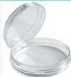
The shallow dish shown here is the container that changed routine culturing. Because the cover fits loosely, dust and contaminants are kept out while air can still reach the culture inside — exactly the balance a culture vessel needs after Pasteur had shown that air carries microbes.
Julius Richard Petri introduced the dish in 1887, at a time when solid media had already replaced broth for isolating bacteria. The shallow, covered form made it practical to inspect colonies without opening the culture.
Fig. 6.1: vertical and horizontal autoclaves
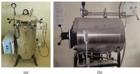
The autoclave is the most common sterilisation instrument in the chapter, and the photograph shows its two designs — (a) vertical and (b) horizontal. The principle is the same: water boils in a closed air-tight chamber, the pressure rises to 15 psi, and the boiling temperature rises with it to 121°C. Articles are exposed to steam at this temperature.
Two distinctions matter. A jacketed autoclave keeps steam in the jacket so articles are not directly exposed; a non-jacketed one exposes them directly. The holding time is the time the whole load is exposed — it depends on the size and type of load and generally exceeds 30 minutes.
Fig. 6.2: the streak plate method
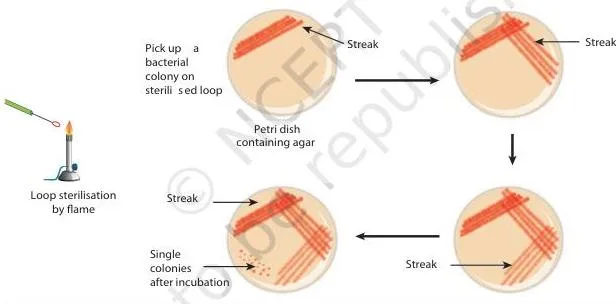
The diagram shows an inoculating loop streaking a drop of bacterial suspension across solid agar in a pattern that progressively dilutes the cells. After incubation at the right temperature, cell division produces visible colonies — and near the end of the streak, where the cells are most dilute, the colonies are isolated.
This is the book’s way of showing why the end of the streak is where single strains separate: on a solid surface daughter cells cannot move away, so each clump of deposited cells grows into one visible colony.
Fig. 6.3: the pour plate method
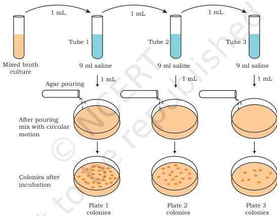
The figure shows the alternative route: here the dilution happens in the tube before plating, not on the agar. The original sample is diluted several times, small volumes (1 mL) of each dilution go into a Petri dish, and melted agar is poured in and mixed gently with a circular motion.
After incubation, isolated cells grow into colonies that can be used to establish pure cultures. Contrast this with the streak plate: in the pour plate, colonies can form within the agar as well as on its surface.
Fig. 6.4: the spread plate technique
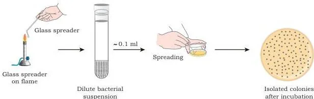
The third isolation route is the simplest to picture: a small volume of diluted bacterial mixture is dropped at the centre of a solidified agar plate and spread evenly over the surface with a sterile, L-shaped glass spreader.
The figure’s point is the tool. The glass spreader is sterilised by dipping it in alcohol and flaming to burn the alcohol off, and after incubation some of the dispersed cells develop into isolated colonies on the surface.
Fig. 6.5: the microbial growth curve
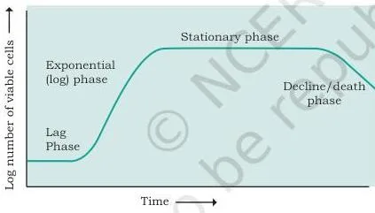
Read the axes first: the vertical axis is the logarithm of the number of viable cells, and the horizontal axis is incubation time. The log scale is what lets all four phases be seen on one plot, because growth is exponential.
Walk the curve left to right: the flat lag phase, the steeply rising exponential phase where doubling happens, the horizontal stationary phase where division and death balance, and the falling death phase. Exercise question 3 asks for exactly this account, so a student who can read this figure can answer it.
Key definitions from this chapter
These are the terms the chapter and its exercises assume you know. Each definition is one line so you can check them fast.
| Term | One-line definition |
|---|---|
| Microbiology | The study (logos) of small (micro) organisms (bios) |
| Pure culture | A culture consisting entirely of one strain of organism |
| Colony | Pure, discrete growth of bacteria on solid media; the term was coined by Koch |
| Sterilisation | Killing or removing all living microorganisms, including bacterial spores |
| Disinfection | Surface treatment by UV radiation or chemicals |
| Pasteurisation | Heating milk to kill pathogenic microorganisms; it does not fully sterilise the milk |
| Synthetic (chemically defined) medium | Medium in which every chemical component is known |
| Complex medium | Medium containing some constituents of unknown chemical composition |
| Selective medium | Favours growth of one type of microorganism and suppresses others |
| Differential medium | Distinguishes microorganism groups by their appearance and biological characteristics |
| Enrichment medium | Nutritional environment adjusted to enhance growth of a target group in a mixed inoculum |
| Mesophile | Microorganism with optimal growth in the range 20–45°C |
| Generation time | Time required for cell mass or cell number to double during balanced growth |
Exercise question 5 asks for three terms, but the chapter defines two of them: sterilisation and disinfection. The third, “sanitisation”, is not separately defined in this chapter, so answer the two the book covers and note that the third is not settled here.
Common mistakes in this chapter
Most errors here are concept slips, not calculation slips. Each mistake below comes with its correction and a way to check yourself.
| Mistake | Correct rule | How to check your answer |
|---|---|---|
| Treating pasteurisation as sterilisation | Pasteurisation kills pathogenic microorganisms but leaves many living organisms and spores alive; sterilisation removes all living microorganisms including spores | Ask whether the method destroys spores. Milk after pasteurisation is not sterile |
| Thinking agar feeds the microbes | Agar is a sulphated polymer from red algae that provides solid support, not nutrients; most microorganisms cannot degrade it | Remember the history: agar replaced gelatin partly because it is not digested by the culture |
| Autoclaving every component of a medium | Heat-labile substances such as the antibiotic ampicillin are destroyed by autoclaving and must be filter-sterilised with membrane filters | If an autoclaved selective medium lets both resistant and sensitive strains grow, the selective agent was destroyed |
| Dropping the 2.303 factor in \( \mu = 2.303 \frac{(\log X_t – \log X_0)}{t} \) | The formula uses log base 10, and ln(10) = 2.303, so each tenfold change in cell number adds 2.303 to the numerator | Count the tenfold rises: \( 10^3 \) to \( 10^9 \) is six rises, so the numerator is \( 6 \times 2.303 \) |
| Reading the lag phase as inactivity | There is no net increase in mass, but the cell is synthesising the components required for division | A short lag follows a young inoculum, a long lag follows an old or refrigerated one — both show the cells are active |
| Confusing selective and differential media | Selective media favour one type and suppress others; differential media distinguish groups by appearance. MacConkey agar is both | On MacConkey agar, lactose fermenters give red colonies and non-fermenters give colourless ones |
| Mixing up Spallanzani, Pasteur and Koch | Spallanzani’s boiled broth contradicted spontaneous generation; Pasteur disproved it and postulated the germ theory; Koch proved it with Bacillus anthracis and developed colonies on solid media | Match each scientist to one landmark: boiled broth, S-shaped flask, anthrax with colonies |
How the exercises in this chapter test you
The exercise set checks two things: recall of the chapter’s facts, and the ability to apply the growth equations and sterilisation logic. Each question maps to a specific section, as the table below shows. For revision of the same ground, the Class 12 Biotechnology notes collect the points chapter by chapter.
| Question | Format | Section it tests | What a complete answer needs |
|---|---|---|---|
| 1 | Descriptive | 6.2 | Macronutrients, micronutrients, carbon, nitrogen, phosphorus and sulphur sources, growth factors, anti-foams, water |
| 2 | Descriptive | 6.2 | Define culture medium, then classify by composition, consistency and function |
| 3 | Descriptive | 6.6 | Describe the log viable cells versus time plot and all four phases |
| 4 | Descriptive | 6.4 | Any two of streak, pour, spread — how dilution is achieved and why colonies form |
| 5 | Descriptive | 6.3 | Define sterilisation and disinfection; the chapter does not separately define sanitisation |
| 6 | Descriptive | 6.3 | Heat, radiation, filtration and chemicals, with an example of each |
| 7 | Single choice | 6.1, 6.2 | The chapter’s categories point to (b) Prototroph: heterotrophs need organic carbon, this strain does not |
| 8 | Single choice | 6.1 | (b) Robert Koch — he coined the term colony for pure, discrete growth on solid media |
| 9 | Single choice | 6.3 | (a) Pasteurisation — HTST and UHT are named among its methods |
| 10 | Single choice | 6.1 | The chapter says scientists of the time believed in spontaneous generation; Spallanzani’s boiled broth contradicted it |
| 11 | Single choice | 6.1 | (d) Louis Pasteur — he postulated the germ theory after disproving spontaneous generation |
| 12 | Calculation | 6.6 | Log difference 3 in 4 hours; \( \mu \) comes out near 1.72 per hour and \( t_d \) near 0.4 h (24 min), as printed with the question |
| 13 | Assertion-reason | 6.6 | (a) — alcohol is a metabolic waste product; its accumulation matches the death phase account |
| 14 | Assertion-reason | 6.3 | (a) — autoclaving destroys the antibiotic, so both strains grow; filter-sterilise instead |
| 15 | Assertion-reason | 6.5 | Both true; the extreme-thermophile fact is the chapter’s evidence for the wide range, so (a) |
Four formats are used: descriptive answers (questions 1–6), single-choice questions (7–11), one calculation (12) and assertion-reason items (13–15). For an assertion-reason item, test the reason on its own merits first, then decide whether it explains the assertion. Question 14 shows the method:
Assertion: a selective medium containing ampicillin is sterilised by autoclaving, and both amp\(^S\) and amp\(^R\) microbes grow.
Test the reason first: ampicillin is heat labile — the chapter puts antibiotics among the substances that must be sterilised by filtration, not heat.
So the reason (filter-sterilise the antibiotic before adding it) is true.
Then check the explanation: autoclaving destroys the ampicillin, so the medium no longer contains a working selective agent and both resistant and sensitive strains grow.
The reason explains the assertion.
Answer: (a) both true, reason is the correct explanation.
Question 12 is the only exercise with a numerical answer printed in the book — a specific growth rate near 1.72 per hour and a generation time near 0.4 hours (24 minutes). The formulas section below shows the full method with different numbers.
Question 10 needs care. The book does not name a single scientist who “suggested” spontaneous generation; it says scientists of the time believed microbes arise from lifeless matter, and that Spallanzani’s boiled broth contradicted that belief. Answer from the chapter’s account.
Textbook contents and the examinable syllabus are not always identical — check the current official syllabus before the exam.
Growth rate formulas: symbols, units and a worked example
Exponential growth is described by two linked quantities: the specific growth rate and the generation time. Every symbol used in Section 6.6 is defined below.
| Symbol | Meaning | Typical unit |
|---|---|---|
| \( \mu \) | Specific growth rate | Per hour |
| \( t_d \) | Doubling or generation time | Hours or minutes |
| \( X_t \) | Cell concentration at time \( t \) | Cells per mL |
| \( X_0 \) | Initial cell concentration | Cells per mL |
| \( t \) | Time taken | Hours |
| \( n \) | Number of generations | Count |
The chapter gives two equivalent forms for doubling time, and one for the specific growth rate:
\[ t_d = \frac{0.693}{\mu} \]
\[ \mu = 2.303 \frac{(\log X_t – \log X_0)}{t} \]
\[ t_d = \frac{t}{n} \]
Why the 2.303? The formula uses log base 10, and ln(10) = 2.303. So every tenfold increase in cell numbers adds exactly 2.303 to the numerator — count the tenfold rises, multiply by 2.303, divide by the time.
Question (original numbers): a culture grows from \( 10^3 \) cells per mL to \( 10^9 \) cells per mL in 6 hours of exponential growth.
Find the specific growth rate and the generation time.
Step 1: the log difference is \( 9 – 3 = 6 \) — that is six tenfold rises.
Step 2: substitute into the formula for \( \mu \).
\[ \mu = 2.303 \times \frac{6}{6} = 2.303\ \text{h}^{-1} \]
Step 3: convert to generation time.
\[ t_d = \frac{0.693}{2.303} = 0.30\ \text{h} = 18\ \text{min} \]
Check: 6 hours ÷ 0.30 hours per generation = 20 generations, and \( 10^3 \times 2^{20} \approx 1.05 \times 10^9 \), which matches the final count.
Final answer: \( \mu = 2.303 \) per hour and \( t_d = 0.30 \) hours (18 minutes).
The chapter closes the section with its industrial point: on the basis of the values of \( \mu \) and \( t_d \), the media composition and fermentation batch time are decided to optimise production in commercial fermenters. These two numbers are design parameters, not just exam arithmetic.
What to remember from Chapter 6
These are the chapter’s own summary points, in short form, for a quick check before an exam.
- Microbiology is the study of microorganisms — small life forms found everywhere.
- Every microbe needs a source of energy, carbon, nitrogen, oxygen, iron, other minerals, micronutrients and water.
- Prior knowledge of the microbe’s habitat is the best guide to choosing a culture medium.
- Media are classified by chemical composition (synthetic versus complex), by consistency (solid, liquid, semi-solid) and by function (selective, differential, enrichment).
- Sterilisation — killing or removing all living microorganisms including spores — is essential for any microbial study, done by physical or chemical methods.
- Temperature, pH, oxygen and other physical factors shape microbial growth.
- Plotting the logarithm of viable cell number against incubation time gives a four-phase growth curve: lag, exponential, stationary and death.
Related chapters and resources
This listing is maintained for the 2026-27 academic session using the NCERT textbook information available to us. NCERT remains the authority for confirming the latest edition.
Chapter 6 is the first chapter of a five-chapter unit in the Class 12 Biotechnology NCERT book. The unit continues with Plant Tissue Culture (Chapter 7), Animal Cell Culture (Chapter 8), Stem Cell Culture and Organ Culture (Chapter 9), and Bioprocessing and Biomanufacturing (Chapter 10).
For the rest of the book and every other subject in the same class, use the Class 12 NCERT books hub. The next chapter, Plant Tissue Culture (Chapter 7), carries the culture ideas of this chapter across to plants.
When you move from the PDF to revision, the Class 12 Biotechnology notes, the Class 12 notes hub and the full CBSE notes index collect the key points chapter by chapter. Notes on genome engineering and plant tissue culture cover other chapters of the same book.
Sources and data verification
This page describes Chapter 6 of the NCERT Biotechnology textbook for Class XII, whose printed chapter title is “Microbial Culture”, the opening chapter of the unit “Microbial, Plant, Animal Cell, Organ Cultures and Bioprocessing”. The section names, figures, summary points and exercise questions above come from the current NCERT edition of this book.
The official chapter PDF is published by NCERT on ncert.nic.in and is linked at the top of this page; the book’s page on the NCERT textbook portal is available here.
This listing is maintained on the basis of the current NCERT book, as published by NCERT.
NCERT settles textbooks, editions and PDFs; CBSE settles the curriculum, syllabus and examinations. Textbook contents and the examinable syllabus are not always identical, so check the current official syllabus before relying on this chapter for exam preparation.
Frequently asked questions
What is a pure culture and why do we need one?
A pure culture is a culture consisting of entirely one strain of organism. Microbes in nature live in mixed communities, so isolating a single strain — through the streak, pour or spread plate methods — is the only way to study that particular microbe’s properties and behaviour (Section 6.4).
Does agar provide nutrients to growing microbes?
No. Agar is a sulphated polymer extracted from red algae that provides solid support for the medium, and most microorganisms cannot degrade it. That is exactly why it replaced gelatin: it holds the medium firm without feeding the culture (Section 6.2).
Why is pasteurisation not the same as sterilisation?
Pasteurisation heats milk to kill pathogenic microorganisms but does not fully sterilise it — many living organisms, including spores, survive. Sterilisation means killing or removing all living microorganisms, including bacterial spores (Section 6.3).
How do I calculate the generation time of a bacterial culture?
Use \( t_d = 0.693/\mu \) when you know the specific growth rate, or \( t_d = t/n \) when you know the time and the number of generations. For a culture growing at \( \mu = 2.303 \) per hour, \( t_d = 0.693/2.303 = 0.30 \) hours = 18 minutes (Section 6.6).
What is the difference between selective and differential media?
Selective media favour one type of microorganism and suppress the rest; differential media distinguish groups by their appearance or biological characteristics. MacConkey agar does both — its dye suppresses Gram-positive bacteria, and lactose fermenters form red colonies while non-fermenters stay colourless (Section 6.2).
Who proved the germ theory of disease, Pasteur or Koch?
Pasteur postulated the germ theory after his experiments with S-shaped flasks disproved spontaneous generation. Koch then proved it by injecting pure cultures of Bacillus anthracis into mice and showing that the bacilli caused anthrax (Section 6.1).
Reference: NCERT Class 12 Biotechnology textbook, chapter 6, official edition on ncert.nic.in.
Explore Class 12 Biotechnology Books
Related chapters: