
Gene cloning class 12 is the third chapter of the NCERT Biotechnology textbook, and the official chapter PDF is right here.
This page gives you the downloadable file plus a plain-language walkthrough of what the chapter teaches — identifying a candidate gene, isolating nucleic acids, cutting and joining DNA, screening recombinants, PCR and DNA libraries — so you can read the book with the hard parts already explained.
Download Gene Cloning Class 12 NCERT Chapter 3 PDF
The full chapter as published by NCERT — sections 3.1 to 3.8, the summary and the exercises — is available in one official file that opens in any PDF reader.
Download the official NCERT Class 12 Biotechnology Chapter 3 Gene Cloning PDF directly from the NCERT portal at ncert.nic.in; this is the current NCERT edition, identical to the printed pages of the book, and it is free to download and read offline.
| What the chapter holds | Count | Where it is used |
|---|---|---|
| Printed pages | 40 | |
| Sections in the chapter | 8 | |
| Figures with NCERT captions | 24 | |
| Tables | 2 | |
| Exercise questions | 19 | answered in our NCERT Solutions |
| Official NCERT PDF | Download the chapter PDF | the chapter exactly as NCERT publishes it |
Chapter at a Glance: Gene Cloning Class 12
The table below shows what is inside the official chapter file — its sections, figures and exercise questions.
The chapter teaches cloning as one continuous experiment. It opens at Section 3.1 (Identification of Candidate Gene), works through Section 3.2 (Isolation of Nucleic Acids) through to Section 3.8 (DNA Libraries), and ends with a chapter summary and the exercise set. Everything a gene-cloning experiment does — find, isolate, cut, join, transfer, screen, confirm, copy, store — appears as a section in this chapter.
Gene Cloning Chapter Contents: The Cloning Workflow
Gene cloning is taught in the order the experiment actually happens. The table below maps each chapter section to the stage of a real cloning run, so you always know where in the workflow a section sits.
| Chapter section | What happens at this stage | Why it matters |
|---|---|---|
| 3.1 Identification of Candidate Gene | Choose the gene you want and work out its DNA sequence, or build cDNA from its tissue-specific mRNA. | You cannot clone a gene you cannot recognise. |
| 3.2 Isolation of Nucleic Acids | Break cells open and extract clean DNA or RNA in four steps. | Every later step needs pure starting material. |
| 3.3 Enzymes Used for Recombinant DNA Technology | Cut the gene and the vector, join them, and modify the ends of DNA molecules. | Enzymes are the tools that build the recombinant DNA molecule. |
| 3.4 Modes of DNA Transfer | Move the recombinant DNA into a host cell, bacterial or eukaryotic. | The host is what multiplies the gene into many copies. |
| 3.5 Screening and Selection | Find the rare host cells that actually carry the recombinant DNA. | Transformation works on very few cells; the rest must be eliminated. |
| 3.6 Blotting Techniques | Confirm the gene is present by detecting its DNA, its RNA or its protein. | Proof that the clone is real and being expressed. |
| 3.7 Polymerase Chain Reaction (PCR) | Amplify a tiny amount of DNA into millions of copies by thermal cycling. | Experiments need far more DNA than cloning alone produces. |
| 3.8 DNA Libraries | Store the whole genome — or only its expressed genes — as collections of clones. | A library lets you isolate any gene again later. |
This layout is the chapter’s own: the book presents sections 3.1 to 3.8 in exactly this order.
Key Concepts in Gene Cloning Class 12, Explained Simply
Think of this chapter as a recipe for making many identical copies of one gene. The recipe has a fixed sequence, and every chapter section is one step of it.
- Identify the candidate gene you want to copy.
- Isolate the nucleic acids (DNA or RNA) that carry it.
- Cut and join DNA using enzymes to build a recombinant molecule.
- Transfer the recombinant DNA into a host cell.
- Screen and select the cells that took it up.
- Confirm the gene is there by blotting.
- Amplify the copies by PCR.
- Store the clones in a DNA library.
The subsections below follow this same order, one per chapter section. Keep the recipe in mind and no single technique in the chapter feels unrelated.
How a Candidate Gene Is Identified
The chapter calls finding the gene “the first and most formidable problem” in cloning. A candidate gene is chosen because it matters for health, the economy, metabolism or evolution — its examples include diabetes from insulin deficiency, chlorosis from iron deficiency, salinity tolerance, milk proteins and blood-clotting factors.
Why is the search so hard? A haploid human genome holds roughly 3.2 billion base pairs, while a typical candidate gene is only about 3000 to 3500 bp — roughly one-millionth of the genome. The chapter says this is more difficult than looking for a needle in a haystack.
Two routes lead to the candidate gene (Section 3.1):
- Deduce the DNA sequence from the known amino acid sequence of the protein the gene codes for.
- Build the cDNA: isolate the gene’s mRNA from the specific tissue where it is expressed, copy it into single-stranded cDNA with reverse transcriptase, then make it double-stranded. This cDNA is the candidate gene and can be cloned directly.
Isolating DNA and RNA: The Four Steps
Before any molecular biology experiment you need clean nucleic acids, and the chapter gives two reasons this is hard: DNA and RNA occur in very small amounts compared with proteins, carbohydrates and lipids, and their great length makes them easy to break by physical or chemical stress.
Extraction always follows the same four steps (Section 3.2):
- Break open the cells — mechanically (grinding, homogenisation, sonication, blending) or enzymatically — to release the nucleic acids.
- Protect the nucleic acids from nucleases (DNases and RNases) that are released at the same time.
- Separate the nucleic acids from proteins, polysaccharides and other molecules.
- Precipitate and concentrate them by adding ethanol or isopropanol.
The reagents differ with the organism because the cell boundary differs:
- Bacteria: lysozyme digests the peptidoglycan of the cell wall, and SDS (sodium dodecyl sulphate), an anionic detergent, lyses the lipid membrane.
- Plants: tough walls are broken in a blender; the cationic detergent CTAB separates DNA from polysaccharides (their solubility in CTAB depends on ionic strength), and PVP neutralises the phenols that contaminate plant DNA.
- The extraction buffer is mildly alkaline with low ionic strength (0.05 M) and contains EDTA, a chelating agent. EDTA binds the divalent cations \( Mn^{2+} \) and \( Mg^{2+} \) that nucleases need, so it blocks their action; the alkaline pH also weakens the binding of DNA to basic proteins such as histones.
- Deproteinisation uses chloroform and isoamyl alcohol: chloroform denatures proteins and isoamyl alcohol prevents foaming. After centrifugation the denatured proteins settle in the lower organic layer while the aqueous upper layer holds the nucleic acids.
- Pure DNA: ribonuclease A digests the RNA that would otherwise contaminate the DNA.
RNA is harder to isolate because it is single-stranded and RNases — the enzymes that degrade it — are abundant in the environment.
The chapter’s method uses GITC (guanidinium isothiocyanate)-phenol-chloroform. GITC is an acidic chaotropic reagent that disrupts hydrogen bonds and denatures proteins and RNases; phenol denatures proteins; chloroform solubilises lipids.
After centrifugation the RNA stays in the aqueous phase, while DNA and denatured proteins remain in the interface or organic phase. The RNA is then recovered from the aqueous phase using isopropanol.
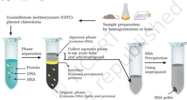
Fig. 3.2(b) shows the extraction after this separation — the RNA-containing aqueous phase from which RNA is precipitated with isopropanol. The parallel flow chart for DNA isolation is Fig. 3.1, read diagram by diagram below.
Box 1 — plasmid vs genomic DNA: cloning experiments isolate both. Boiling a bacterial lysate denatures the large chromosomal DNA irreversibly, so it forms a gel that is centrifuged away, while the small circular plasmid DNA renatures and stays soluble.
In the alternative method, SDS and NaOH denature the contents; after sodium ions are replaced with potassium ions, the clumped chromosomal DNA and proteins precipitate, and the plasmid DNA is isolated from the supernatant by ethanol precipitation.
Enzymes That Cut and Join DNA
Manipulating DNA is mostly cutting and joining: the gene of interest must be cut out of the genome, the vector must be cut open, and the two must be sealed together. This section introduces the enzyme toolkit of recombinant DNA technology, and one rule dominates it — only Type II restriction enzymes are used in rDNA technology.
Nucleases. Nucleases cleave nucleic acids by hydrolysing the phosphodiester bond between adjacent nucleotides. An exonuclease removes nucleotides one at a time from the 3′ or 5′ end; an endonuclease breaks internal phosphodiester bonds (Section 3.3).
Restriction enzymes. Restriction endonucleases are endonucleases that cut at a specific DNA sequence called the recognition site, usually 4 to 8 bp long. They are mostly found in bacteria and archaea, where they defend against invading bacteriophages.
There are three types (Table 3.1). Type I cuts at random positions about 1000 bp from the recognition site and needs SAM, \( Mg^{2+} \) and ATP; Type III cuts 24 to 26 bp away and needs the same cofactors.
Type II cuts within the recognition site itself, needs only \( Mg^{2+} \), and has only cleavage activity — that precise, predictable cut is why Type II enzymes such as EcoRI, BamHI and HindIII are the ones used to build recombinant DNA.
Type II recognition sites are palindromic sequences: the sequence on one strand reads the same as the complementary strand read backwards. A staggered cut leaves short single-stranded overhangs called sticky (cohesive) ends; a straight cut through both strands at the same point leaves blunt ends.
Naming enzymes (Table 3.2): the name records the source microorganism. EcoRI = genus Escherichia (E) + species coli (co) + strain Ry13 (R) + first endonuclease identified (I). Similarly, HindIII = Haemophilus influenzae strain Rd, third enzyme.
The full toolkit, with one role per enzyme:
| Enzyme | What it does | Role in rDNA technology |
|---|---|---|
| Exonuclease | Removes nucleotides one at a time from the 3′ or 5′ end of a polynucleotide chain. | Trimming the ends of DNA. |
| Endonuclease / restriction enzyme | Cuts internal phosphodiester bonds; restriction enzymes cut at a specific 4–8 bp recognition sequence. | Type II REs (EcoRI, BamHI, HindIII) cut the vector and the insert to create matching ends. |
| DNA ligase | Catalyses the formation of a phosphodiester bond, joining two DNA strands together. | Seals the insert into the vector; T4 ligase joins sticky and blunt ends, E. coli ligase joins sticky ends. |
| DNA polymerase | Synthesises a new DNA strand in the 5’→3′ direction from dNTPs on a template; needs a primer with a free 3′-OH. | Copying and filling in DNA strands. |
| Alkaline phosphatase | Removes the terminal phosphate group from the 5′ end of DNA. | Prevents unwanted self-ligation of vector ends. |
| Polynucleotide kinase | Adds a phosphate group onto a free 5′-OH end. | Labels or prepares ends for ligation — the reverse effect of alkaline phosphatase. |
| Terminal transferase | Adds similar nucleotide residues onto the 3′ end of DNA without requiring a template. | Builds homopolymer tails on DNA ends. |
| Reverse transcriptase | Generates a complementary DNA (cDNA) strand from an RNA template — reverse transcription. | Builds cDNA from mRNA for candidate genes, cDNA libraries and RT-PCR. |
| Poly A polymerase | Adds adenine residues to the 3′ end of RNA. | Tailing or labelling RNA molecules. |
Box 2 of the chapter records that Werner Arber, Daniel Nathans and Hamilton Smith shared the 1978 Nobel Prize in Physiology or Medicine for discovering restriction enzymes; HindII, isolated by Hamilton Smith, was the first restriction enzyme found.
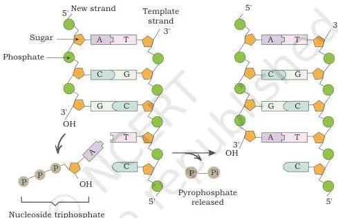
Fig. 3.6 shows why every polymerase reaction needs a primer: the new chain grows only from the free 3′-OH end of an existing strand, and the polymerase cannot start a new chain by itself.
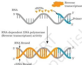
Fig. 3.7 is reverse transcription: reverse transcriptase reads the RNA template and builds a complementary DNA strand. This single step is the basis of cDNA candidate genes, cDNA libraries and RT-PCR.
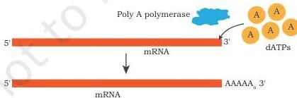
Fig. 3.8 shows poly A polymerase incorporating adenine residues onto the 3′ end of RNA — a small step, but it is how RNA molecules are tailed or labelled. The cutting-and-sealing diagrams of Fig. 3.3, 3.4 and 3.5 are walked through below.
Modes of DNA Transfer into a Host
A recombinant DNA molecule is useless until it reaches a host cell. The chapter first shows how bacteria naturally pick up foreign DNA by three routes, then lists the laboratory methods used in rDNA technology (Section 3.4).
- Transformation — a cell takes up free exogenous DNA directly from its surroundings through the cell membrane. The recipient cells are called transformants.
- Transduction — a bacteriophage carries the DNA. In the lysogenic cycle the phage genome integrates into the bacterial chromosome; when it later excises, it occasionally takes bacterial DNA along, packages it into new phage particles, and delivers it to the next bacterium it infects. The recipients are called transductants.
- Conjugation — direct cell-to-cell transfer of DNA from a donor to a recipient through a tube called a pilus, mediated by F plasmids. Relaxase nicks one strand of the F plasmid in the donor, that strand moves through the pilus, and both cells replicate it into a double-stranded copy.
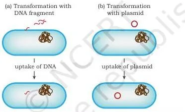
Fig. 3.9 draws the simplest route: the bacterial cell takes up a foreign DNA molecule directly from its surroundings, becoming a transformant.
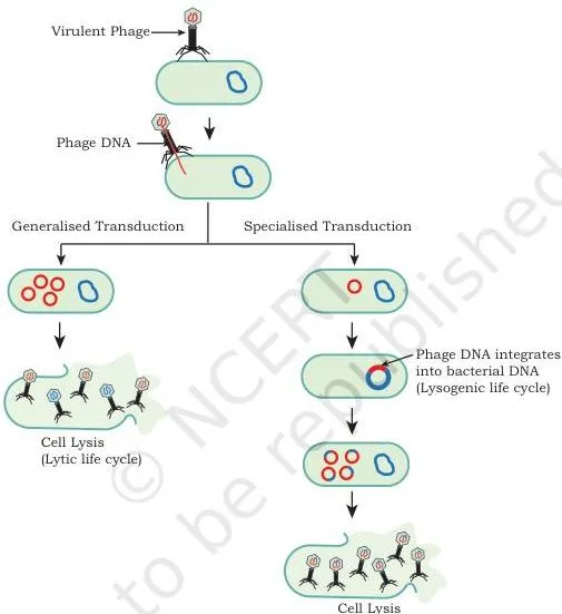
Fig. 3.10 shows transduction — the bacteriophage acts as the delivery vehicle, carrying bacterial DNA from one host into the next.
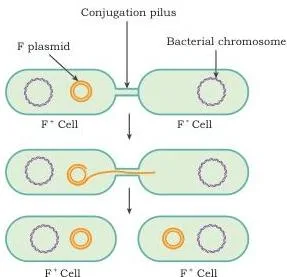
Fig. 3.11 shows conjugation: the donor and recipient touch through the pilus, and a single strand of the F plasmid crosses before both cells rebuild the double-stranded plasmid.
In the laboratory, foreign DNA is introduced by five common methods — calcium chloride with heat shock, electroporation, lipofection, microinjection and the biolistic gene gun. They are drawn together in Fig. 3.12, walked through below.
Screening and Selection of Recombinants
Screening is the step students find hardest, so the reason comes first: transformation success is very low. Most bacteria never take up a plasmid at all; of those that do, some carry the plasmid without the insert, and only a few carry the recombinant plasmid with your gene. Screening finds those few (Section 3.5).
The whole method rests on a difference in a biological trait — antibiotic resistance, an enzyme such as \( \beta \)-galactosidase, a fluorescent protein like GFP, or a nutritional requirement such as leucine.
Direct selection uses one trait, usually antibiotic resistance. Bacteria that took up a plasmid carrying the resistance gene survive on antibiotic medium; bacteria without the plasmid die. But direct selection shows only that the plasmid is present, not that the insert is inside it — it identifies transformants, not recombinants.
Insertional inactivation is more efficient: the vector carries two markers, and the insert is deliberately placed inside one of them, destroying that marker.
- The \( amp^R \)/\( tet^R \) example (Fig. 3.13): the insert is cloned into the \( amp^R \) gene. Plating on tetracycline kills non-transformed cells, leaving two kinds of transformed colonies on the master plate. Replica-plating those colonies onto ampicillin lets only non-recombinants grow. Colonies present on the master plate but absent from the ampicillin replica plate carry the insert.
- Blue-white selection (Fig. 3.14): the second marker is lacZ, which codes for \( \beta \)-galactosidase. This enzyme cleaves X-gal, a colourless chromogenic analog of lactose, into an insoluble blue pigment. If the insert sits inside lacZ, no active enzyme is made, X-gal is not hydrolysed, and the colony stays white. On ampicillin + X-gal + IPTG medium, blue colony = non-recombinant, white colony = recombinant.
- GFP alternative: the same inactivation logic with green fluorescent protein — an insert that disrupts the GFP coding sequence leaves non-fluorescing bacteria.
Blotting Techniques: DNA, RNA and Protein
Blotting answers one question: is my gene actually there, and is it being expressed? The three main blots share one workflow — separate molecules by size on a gel, transfer them onto a membrane, then detect the one you want with a probe or antibody (Section 3.6).
A probe is a single strand of DNA complementary to the target sequence, labelled with a detectable tag. The detection tags the chapter lists are radioactivity, fluorescence, chemiluminescence and chromogenic (colour) detection.
| Technique | Molecule detected | Gel used | Detection tool |
|---|---|---|---|
| Southern blot | DNA | Agarose gel, after restriction digestion | Labelled single-stranded DNA probe |
| Northern blot | RNA | Agarose gel | Labelled probe (radioactive or fluorescent) |
| Western blot | Protein | SDS-PAGE gel | Specific antibody |
| Eastern blot (Box 3) | Post-translational modifications of proteins | Gel electrophoresis | Specific substrates (e.g. cholera toxin, concanavalin) or antibodies |
Mnemonic — use the central dogma: DNA → RNA → protein → modification. Southern blot detects DNA, Northern blot detects RNA, Western blot detects protein, and Eastern blot detects post-translational modification, in exactly that order. The order also matches a working experiment: Southern finds the gene, Northern finds its transcript, Western finds the final protein.
Southern blot details: DNA is cut into fragments by a restriction enzyme, separated on an agarose gel by electrophoresis, transferred by capillary action to a nitrocellulose or nylon membrane, baked to fix it, and then detected with a labelled probe — for example by autoradiography on X-ray film.
Northern blot details: total RNA is separated on an agarose gel and transferred to a positively charged nylon membrane. The transfer buffer contains formamide, which lowers the annealing temperature of probe-RNA binding and so protects the fragile RNA; the RNA is fixed by UV light or heat before probing.
Western blot details: proteins are separated on an SDS-PAGE gel by their electrophoretic mobility (charge, size and structure), transferred to a nitrocellulose membrane, and the desired protein is detected with a specific antibody.
PCR, RT-PCR and Real-Time PCR
Cloning gives a few copies of a gene; PCR (Polymerase Chain Reaction) turns a few copies into millions. Developed by Kary B. Mullis, who received the Nobel Prize in Chemistry in 1993, it is often called “molecular photocopying”. A machine called a thermocycler repeats cycles of heating and cooling (Section 3.7).
Each cycle has three temperature steps:
- Denaturation (about 94–95°C): heat breaks the hydrogen bonds between the two strands, so each strand can act as a template.
- Annealing (about 50–55°C): the temperature is lowered so the forward and reverse primers bind to their complementary sites on the template.
- Extension (about 72°C): a heat-stable DNA polymerase adds dNTPs onto the 3′-OH ends of the annealed primers, growing new strands in the 5’→3′ direction.
Why a heat-stable enzyme? The denaturation step is hot enough to destroy ordinary polymerases. Taq polymerase comes from Thermus aquaticus, a bacterium that lives in hot springs, and Pfu polymerase, from Pyrococcus furiosus, copies DNA with higher fidelity. Primers are synthetic single-stranded oligonucleotides designed from the known sequence flanking the target region.
The DNA copies produced by PCR are called amplicons.
Worked example — the doubling rule. Every cycle doubles the DNA, so after \( n \) cycles one template molecule gives \( 2^n \) copies. Start with one template and run 30 cycles:
\[ 2^{30} = (2^{10})^3 \approx (10^3)^3 = 10^9 \]
Since \( 2^{10} = 1024 \), the exact value is \( 2^{30} = 1,073,741,824 \) — about 1.07 billion amplicons. If the reaction starts with 10 template molecules instead of one, every cycle amplifies all of them, so the final yield is about ten times larger — roughly 10.7 billion amplicons. This is why PCR can work even on a minute DNA sample from a crime scene or a fossil.
A typical reaction runs 25 to 30 cycles.
Three flavours of PCR matter in this chapter:
- Semi-quantitative PCR: ordinary end-point PCR — the amplified product is run on a gel at the end and the intensity of the DNA band is measured.
- RT-PCR (reverse transcription PCR): mRNA is first converted to cDNA by reverse transcriptase, and that cDNA is then amplified by PCR. This is how RNA is studied.
- Real-time quantitative PCR (qPCR): a fluorescent dye such as SYBR green binds double-stranded DNA, and the fluorescence — proportional to the amount of PCR product — is measured after every cycle. No gel is needed; the thermocycler carries a fluorescence detector.
COVID-19 connection (Box 5): the standard test is real-time reverse transcription PCR (rRT-PCR). RNA extracted from a respiratory sample is converted to cDNA and amplified with primers and probes for the SARS-CoV-2 N gene, ORF1a/b and a human RNase P control.
Each probe has a reporter dye at its 5′ end and a quencher at its 3′ end; when Taq polymerase’s 5’→3′ exonuclease activity cuts the probe during amplification, the reporter separates from the quencher and fluoresces.
The Ct value is the cycle number at which the fluorescence crosses the threshold — the number of cycles needed to amplify the viral RNA to a detectable level. The Ct value is inversely proportional to the amount of target RNA: a low Ct means a lot of viral RNA in the sample.
Genomic DNA Libraries and cDNA Libraries
Genomes are enormous and the genes in them are arranged randomly, and most of a genome is never transcribed. Isolating one unknown gene from all that DNA is a big task, so scientists store DNA as libraries — collections of clones, each holding a fragment, from which any gene can later be pulled out (Section 3.8).
A genomic library is a collection of clones whose fragments together represent the complete genome of an organism, including sequences that are never transcribed. It is built by digesting genomic DNA with a restriction enzyme, ligating the fragments into vectors, transforming bacteria, and growing them into colonies.
A cDNA library is built from the mRNA of one tissue or cell type. Because gene expression is tissue-specific — insulin genes are expressed in the beta cells of the pancreas, albumin genes in liver cells — a cDNA library contains only the genes active in that tissue at that state. The mRNA is converted to cDNA by reverse transcriptase before cloning.
| Feature | Genomic DNA library | cDNA library |
|---|---|---|
| Starting material | Total genomic DNA of the organism | mRNA from a specific tissue or cell type |
| What it represents | The complete genome, including untranscribed and non-coding sequences | Only the genes expressed in that tissue at that time |
| Key construction step | Restriction digestion of genomic DNA, then ligation into vectors | Reverse transcription of mRNA into cDNA, then ligation |
| Same for every tissue? | Yes — the genome is the same in all cells | No — it changes with tissue type and physiological state |
| Applications | Whole-genome sequencing; searching for genes not expressed; evolutionary studies; comparing healthy and diseased tissues to identify genetic aberrations | Finding which genes are active in a tissue; isolating a specific gene; cDNA as probes to screen genomic libraries |
Figure Walkthrough: The NCERT Diagrams That Explain Gene Cloning

These are the diagrams the chapter uses to teach the cloning workflow. Each is shown with its NCERT caption, and the notes read the diagram so you can follow it with your finger on the book.
Fig 3.1: DNA Isolation as a Four-Step Flow Chart
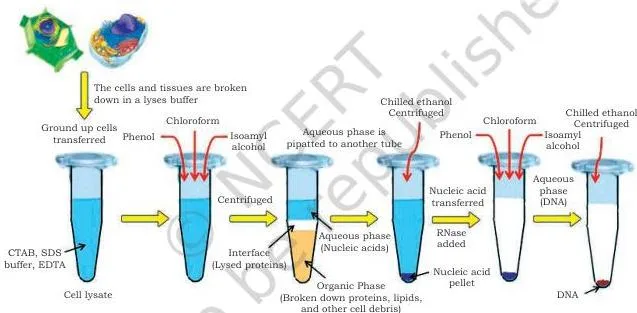
The flow chart runs top to bottom in four boxes. The first box is cell lysis and dissolution — breaking the cells open. The second is the protection step: chemicals such as EDTA and SDS stop the nucleases released along with the DNA.
The third box separates the nucleic acids from other molecules, and the fourth precipitates them with ethanol or isopropanol. Reproduce these four boxes and you have the whole DNA isolation procedure, which is what most isolation questions test.
Fig 3.3, 3.4 and 3.5: How DNA Is Cut and Sealed
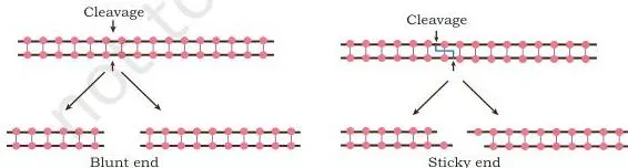
Fig. 3.3(a) shows the exonuclease working at the ends of the molecule — nucleotides are clipped off one at a time from the 3′ or 5′ end. Part (b) of the same figure, which the book draws alongside, shows the endonuclease cutting an internal phosphodiester bond. The distinction to see is location: ends versus interior.
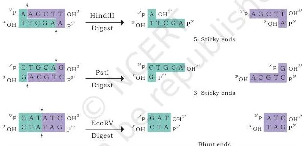
Fig. 3.4 shows why Type II enzymes are so useful. A staggered cut through the two strands at different positions leaves short single-stranded overhangs — the sticky or cohesive ends that can base-pair with a matching partner. A straight cut through both strands at the same place gives blunt ends. The two outcomes are drawn side by side for comparison.
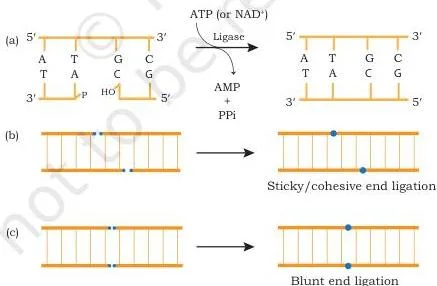
Fig. 3.5 shows ligation in three panels. Panel (a) is the chemistry: a covalent phosphodiester bond forms between the 5′-phosphate group of one chain and the 3′-hydroxyl group of the adjacent chain. Panels (b) and (c) show sticky ends and blunt ends being sealed — T4 DNA ligase can join both.
Fig 3.12: Five Laboratory Methods of DNA Transfer
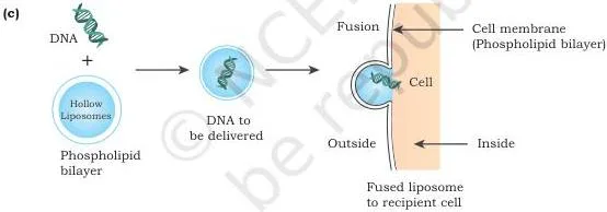
The five panels are five ways to get DNA into a cell. (a) CaCl₂: calcium ions form a complex with the negatively charged DNA, and the cells take it up during heat shock. (b) Electroporation: a mild electric current creates transient micropores in the membrane and DNA enters through them.
(c) Lipofection: a liposome — a phospholipid vesicle — fuses with the cell membrane and delivers its DNA cargo. (d) Microinjection: a fine needle injects DNA directly into the nucleus. (e) Biolistic: a gene gun fires microscopic particles of gold, nickel or tungsten coated with DNA into the cell.
Fig 3.13 and 3.14: How Recombinants Are Detected
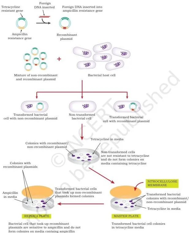
Fig. 3.13 follows the insertional inactivation logic. The master plate (tetracycline) shows all the transformed colonies — recombinants and non-recombinants together. Cells are lifted onto a nitrocellulose membrane and replica-plated onto ampicillin, where only non-recombinants grow. Compare the two plates: colonies on the master plate that are absent from the replica plate carry the insert in their \( amp^R \) gene.
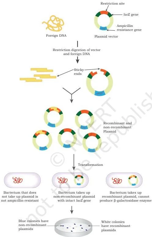
Fig. 3.14 is the blue-white plate: some colonies are blue and some are white. Blue colonies have an intact lacZ gene whose enzyme hydrolyses the colourless X-gal into a blue pigment. White colonies have lacZ disrupted by the inserted DNA, so no enzyme is made and X-gal stays colourless. The white colony is the recombinant.
Fig 3.15, 3.16 and 3.17: Southern, Northern and Western Blots
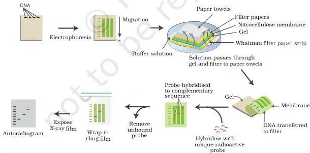
Fig. 3.15 (Southern) shows the complete workflow: DNA fragments are separated on an agarose gel, transferred by capillary action onto a nitrocellulose or nylon membrane, and the desired fragment is detected with a labelled probe. Because the membrane is a copy of the gel, the position of the band tells you the size of the DNA you found.
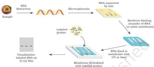
Fig. 3.16 (Northern) repeats the same idea for RNA: total RNA separated on a gel is transferred to a positively charged nylon membrane and hybridised with a probe specific to the RNA of interest. Dark bands on X-ray film (autoradiography) mark the target RNA; formamide in the transfer buffer protects the fragile RNA.
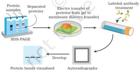
Fig. 3.17 (Western) swaps the probe for an antibody. Proteins separated on an SDS-PAGE gel are transferred to a nitrocellulose membrane, and a specific antibody binds only the protein of interest, exposing its band. Southern = DNA, Northern = RNA, Western = protein.
Fig 3.18 and 3.19: PCR and RT-PCR Cycles
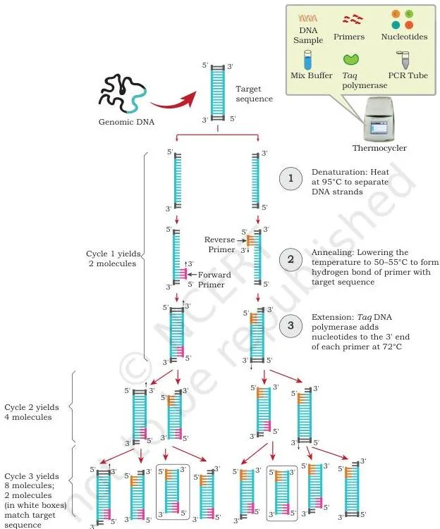
Fig. 3.18 draws the PCR cycle as a loop. Denaturation at about 94–95°C splits the double helix; annealing at about 50–55°C lets the primers attach; extension at about 72°C builds new strands. The diagram shows the two double helices produced at the end of one cycle — that is the doubling: one cycle makes two copies, and the next cycle doubles again.
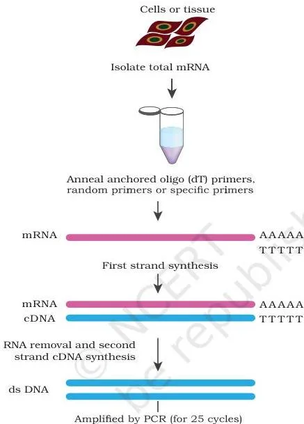
Fig. 3.19 adds the step that makes it RT-PCR: an RNA strand is first copied into cDNA by reverse transcriptase, and only then does the PCR cycling begin. This is exactly the chemistry used in the COVID-19 rRT-PCR test.
Fig 3.20 and 3.21: Building DNA Libraries
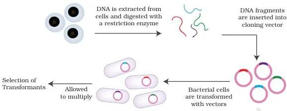
Fig. 3.20 follows genomic DNA across the page: the full genome is cut with a restriction enzyme into fragments, each fragment is inserted into a vector with DNA ligase, the recombinant vectors transform host bacteria, and the colonies that grow collectively hold the complete genome. The whole plate of colonies is the genomic library.
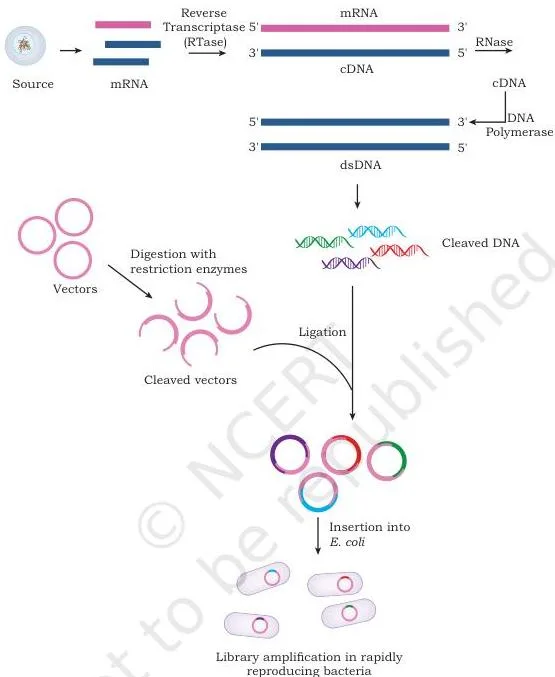
Fig. 3.21 follows mRNA instead: mRNA from a tissue is converted to single-stranded cDNA by reverse transcriptase, RNase removes the mRNA, DNA polymerase builds the second strand, and the double-stranded cDNA is cloned into vectors and transformed into bacteria. Because the mRNA came from one tissue, this library represents only the genes that tissue was expressing.
Gene Cloning Class 12: Key Terms and Definitions
These are the working vocabulary of the chapter, each defined in plain words for quick revision.
| Term | Plain meaning |
|---|---|
| Candidate gene | The gene chosen for cloning because of its biomedical, economic or evolutionary significance. |
| Recognition sequence (site) | The specific 4–8 bp DNA sequence that a restriction enzyme binds and cuts. |
| Palindromic sequence | A sequence that reads the same on the forward strand as on the complementary strand read backwards. |
| Sticky (cohesive) ends | Short single-stranded overhangs left by a staggered cut in DNA. |
| Blunt ends | Cut ends where both strands finish at the same position. |
| Transformants | Recipient bacterial cells that have taken up foreign DNA directly. |
| Transductants | Recipient cells that received bacterial DNA carried by a bacteriophage. |
| Insertional inactivation | Destruction of a marker gene by inserting foreign DNA into it. |
| Probe | A labelled single strand of DNA complementary to the target sequence, used to detect it. |
| Amplicons | The DNA copies produced by PCR. |
| Reverse transcription | Making a cDNA strand from an RNA template using reverse transcriptase. |
| Thermocycler | The automated machine that cycles a PCR reaction through its three temperatures. |
| Genomic library | A clone collection whose fragments together represent the complete genome. |
| cDNA library | Clones made from the mRNA of a particular tissue or cell type. |
| Ct value | The cycle at which PCR fluorescence crosses the threshold; inversely proportional to the amount of target. |
Common Mistakes in Gene Cloning Class 12 and How to Avoid Them
These are the points where students most often slip in this chapter. Each row names the wrong idea, states the correct rule, and gives a quick way to check yourself.
| Mistake | Correct rule | How to check your answer |
|---|---|---|
| “Any restriction enzyme can be used in rDNA technology.” | Only Type II restriction enzymes are used because they cut within the recognition site. Type I cuts about 1000 bp away; Type III cuts 24–26 bp away (Table 3.1). | For every RE you name, state where it cuts: within the recognition site (Type II) or away from it (Types I and III). |
| “The blue colony is the recombinant.” | Blue = intact lacZ, so \( \beta \)-galactosidase hydrolyses X-gal to a blue pigment. White = lacZ inactivated by the insert, so no enzyme is made. The recombinant is white. | Ask what happened to the marker gene. If the insert is inside lacZ, the marker is destroyed → no colour → white colony. |
| “Southern detects RNA, Northern detects DNA.” | Southern = DNA, Northern = RNA, Western = protein, Eastern = post-translational modification of proteins. | Run the central dogma in your head: DNA → RNA → protein → modification. That is the blot order. |
| “Annealing is the hottest PCR step and extension is cooler.” | Denaturation ≈ 94–95°C (hottest), extension ≈ 72°C, annealing ≈ 50–55°C (coolest of the three). | Remember the physical logic: melt the strands hottest, cool to let primers bind, then warm up for copying. |
| “A cDNA library is the same as a genomic library.” | Genomic = whole genome including untranscribed DNA, identical for all tissues. cDNA = mRNA of one tissue, so it changes with tissue type and physiological state. | Would the library contain untranscribed DNA? Genomic yes, cDNA no. Would it differ between pancreas and liver? cDNA yes. |
| “Direct selection proves the insert is present.” | Direct selection (antibiotic resistance) proves only that the plasmid is present — it identifies transformants. Insertional inactivation or blue-white selection proves the insert is present. | Check the marker logic: one marker = transformants only; an insert placed inside a second marker = recombinants. |
How the End-of-Chapter Exercises Test Gene Cloning
The chapter’s exercise set follows the same order as the sections, so it tests every stage of the workflow. The table maps each group of questions to the concept it checks and the structure a full answer needs.
| Questions | Concept tested | What a full answer must include |
|---|---|---|
| 1–3, 5, 7 | Nucleic acid isolation — methods, biological detergents, plant vs bacterial isolation, challenges, chelating agents | The four steps by name; the right reagent per organism (lysozyme and SDS for bacteria; CTAB and PVP for plants); EDTA chelates \( Mn^{2+} \) and \( Mg^{2+} \) to block nucleases. |
| 4 | Restriction enzymes and rDNA technology | Three types, then the justification: Type II cuts within the recognition site and has only cleavage activity, so it is the type used in rDNA technology. |
| 6 | Roles of alkaline phosphatase, DNA ligase, terminal transferase | One line each: removes a 5′ phosphate; joins strands by forming a phosphodiester bond; adds a homopolymer tail at the 3′ end. |
| 8 | Modes of DNA transfer | The three natural routes (transformation, transduction, conjugation) plus laboratory methods (CaCl₂, electroporation, lipofection, microinjection, biolistic), with the recipient names transformants and transductants. |
| 9, 16, 17 | Recall MCQs — blue-white selection, genome size, matched pairs | Blue-white is based on lacZ expression; the diploid human genome is about \( 6.4 \times 10^9 \) bp (twice the haploid \( 3.2 \times 10^9 \) of Section 3.1); EcoRI is Type II, not Type I. |
| 10, 11 | Matched-pair MCQs — blots and polymerases | Southern = DNA, Northern = RNA, Western = protein; Taq = Thermus aquaticus, Pfu = Pyrococcus furiosus. In Q11 the chapter confirms (a), (b) and (c), so the incorrect pair is (d) by elimination. |
| 12 | Screening recombinants in detail | Direct selection vs insertional inactivation; the \( amp^R \)/\( tet^R \) replica-plating logic; blue-white selection with X-gal and IPTG. |
| 13 | Differentiating the blots | For each blot: what is detected, which gel is used, which detection tool (probe vs antibody), and one application. |
| 14 | PCR in detail | Three steps with temperatures; Taq/Pfu polymerase; primers; the \( 2^n \) doubling rule; amplicons; gel-based end-point analysis. |
| 15 | Genomic vs cDNA libraries | A comparative account: source (genomic DNA vs mRNA), content, tissue dependence, construction, and applications of each. |
| 18, 19 | Assertion–reason items on PCR and transformation | Judge assertion and reason separately, then test whether the reason explains the assertion — for PCR, Taq polymerase is what makes amplification possible at high temperature. |
Answer structure tip: for every “describe”, “differentiate” or “comparative account” question, write the definition first, then the key differences (source, target, tool), then one application. For matched-pair MCQs, eliminate the option that is a true fact attached to the wrong term — that is how the blot pairs (Q10) and enzyme pairs (Q11, Q17) are designed.
Gene Cloning Class 12: Revision Summary
This is the night-before version of the chapter’s own summary points, compressed. If you read nothing else, read this list and the mistakes table above.
- DNA isolation has four steps: break cells open → protect nucleic acids from nucleases → separate from other molecules → precipitate with ethanol or isopropanol.
- Bacteria are lysed with lysozyme plus SDS; plant cells use CTAB (with PVP for phenols); the buffer contains EDTA to chelate \( Mn^{2+} \) and \( Mg^{2+} \); ribonuclease A removes RNA.
- RNA isolation uses GITC-phenol-chloroform because RNases are everywhere; RNA stays in the aqueous phase and is precipitated with isopropanol.
- Nucleases cut DNA, ligase joins it, polymerases copy it 5’→3′ from a primer, alkaline phosphatase and polynucleotide kinase remove and add 5′ phosphates, terminal transferase tails 3′ ends, reverse transcriptase makes cDNA, and poly A polymerase tails RNA.
- Only Type II restriction enzymes are used in rDNA technology — they cut within their palindromic recognition site, producing sticky or blunt ends.
- Natural DNA transfer: transformation (transformants), transduction (transductants), conjugation (F plasmid, pilus). Laboratory methods: CaCl₂ + heat shock, electroporation, lipofection, microinjection, biolistic gene gun.
- Screening: direct selection (antibiotic resistance → transformants only) and insertional inactivation (insert destroys a marker — \( amp^R \)/\( tet^R \) replica plating, or blue-white with lacZ and X-gal; blue = non-recombinant, white = recombinant).
- Blotting: separate → transfer → probe/antibody → detect. Southern = DNA, Northern = RNA, Western = protein, Eastern = post-translational modification.
- PCR: denaturation about 94–95°C, annealing about 50–55°C, extension about 72°C, using Taq (Thermus aquaticus) or Pfu (Pyrococcus furiosus) polymerase; each cycle doubles the DNA, so \( n \) cycles give \( 2^n \) copies.
- RT-PCR converts RNA to cDNA before amplification; real-time qPCR measures fluorescence every cycle; in the COVID-19 rRT-PCR test, a low Ct value means more viral RNA.
- A genomic library represents the whole genome; a cDNA library represents the genes expressed in one tissue at one physiological state.
Related NCERT Biotechnology Chapters
Chapter 3 sits inside the NCERT Class 12 Biotechnology textbook, between the host-vector system of Chapter 2, which explains the plasmids and vectors that carry the insert, and Chapter 4, the applications of recombinant DNA technology, which shows what cloned genes are used for.
From here you can also go back to the NCERT Class 12 Biotechnology book page, the Class 12 hub, or the main CBSE notes index.
Note that textbook contents and the examinable syllabus are not always identical — check the current official syllabus for what is examinable this session.
Sources and Data Verification
This listing is maintained for the 2026-27 academic session using the NCERT textbook information available to us. NCERT remains the authority for confirming the latest edition.
This page describes the NCERT Class 12 Biotechnology textbook, Chapter 3, Gene Cloning — the official edition published on ncert.nic.in. The sections, figures, tables and exercise questions described above come from that chapter file.
- This page covers that one chapter, not the full CBSE syllabus; NCERT settles textbooks, editions and PDFs, while CBSE settles curriculum, syllabus and examinations.
- The chapter file and the examinable syllabus are not the same thing — confirm the current syllabus with the board before deciding what to study.
- This listing is maintained for the current academic session using the NCERT information available to us.
Reference: NCERT Class 12 Biotechnology textbook, chapter 3, official edition on ncert.nic.in.
Gene Cloning Class 12: Frequently Asked Questions
Where can I download the official NCERT Class 12 Biotechnology Chapter 3 Gene Cloning PDF?
From the NCERT portal — download the official NCERT Class 12 Biotechnology Chapter 3 Gene Cloning PDF directly from ncert.nic.in. This is the same file that appears in the printed NCERT Class 12 Biotechnology book, and it is free to download.
Which enzymes are used in recombinant DNA technology and what does each one do?
Nucleases cut DNA (exonucleases from the ends, endonucleases internally; Type II restriction enzymes cut at specific recognition sites), DNA ligase joins strands with a phosphodiester bond, DNA polymerase copies DNA 5’→3′ from a primer, alkaline phosphatase removes a 5′ phosphate, polynucleotide kinase adds one, terminal transferase adds a homopolymer tail at the 3′ end, reverse transcriptase makes cDNA from RNA, and poly A polymerase adds adenines to the 3′ end of RNA.
The full enzyme table is in the enzyme section above.
Why is the white colony the recombinant one in blue-white selection?
Because the inserted DNA is placed inside the lacZ gene. An intact lacZ makes \( \beta \)-galactosidase, which hydrolyses the colourless X-gal into a blue pigment, so a blue colony has an unbroken lacZ and is non-recombinant. A white colony has a disrupted lacZ, makes no \( \beta \)-galactosidase, cannot hydrolyse X-gal, and therefore carries the insert — it is the recombinant.
What is the difference between RT-PCR and real-time PCR?
They answer different questions. RT-PCR (reverse transcription PCR) is about the starting material: RNA is first converted to cDNA by reverse transcriptase and then amplified — this is how RNA viruses such as SARS-CoV-2 are detected. Real-time PCR is about the measurement: fluorescence is recorded after every cycle, so the amount of product is tracked as it accumulates.
The COVID-19 test combines both as rRT-PCR because it starts from RNA and measures fluorescence each cycle through the Ct value.
What is the difference between a genomic DNA library and a cDNA library?
A genomic library is built from the organism’s total genomic DNA and represents the complete genome, including sequences never transcribed; it is the same for every tissue. A cDNA library is built from the mRNA of one tissue or cell type, so it contains only the genes that tissue was expressing at that physiological state.
Insulin genes appear in a cDNA library from pancreatic beta cells and albumin genes in one from liver cells, while both genes are present in any genomic library.
Which blotting technique detects DNA, RNA and proteins?
Southern blotting detects DNA, Northern blotting detects RNA, Western blotting detects protein, and Eastern blotting detects post-translational modifications of proteins. The order matches the central dogma: DNA → RNA → protein → modification.
Explore Class 12 Biotechnology Books
Related chapters:
- Recombinant DNA Technology
- Genome Engineering
- Microbial, Plant, Animal Cell, Organ Cultures and Bioprocessing