
Animal cell culture class 12 — Chapter 8 of the official NCERT Biotechnology textbook — explains how scientists grow animal cells outside the living body: the media that feed them, the equipment that keeps them sterile, and the medicines this technology has produced.
It is a 24-page chapter. The official NCERT PDF is right below, and the sections under it walk through the chapter’s ideas in plain language — the definitions, the classifications, the equipment and the applications — so you can study without hunting.
Download the NCERT Class 12 Biotechnology Chapter 8 Animal Cell Culture PDF
This is the official PDF of the chapter as published by NCERT, the same file the book is printed from and the same edition used in schools.
Open the NCERT Class 12 Biotechnology Chapter 8 Animal Cell Culture PDF to read the chapter itself; the breakdown below explains what each section contains and what each figure shows, so you can move straight to the part you need.
| What the chapter holds | Count | Where it is used |
|---|---|---|
| Printed pages | 24 | |
| Sections in the chapter | 8 | |
| Figures with NCERT captions | 11 | |
| Tables | 4 | |
| Exercise questions | 21 | answered in our NCERT Solutions |
| Official NCERT PDF | Download the chapter PDF | the chapter exactly as NCERT publishes it |
Animal Cell Culture Class 12: Chapter at a Glance
The chapter totals are listed in the table above. This table is a reading map: it shows what each of the numbered sections 8.1 to 8.8 teaches, so you know the chapter’s shape before you open the file.
| Section | What it does |
|---|---|
| 8.1 Historical Perspective | How the technique became routine in the 1950s, the HeLa cell line, and the discovery of growth factors |
| 8.2 Culture Media | Natural vs synthetic media, what a typical medium contains, and why serum is both essential and problematic |
| 8.3 Physical Environment | Temperature, osmolality, pH, and the two buffering systems that keep pH stable |
| 8.4 Equipment Used for Cell Culture | Laminar flow hood, CO₂ incubator and inverted microscope — the lab’s three essential instruments |
| 8.5 Types of Animal Cell Cultures and Cell Lines | Primary and secondary cultures, finite vs continuous lines, adherent and suspension growth, subculture and cryopreservation |
| 8.6 Cell Viability Determination | Dye-exclusion assays and metabolic assays such as the MTT test |
| 8.7 Advantages of Animal Cell Culture | The benefits of the technique and its honest limitations |
| 8.8 Applications of Animal Cell Culture | Model systems, virus research, vaccines, and pharmaceutical proteins from EPO to monoclonal antibodies |
What the Animal Cell Culture Chapter Covers
8.1 Historical Perspective. This section explains where the technique came from. Animal cell culture became a routine laboratory method only in the 1950s, after George Gey established the first human cell line, HeLa, from the cervical cancer cells of Henrietta Lacks.
The demand for viral vaccines pushed culture to large scale, and the discovery of growth factors — followed by serum-free media in 1976 — made cells easier to grow.
8.2 Culture Media. The medium is the cells’ food, water and home, so choosing it is the most critical step. A typical medium contains vitamins, amino acids, glucose, inorganic salts, serum and hormones, and it also holds pH and osmolality steady. Media divide into natural media (plasma, serum, tissue extracts) and synthetic media, which pair a basal medium with supplements.
8.3 Physical Environment. Cells are picky about their surroundings. This section covers the conditions a culture must control: temperature around 37 °C for mammalian cells, osmolality near 300 mOsmol, and pH, managed by two buffering systems — the natural CO₂/bicarbonate system and chemical buffering with HEPES. Phenol red in the medium reports pH by colour.
8.4 Equipment Used for Cell Culture. Sterility is the whole game. The section describes the laminar flow hood that filters air through HEPA, the CO₂ incubator that holds temperature, gas and humidity, and the inverted microscope for checking cell health.
8.5 Types of Animal Cell Cultures and Cell Lines. This is the chapter’s core classification. Section 8.5 follows a culture from its origin in tissue (primary culture), through sub-culturing (secondary culture), into two possible life spans — finite and continuous cell lines. It also contrasts adherent and suspension cells, and explains subculture and cryopreservation.
8.6 Cell Viability Determination. A culture is only useful if you know its cells are alive. This section presents the two families of viability tests: dye-exclusion assays and metabolic assays such as the MTT test.
8.7 Advantages of Animal Cell Culture. This section answers why researchers bother. Cells grow under controlled conditions, proliferate into a homogenous genetic population, provide adequate numbers for chemical studies, and support easy production of biopharmaceuticals — although the technique is highly sensitive and may not fully represent the in vivo state.
8.8 Applications of Animal Cell Culture. This is the payoff of the chapter. Cell culture serves as a model system for disease and drug studies, as a tool for virus research and large-scale vaccine production, and as a production house for pharmaceutical proteins such as EPO, Factor VIII, tPA and monoclonal antibodies.
Key Concepts in Animal Cell Culture
This is the teaching core of the page. Each idea below is explained in the order you should learn it, with the plain question it answers stated first.
What animal cell culture is, and the HeLa cells that started it
Animal cell culture is the in vitro maintenance and proliferation of animal cells — growing cells outside the living organism in a laboratory container, provided they get appropriate nutrients and growth conditions. In vitro means performed outside the living body; in vivo means within the living organism.
A clone is a homogenous population derived from a single parental cell, so all cells in a clonal population are genetically identical.
Animal cells divide slowly — usually 18 to 24 hours per division — which is why contamination is dangerous. A small number of bacteria soon outgrows a much larger population of animal cells.
Why does the chapter open with Henrietta Lacks? Because her cells created the field. Lacks, an African-American woman, was diagnosed with terminal cervical cancer in 1951 and treated at Johns Hopkins by George Gey, who removed cells from her cervix without her permission. Gey found those cells could be kept alive and grown indefinitely.
For decades afterwards, her cells — the HeLa line — were used in experiments ranging from the long-term effects of radiation to testing the live polio vaccine.
The ethics point matters. Lacks’ cells were commercialised, and the medical researchers who patented her tissue profited from them, while she had never consented. The chapter records these facts plainly; the lesson for a biology student is that consent and benefit-sharing are central to modern research.
Culture media: what cells are fed, and the role of serum
The culture medium is the most critical choice in cell culture, because the right medium depends on the cell type and on the purpose of the culture — growth, differentiation or production of a desired product. A typical medium contains vitamins, amino acids, glucose, inorganic salts, serum and hormones, and it also maintains pH and osmolality.
Media come in two categories:
- Natural media — biological substances such as plasma, serum and tissue extracts. Their main drawback is poor reproducibility, because the exact composition is never fully known.
- Synthetic (artificial) media — a basal medium plus supplements such as serum, growth factors and hormones. Depending on the supplement, they may be serum-containing, serum-free, xeno-free, protein-free or chemically defined.
Chemically defined media split into balanced salt solutions — PBS, DPBS, HBSS, EBSS, which maintain physiological pH and osmotic pressure with inorganic salts — and complex media such as EMEM, DMEM, RPMI-1640 and Ham’s F-12.
Serum is one of the most important components of a medium. It supplies amino acids, proteins, vitamins, carbohydrates, lipids, hormones and growth factors; it provides binding proteins such as albumin and transferrin that carry other molecules into the cell; and it adds adhesion factors that help cells stick to the substrate before they divide.
Fetal bovine serum is the common choice.
But serum has four real drawbacks:
- It contains insufficient cell-specific growth factors, so it cannot feed cells on its own.
- It may contain cytotoxic compounds and growth inhibitors that slow cell growth.
- It carries a contamination risk — viruses, fungi and mycoplasma.
- It interferes with purification of products such as pharmaceutical compounds.
Antibiotics such as penicillin and streptomycin are commonly added to control bacteria and fungi, although they are not essential for growth.
Physical environment: temperature, osmolality, pH and buffering
Beyond food, cells need a controlled physical environment: temperature, water balance and pH. All three are governed by the medium and the incubator.
Temperature. Mammalian cells grow at 37 °C because that simulates the body temperature of Homo sapiens, and most cultured cells come from warm-blooded animals that grow optimally here. This is why a CO₂ incubator is used.
Osmolality. Commercial media are formulated near 300 mOsmol. Glucose and salts are the main contributors, with amino acids adding significantly. If the outside osmotic pressure is too low, cells swell; if too high, they shrink. An osmometer measures it.
pH and buffering. The natural buffering system balances gaseous CO₂ with the CO₂/HCO₃⁻ content of the medium — more external CO₂ makes the medium more acidic — so these media need air with 5–10% CO₂, supplied by the incubator. Pyruvate in the medium helps cells produce their own CO₂, reducing dependence on the external supply. This system is low-cost and non-toxic.
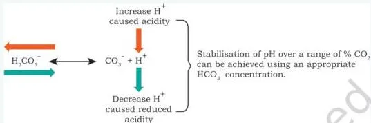
Read the diagram as a balancing act. When metabolism adds extra H⁺ ions, free bicarbonate ions react with them to form carbonic acid, “shifting the reaction to the left” and stabilising pH. When H⁺ falls, the shift goes to the right. The medium resists sudden pH swings as long as CO₂ is available.
The chemical buffering system uses HEPES, a zwitter ion with superior buffering capacity in the pH range 7.2–7.4. It needs no controlled gas atmosphere, but it is relatively expensive and toxic to some cell types at high concentration.
Phenol red is the colour reporter in commercial media:
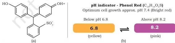
The figure shows the molecule and its colour scale. At acidic pH the medium turns yellow; at alkaline pH it turns pink; at the optimal pH of 7.4 it is bright red. Checking the medium’s colour daily is a quick health check of the culture.
Equipment that keeps cultures alive and sterile
An ideal animal cell culture laboratory needs a laminar hood, a CO₂ incubator, an inverted microscope, and support equipment such as an autoclave and centrifuge. The first three carry most of the work.
Laminar flow hood. All cell culture work is done aseptically here. Air is drawn in under negative pressure, passes a pre-filter that traps fungi, bacteria and dust, then flows continuously through HEPA filters into the enclosed cabinet. The cabinet is kept under constant positive pressure so contaminated room air cannot flow in.
Safety depends on direction. A vertical (down-flow) hood blows sterilised air down on the work area, away from the operator, and is considered safer. A horizontal hood blows air directly towards the operator and offers no protection from the hazards of cultured cells, which is why horizontal hoods are not popular.
CO₂ incubator. The incubator maintains sterility, a constant temperature, a 5–10% CO₂ atmosphere and about 95% relative humidity. The CO₂ cylinder is kept outside the laboratory and the gas is piped in, to keep the handling area clean. The medium is buffered with sodium bicarbonate/carbonic acid and the pH must be strictly maintained.
Inverted microscope. The microscope needs phase-contrast optics and ideally a photographic facility. It is used to monitor cell morphology and signs of stress — granularity, degree of spreading, membrane blebbing, multi-nucleates, vacuolation — and to spot early microbial contamination before irreplaceable material is lost. Choose a long working-distance condenser so flasks and roller bottles can be viewed; a 20× objective is usually sufficient.
Primary and secondary cultures, and finite vs continuous cell lines
This is the classification ladder of the chapter: tissue → primary culture → secondary culture → cell line. Each step changes what the cells look like and how long they can live.
Primary cell culture comes directly from host tissue. The tissue is dissected under sterile conditions and disaggregated either mechanically — chopped into fragments called explants, from which cells grow out — or enzymatically with trypsin or collagenase. The result is mostly heterogeneous, similar to the parent tissue, and cells may grow adherent or in suspension.
Secondary cell culture is what you get when a primary culture is sub-cultured. During passaging the fastest-growing cells come to dominate, giving genotypic and phenotypic uniformity — but repeated sub-culture can make the cells different from the original.
By life span, cell lines fall into two classes:
| Feature | Finite cell lines | Continuous cell lines |
|---|---|---|
| Life span | Limited number of divisions | Indefinite; can be sub-cultured and grown permanently |
| Senescence | Cells lose the ability to proliferate (senescence) | No senescence — cells are immortal |
| Origin | Derived from primary cultures of normal cells | Arise when finite-line cells transform (spontaneously or by chemical/viral induction) |
| Tumour-forming behaviour | Not tumorigenic | Immortal and tumorigenic |
| Growth demands | Normal nutrient needs | Faster growth, fewer nutrients, higher cell density, less adherent |
Read the table carefully: a continuous line is not a separate species of cell. It is a transformed descendant of a finite line that has regained the ability to divide indefinitely.
Adherent and suspension cells, and how subculture works
Cells also differ in how they live inside the vessel. Adherent (anchorage-dependent) cells need a solid or semi-solid substrate and proliferate as a monolayer — one cell thick — held to the vessel by the extracellular matrix. Fibroblasts and epithelial cells are typical, and single layers can be transferred to coverslips for microscopy.
Suspension (anchorage-independent) cells float in the medium. Haematopoietic stem cells (from blood, spleen and bone marrow) and tumour cells are examples. They grow faster, need fewer medium changes and are homogeneous.
Subculture (passaging) answers the practical question “what do you do when the flask is full?” At confluence the monolayer becomes crowded and overgrows, which is unfavourable and leads to cell death. The cells are detached — with trypsin (or trypsin + EDTA), or mechanically with a cell scraper — centrifuged, and re-seeded in fresh medium.
This is commonly called “splitting”, because one flask’s cells are divided between two flasks. The passage number refers to the number of times a cell line has been sub-cultured.
Cryopreservation: freezing cells without killing them
Cryopreservation stores cells long-term at very low temperatures. Storage is at −180 to −196 °C in liquid nitrogen, because ice crystal formation is reduced below −130 °C.
Freezing kills cells through ice crystal damage, changes in electrolyte concentration, dehydration and pH shifts. That is why cryoprotective agents — glycerol or DMSO — are added; a typical freezing medium is 90% serum + 10% DMSO.
The order of steps matters. Memorise the sequence exactly:
- Use healthy cells growing in log phase, and replace the medium 24 hours before freezing.
- Cool slowly from room temperature to −80 °C, so water moves out of the cells before they freeze.
- Transfer the cells to liquid nitrogen for storage.
- Thaw rapidly — place the vial in a 37 °C water bath with moderate shaking, to minimise ice crystal formation.
- Transfer the thawed cells directly into a culture vessel with fresh medium.
Measuring cell viability: dye exclusion and the MTT assay
Viability measurement — deciding whether cells are alive — is needed to judge the effectiveness of a pesticide or insecticide, the damage caused by toxins, and the potential of a drug. There are two families of tests.
Dye-exclusion assays. A dye enters the cell and intercalates with DNA in the nucleus. Live cells have intact membranes and exclude the dye; dead cells have lost membrane integrity, let the dye in and are stained. Examples are trypan blue, propidium iodide, 7-AAD and acridine orange.
Metabolic assays. These measure a biochemical reaction that living cells perform, by absorbance, fluorescence or luminescence. In the MTT assay, yellow, water-soluble MTT is reduced by the mitochondrial dehydrogenase enzymes of live cells to purple, insoluble formazan, which is dissolved in DMSO. The amount of formazan is proportional to the number of cells.
Worked example: a researcher stains a culture with trypan blue and counts 200 cells. 40 cells appear blue.
- Step 1: The blue cells let the dye in, so they are dead: dead cells = 40.
- Step 2: The remaining cells excluded the dye, so live cells = 200 − 40 = 160.
\[ \text{Viability} = \frac{160}{200} \times 100 = 80\% \]
Final answer: the culture is 80% viable — 160 live cells out of 200.
Advantages, limitations and medical applications
Advantages of animal cell culture — the reasons it became a dominant tool in life science research:
- Cells grow under a controlled physico-chemical environment.
- They proliferate into a homogenous genetic population.
- Adequate cell numbers are available for chemical studies.
- Easy production of biopharmaceuticals.
- No ethical clearance is required — the chapter lists this as an advantage, a claim worth reading next to the Henrietta Lacks story.
Limitations: the technique is highly sensitive, so even small changes cut productivity, and cultured cells may not fully represent the in vivo phenotype and genotype.
Applications: as a model system for studying interactions between cells, disease agents and drugs; for virus research; for large-scale vaccine production; and as a production house for pharmaceutical proteins. The chapter’s Table 8.3 lists the key products:
| Protein | Animal cells used | Therapeutic use |
|---|---|---|
| Follicle Stimulating Hormone (FSH) | CHO cells | Infertility |
| Human Growth Hormone (HGH) | CHO cells | GH deficiency |
| Erythropoietin (EPO) | CHO cells | Anaemia |
| Factor VIII | CHO cells | Haemophilia A |
| Factor IX | CHO cells | Haemophilia B |
| Interleukin-2 (IL-2) | CHO cells | Cancer therapy |
| Tissue Plasminogen Activator (tPA) | CHO cells | Stroke |
| Monoclonal antibodies (mAbs) | Hybridoma cells | Cancer therapy and autoimmune diseases |
Note that CHO means Chinese Hamster Ovary — the workhorse cell line for protein drugs; every protein in the table except the monoclonal antibodies is made in CHO cells.
EPO: a glycoprotein hormone secreted by the kidney under hypoxic (low-oxygen) or anoxic conditions. It stimulates the bone marrow to produce red cells and is used for anaemia caused by chemotherapy, in AIDS treatment and in chronic renal failure. Recombinant human EPO from CHO cells beats blood transfusion because no donors, transfusion facilities or transfusion-associated disease risk are involved.
Factor VIII and Factor IX: clotting glycoproteins whose absence causes haemophilia A and haemophilia B (Christmas disease). Both are produced in CHO cells for treatment.
tPA: a serine protease that converts plasminogen to plasmin, which dissolves blood clots. It is approved for certain heart attack and stroke patients and was the first drug produced through mammalian cell culture.
OKT-3 (Muromonab-CD3): a murine monoclonal antibody directed against the CD3 receptor, given intravenously to reverse acute rejection of transplanted kidneys, hearts and livers. Binding CD3 makes the T-cell receptor undergo endocytosis, leaving the T cell inert; the T cells are then eliminated by phagocytosis. OKT-3 was the first monoclonal antibody approved for acute transplant rejection in clinics.
Trastuzumab (Herceptin): a monoclonal antibody for early-stage HER2-positive breast cancer. Cancer cells overexpress HER2 receptors; trastuzumab attaches to the receptors and blocks the growth signals, impairing the cancer’s growth.
Reading the Diagrams of This Chapter
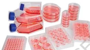
This section reads each diagram in the chapter as a study tool, so the figure becomes something you learn from rather than a picture you skip. Every diagram below states the single point you should take from it.

Fig 8.2 shows where cells actually live. The flask, microplates and Petri dishes are the vessels whose medium colour you check daily, and the scraper is the mechanical way to detach adherent cells during passaging — the alternative to trypsin.
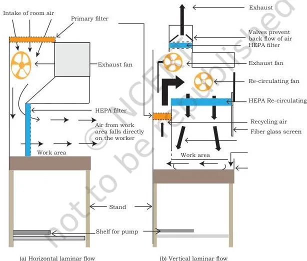
Fig 8.3 compares the two hood designs. In the horizontal hood, sterilised air is directed at the person doing the work; in the vertical down-flow hood, it is directed onto the work surface. The vertical design is safer because the operator is not exposed to air that has passed over the cultures.
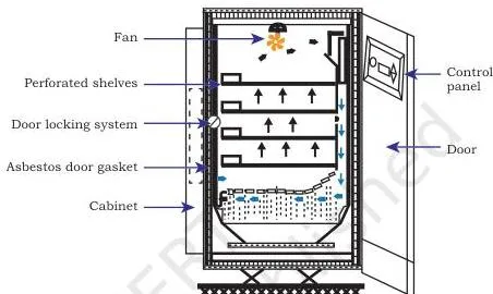
Fig 8.4 shows the incubator that delivers the physical environment: constant temperature, sterility, 5–10% CO₂ and about 95% humidity. The CO₂ cylinder stays outside the laboratory and the gas is piped in.
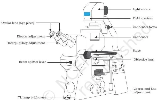
Fig 8.5 is the instrument you use to monitor the culture — cell morphology, granularity, membrane blebbing, vacuolation. Catching a problem early prevents the loss of irreplaceable material and avoids experiments on unhealthy cells.
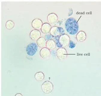
Fig 8.7 is the dye-exclusion assay in action. The blue cells let the dye in through damaged membranes — they are dead. The clear cells excluded it and are alive. Counting blue against clear cells gives the viability of the culture.
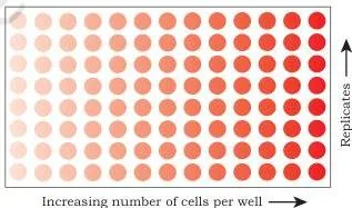
Fig 8.8 shows why metabolic assays are quantitative: the colour is directly proportional to the cell number. The more living cells, the more formazan is produced and the deeper the colour.
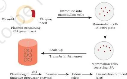
Fig 8.9 follows tPA from production to action. The drug is made by mammalian cell culture, and in the body it converts plasminogen into plasmin, the enzyme that dissolves blood clots in heart attack and stroke patients.
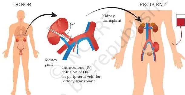
Fig 8.10 shows the clinical logic of OKT-3. In kidney transplantation the antibody binds the CD3 receptor on T cells, leaving them inert and unable to drive acute rejection; the T cells are then cleared by phagocytosis.
The two buffering figures — the Le Chatelier bicarbonate diagram and Fig 8.1 on phenol red — were explained in the physical environment section above; go back and read them with that walkthrough.
Key Terms Defined Simply
Scan this glossary the night before an exam. Each term is defined as the chapter uses it, in plainer words.
| Term | Plain definition |
|---|---|
| Cell culture | Growing cells under laboratory conditions, outside the living organism |
| In vitro | Performed outside the living body, in a container |
| Clone | A homogenous population derived from a single parental cell — all cells genetically identical |
| Explant | A fragment of chopped tissue from which cells grow out in primary culture |
| Primary cell culture | The culture obtained directly from host tissue after mechanical or enzymatic disaggregation |
| Secondary cell culture | The culture produced when a primary culture is sub-cultured |
| Cell line | Cells obtained by sub-culturing a primary culture |
| Finite cell line | A line with a limited number of divisions and a limited life span |
| Continuous cell line | A line whose cells have transformed and divide indefinitely — immortal and tumorigenic |
| Senescence | The loss of the ability to proliferate after repeated passaging |
| Monolayer | Adherent cells covering the vessel surface as a single layer one cell thick |
| Subculture / passaging | Transferring part of a culture to fresh medium in a new vessel; commonly called splitting |
| Passage number | The number of times a cell line has been sub-cultured |
| Cryopreservation | Storing cells at very low temperatures (−180 to −196 °C) in liquid nitrogen |
| Cryoprotective agent | Glycerol or DMSO added to prevent ice damage during freezing |
| Viability | The determination of whether cultured cells are alive or dead |
| Dye-exclusion assay | A viability test in which live cells exclude a dye while dead cells let it enter and bind DNA |
Common Mistakes Students Make in This Chapter
These are the points where readers of this chapter slip most often. Each row gives the mistake, the correct rule, and a quick way to check yourself.
| Mistake | Correct rule | How to check your answer |
|---|---|---|
| Thinking live cells take up trypan blue | Live cells exclude the dye; dead cells with broken membranes let it in, and it binds DNA | In a stained sample, the blue cells are the dead ones — count them as dead |
| Mixing up finite and continuous cell lines | Continuous lines arise when finite-line cells transform and become immortal and tumorigenic; lines from normal cells stay finite | Ask: can this line divide indefinitely, and did it come from normal tissue? |
| Assuming serum alone can feed cells | Serum has insufficient cell-specific growth factors and may carry inhibitors or contaminants | The chapter lists four serum drawbacks — recite them |
| Forgetting that bicarbonate-buffered media need 5–10% CO₂ | The CO₂ incubator supplies the CO₂ that keeps the pH stable | Why is a CO₂ cylinder piped into the incubator from outside? |
| Reversing phenol red colours | Yellow = acidic, pink = alkaline, bright red = pH 7.4 | Which colour means a healthy culture at optimum pH? Red |
| Thinking horizontal laminar hoods are safer | Vertical down-flow blows air away from the operator; horizontal blows it at them | Which direction does sterilised air travel in each hood? |
| Mixing up the cryopreservation order | Cool slowly to −80 °C → liquid nitrogen → thaw rapidly at 37 °C | Recite the five steps of the cryopreservation protocol above |
| The assertion–reason trap of Q21 | The reason describes transformation into continuous lines, so it cannot explain why normal-cell lines are finite | Does the reason explain the assertion, or does it state a related fact? (Worked fully in the exercise section) |
How the Chapter’s Exercises Test You
The end-of-chapter exercises reveal the skills NCERT expects from this material. Questions 1 to 9 are descriptive, questions 10 to 19 are objective-style, and questions 20 and 21 are assertion–reason pairs that test the same facts in a harder format. The table maps every question to the section that answers it.
| Question | What it tests | Section that answers it |
|---|---|---|
| 1 | The definition of animal cell culture | Opening of the chapter |
| 2 | Culture media and their types | 8.2 |
| 3 | Advantages and disadvantages of serum | 8.2 |
| 4 | Chemically synthesised media | 8.2 (Box 3) |
| 5 | Primary cell culture and how it is developed | 8.5 |
| 6 | Subculture or passaging | 8.5 |
| 7 | Finite vs continuous cell lines | 8.5 |
| 8 | Cell viability measurement | 8.6 |
| 9 | Applications of cell culture | 8.8 |
| 10 | Example of an animal cell culture medium | 8.2 |
| 11 | The culture prepared directly from tissue | 8.5 |
| 12 | Role of sodium bicarbonate in the medium | 8.3 |
| 13 | Component NOT present in the growth medium | 8.2 |
| 14 | Methods of cell disaggregation | 8.5 |
| 15 | The term for producing proteins from transferred genes in animals | 8.8 |
| 16 | Protein-free animal cell culture media | 8.2 |
| 17 | Purpose of the MTT assay | 8.6 |
| 18 | Meaning of passaging | 8.5 |
| 19 | NOT a major function of serum | 8.2 |
| 20 | Assertion–reason on serum | 8.2 |
| 21 | Assertion–reason on finite cell lines | 8.5 |
Correct options for the objective-style questions 10 to 19, with the one-line reason for each:
- Q10: (a) — DMEM is named among complex media in the chapter; MS and LB media are not animal cell culture media.
- Q11: (a) — primary cell culture is inoculated directly from the tissue of an organism.
- Q12: (c) — sodium bicarbonate maintains the correct pH when CO₂ is present, through the bicarbonate buffering system.
- Q13: (d) — the chapter’s medium contains vitamins, amino acids, glucose, salts, serum and hormones; starch is not among them.
- Q14: (d) — disaggregation may be physical (chopping), enzymatic (trypsin, collagenase) or chelating (trypsin + EDTA).
- Q15: (b) — molecular pharming: genes transferred into animals so the encoded proteins appear in milk, blood, etc.
- Q16: (d) — none of the above; RPMI-1640 is a complex medium, and MS and LB media are not animal cell culture media.
- Q17: (a) — MTT is a cell viability test based on the metabolic activity of live cells.
- Q18: (a) — passaging is sub-culturing of the cells.
- Q19: (c) — promoting tuber and bulb formation is a plant function; serum enhances attachment, stimulates growth and provides transport proteins.
The assertion–reason questions 20 and 21 need a three-step method. Test the assertion alone, test the reason alone, then ask whether the reason actually explains the assertion.
Q20, Step 1: The assertion says serum is the most important component of culture media.
Section 8.2 calls serum one of the most important components, so the assertion is true.
Step 2: The reason says serum is a good source of nutrients and helps in cell proliferation and cell-matrix attachment.
All three statements are given in Section 8.2, so the reason is true.
Step 3: Does the reason explain the assertion?
Yes — it states the functions that make serum so important.
Answer: (a) both true, reason is the correct explanation.
Q21, Step 1: The assertion says cell lines from primary cultures of normal cells are finite.
Section 8.5 states exactly this, so the assertion is true.
Step 2: The reason says some finite-line cells transform and retain the ability to divide indefinitely.
This is also true — but it describes how continuous cell lines arise.
Step 3: Does the reason explain the assertion?
No — transformation explains the immortality of continuous lines, not why normal-cell lines stay finite.
Answer: (b) both true, but the reason is not the correct explanation.
One honest note: textbook contents and the examinable syllabus are not always identical. Check the current official CBSE syllabus for the exact scope of this chapter in your session.
The Chapter’s Own Recap, Explained Simply
The chapter closes with its own list of takeaways, restated here in plainer words. Use this as a checklist: if you can explain each bullet in your own words, you are ready to move on.
- Animal cell culture is the in vitro maintenance and proliferation of animal cells in an appropriate nutrient medium.
- Optimal growth needs the right temperature, pH and growth medium.
- Media fall into two major categories: natural media (plasma, serum, tissue extracts) and artificial or synthetic media.
- Synthetic media are modified according to purpose and divided into serum-containing, serum-free, chemically defined and protein-free media.
- Cultures are classified as primary (from tissue directly) or secondary (after sub-culturing); primary cells grow as adherent monolayers or in suspension.
- By life span, cell lines are finite or continuous.
- Viability is measured by dye-exclusion assays and metabolic assays.
- Applications span molecular genetics, immunological analysis, gene therapy, bioengineering and the pharmaceutical industry.
- Cell culture supports drug research and treatment of diseases such as cancer and genetic disorders.
Related Chapters and Resources
This listing is maintained for the 2026-27 academic session using the NCERT textbook information available to us. NCERT remains the authority for confirming the latest edition.
If this chapter is part of your revision plan, these pages will help you place it in the larger syllabus.
- Class 12 Biotechnology chapters — the full book page, where every chapter of the NCERT Biotechnology textbook is listed together.
- Plant tissue culture — the chapter that applies the same culture concepts to plant cells.
- Stem cell culture and organ culture — the chapter that extends culture techniques further.
- NCERT Class 12 books — all Class 12 subjects in one directory.
- NCERT books for all classes — the main NCERT books directory for every class.
Sources and data verification
All figures, section contents and exercise details on this page describe the NCERT Class 12 Biotechnology textbook, Chapter 8 “Animal Cell Culture”, official edition. This page covers that single chapter of that book, and is maintained for the current academic session using the edition NCERT publishes.
NCERT settles textbooks, editions and official PDFs; CBSE settles the curriculum, syllabus and examinations. You can confirm the current edition of the book on the official NCERT e-textbook portal.
Reference: NCERT Class 12 Biotechnology textbook, chapter 8, official edition on ncert.nic.in.
Frequently Asked Questions on Animal Cell Culture
What is animal cell culture?
Animal cell culture is the in vitro maintenance and proliferation of animal cells — keeping cells alive and dividing outside the living organism, provided they are given appropriate nutrients, temperature, pH and growth conditions.
What is the difference between primary and secondary cell culture?
A primary cell culture comes directly from host tissue after mechanical or enzymatic disaggregation, and is mostly heterogeneous. A secondary culture is produced when a primary culture is sub-cultured in fresh medium; the fastest-growing cells dominate, giving genotypic and phenotypic uniformity — although the cells may become different from the original.
Why is serum added to animal cell culture media and why is it a problem?
Serum supplies amino acids, proteins, vitamins, carbohydrates, lipids, hormones and growth factors, plus binding proteins and adhesion factors. It is a problem because it contains insufficient cell-specific growth factors to work alone, may carry cytotoxic compounds and inhibitors, risks contamination with viruses, fungi or mycoplasma, and interferes with the purification of culture products.
How does the trypan blue viability test work?
Trypan blue enters cells and intercalates with DNA in the nucleus. Live cells have intact membranes and exclude the dye, so they appear clear; dead cells have lost membrane integrity, let the dye in and appear blue. Viability is the proportion of cells that stayed clear.
Why is DMSO added while freezing cultured cells?
DMSO is a cryoprotective agent. Freezing kills cells through ice crystal formation, electrolyte changes, dehydration and pH shifts; DMSO (or glycerol) is added — typically 90% serum with 10% DMSO — to protect the cells through slow cooling and storage in liquid nitrogen.
What are finite and continuous cell lines?
Finite cell lines come from primary cultures of normal cells, have a limited number of divisions and eventually lose the ability to proliferate (senescence). Continuous cell lines arise when some finite-line cells transform — spontaneously or by chemical or viral induction — and divide indefinitely; they are immortal, tumorigenic, grow faster, need fewer nutrients and are less adherent.
Explore Class 12 Biotechnology Books
- Previous: Plant Tissue Culture
- Next: Stem Cell Culture and Organ Culture
Related chapters: