These Tissues in Action class 9 notes compress the whole NCERT Exploration chapter into one revision sheet: meristematic and permanent plant tissues, the four animal tissues, the musculoskeletal system, joints and totipotency — with comparison tables, worked examples and examiner’s notes.
Use them after a first reading or the night before a test; the Table of Contents below jumps you straight to any weak spot.
Everything here follows the Class 9 Science textbook chapter ‘Tissues in Action’ (chapter 3) and builds directly on Chapter 2, where the cell was introduced as the basic unit of life — if organelles and cell division feel hazy, revise those cell notes first. You can also verify any detail against the official NCERT chapter PDF on the NCERT website.
From Cells to Tissues: The Idea of Division of Labour
A tissue is a group of cells, similar in structure, that work together to perform a specific function (NCERT, p. 1). In a multicellular organism the cells are organised in a strict hierarchy:
- Cell — the basic unit of life.
- Tissue — similar cells grouped for one function.
- Organ — more than one tissue type working together.
- Organ system — several organs cooperating.
- Organism — the complete living body.
In a unicellular organism such as an amoeba, a single cell carries out every life process. Multicellular plants and animals instead split the work between specialised groups of cells. That split is the division of labour — it raises efficiency and lets the body run complex life processes like movement, transport and coordination (NCERT, p. 1).
Plant and animal tissues are not built the same way. There are four reasons (NCERT, p. 2):
- Movement — animals move, so their cells must change shape easily; plants are fixed in place, and the cell wall gives them the rigidity needed to stand upright.
- Nutrition — animals digest food from outside sources; plants use solar energy to photosynthesise their own food.
- Transport — both groups need distinct tissues to move water, food and other substances.
- Growth — plants and animals grow by different tissues, so their growth patterns differ.
Plant Tissues for Growth: The Three Meristems
Plants grow in three distinct ways (NCERT, p. 2): increase in length (taller stems, deeper roots), increase in girth (thicker stems), and regrowth after cutting or grazing. Each job belongs to a different meristematic tissue — tissue of actively dividing cells.
The onion-root experiment: where length growth happens
Activity 3.1 compared two onion bulbs in jars of water. Jar A’s roots kept growing; Jar B’s stopped growing once its root tips were cut by about 1 cm (NCERT, p. 2). The inference: roots grow only from their tips, because the tips contain cells that divide continuously — the same actively dividing cells you saw in mitosis in Chapter 2.
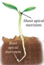
The growth zones at the tips of roots and shoots are the apical meristems, and they produce growth in length (NCERT, p. 2).
Lateral meristem: growth in girth
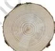
Cut a tree trunk open and you see ring-like patterns — annual growth rings (NCERT, p. 3). Wide rings record favourable growing conditions and narrow rings unfavourable ones; counting the rings gives the tree’s age.
The girth itself grows because actively dividing cells arranged in a ring in the stem keep producing new cells inside and outside in a concentric manner. This tissue is the lateral meristem (NCERT, p. 3).
Intercalary meristem: regrowth after cutting
Cut a young stem’s tip and it stops growing in length, but new branches arise from the nodes — the points where branches or leaves arise. The part of stem between two nodes is the internode.
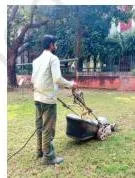
The intercalary meristem sits at the base of the internode, just above the node. It is why a trimmed hedge turns bushy and mown or grazed grass grows back (NCERT, p. 4).
| Meristem | Location | Growth it produces | Example you can see |
|---|---|---|---|
| Apical | Tips of roots and shoots | Length (height, root depth) | Sapling growing taller |
| Lateral | Ring of dividing cells in the stem | Girth (stem diameter) | Tree trunk thickening, annual rings |
| Intercalary | Base of internode, just above the node | Regrowth after cutting or grazing | Hedge turning bushy, grass regrowing |
Memory hook — A-L-I: Apical adds Length, Lateral adds girth from its ring, Intercalary restores growth after injury. Tips = tall, ring = round, node = renewal.
What meristematic cells look like — and why
Meristematic cells are small, with thin cell walls, a large prominent nucleus, dense cytoplasm rich in organelles, no vacuoles, and little or no intercellular space (NCERT, p. 4). The missing vacuole is deliberate: a large water-filled vacuole would push the nucleus aside and inflate the cell, slowing division. A compact cell with a central nucleus divides far faster.
As meristematic cells divide, some stay meristematic while others lose the ability to divide and change structure and function to become permanent tissues, specialised for support, transport or storage. This change is called differentiation (NCERT, p. 4).
Simple Permanent Tissues: Parenchyma, Collenchyma, Sclerenchyma
Look at a transverse section (T.S.) of a sunflower stem and you will see different groups of specialised cells — the permanent tissues (NCERT, p. 4).
Permanent tissues are simple (one cell type) or complex (more than one cell type). Before the supporting tissues, one protective tissue covers everything: the epidermis.
Protective tissue: epidermis
- Epidermis — the tightly packed single layer of flat, rectangular cells that forms the plant’s outer covering (NCERT, p. 5).
- Cuticle — a waxy layer of cutin over the epidermis that limits water loss, resists mechanical injury and blocks parasites.
- Root hair — hair-like projections of epidermal cells in roots that increase the surface area for absorbing water and minerals.
- Stomata — pores in the leaf epidermis for gaseous exchange and transpiration; transpiration creates a pull that helps xylem carry water upward and also removes wastes.
Supporting tissues compared
The three simple supporting tissues differ in cell wall, living or dead status, and the strength they give (NCERT, pp. 5–6).
| Tissue | Cell wall | Living or dead | Function | Example |
|---|---|---|---|---|
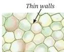
| Parenchyma | Thin | Living | Stores food; performs photosynthesis in green parts; air spaces help aquatic plants float | Green parts of plants; air spaces of aquatic plants |
| Collenchyma | Unevenly thickened corners from pectin | Living | Flexibility — stems and tendrils bend without breaking | Fresh twig; leaf stalks of coriander |
| Sclerenchyma | Thick, lignified | Mostly dead | Hardness and strength | Coconut husk; walnut shell; seed coats |
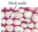
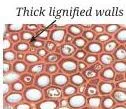
Coconut husk fibres are hard and brittle while coriander leaf stalks are soft and flexible — the first is sclerenchyma thickened with lignin, the second collenchyma stiffened with pectin (NCERT, p. 6).
Conducting Tissues: Xylem and Phloem Side by Side
Two questions drive this section (NCERT, p. 6): how does water reach the leaves of tall trees, and how does food made in leaves reach the rest of the plant? The answers are the complex permanent tissues — xylem and phloem — each built from several cell types working together.
| Feature | Xylem | Phloem |
|---|---|---|
| Job | Transports water and minerals from roots upward; gives the plant strength | Transports food from leaves to other parts |
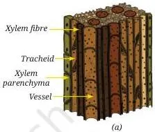
| Components | Tracheids, vessels, xylem parenchyma, xylem fibres | Sieve tubes, companion cells, phloem parenchyma, phloem fibres |
| Living or dead | Only xylem parenchyma is living; tracheids, vessels and fibres are primarily sclerenchymatous | Mostly living cells |
| Special structure | Tracheids and vessels are tubular and thick-walled | Sieve tubes: long tubular cells joined end-to-end by perforated walls; companion cells regulate loading and unloading of sugars |
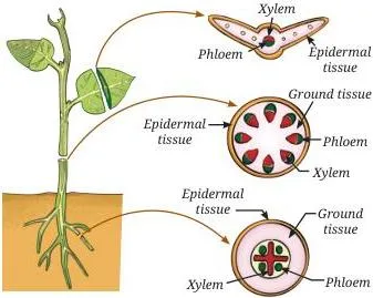
Phloem parenchyma stores food materials, resin, tannins and latex; phloem fibres are sclerenchymatous and add strength. Remember the direction: xylem runs water and minerals up, phloem carries food out of the leaves (NCERT, p. 6).
Tissue Systems: How Plant Tissues Work Together
Plant tissues never work alone; they are organised into three tissue systems (NCERT, pp. 6–7).
| System | What it forms | Components | Main job |
|---|---|---|---|
| Dermal | Outer covering | Epidermis with cuticle | Protects inner parts, reduces water loss |
| Ground | Main body between dermal and vascular tissue | Parenchyma, collenchyma, sclerenchyma | Support and storage |
| Vascular | Conducting strands | Xylem and phloem | Transport of water, minerals and food |
Cork cambium — where bark comes from
In young plants the protective layer is the single-layered epidermis. As the plant ages, some cells just below the epidermis regain the ability to divide and act as lateral meristematic cells to form the cork cambium. Its divisions produce cork cells — dead, compactly arranged, and impermeable to water and gases. Together they form the bark (NCERT, p. 7).
Notice the loop back to the lateral meristem: the same type of tissue that adds girth also builds the protective bark.
Epithelial Tissue: Structure Matches Function
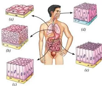
Epithelial tissue forms the skin and lines internal organs — mouth, lungs, blood vessels and intestine. Its cells are packed closely with very little space between them, which keeps germs out, reduces water loss, and helps absorption, secretion and the movement of substances (NCERT, p. 7). The textbook’s Table 3.2 pairs every function with a structure built for it.
| Function | Structure | Location |
|---|---|---|
| Exchange — rapid diffusion of liquids and gases | Single layer of thin, flat cells | Lining of blood vessels and lungs |
| Protection | Many layers; outer cells flat and tightly packed | Skin, mouth, oesophagus |
| Secretion | Cells specialised for releasing substances, cuboidal or columnar | Salivary glands, sweat glands, stomach lining |
| Sensory — smell, taste, sound, balance | Receptor cells with hair-like cilia | Nostrils, taste buds, inner ear |
| Absorption | Single layer of tall, pillar-like cells, often with hair-like structures | Lining of the small intestine |
Why is the lung lining only one or a few cells thick? Exchange works by diffusion, and a thin flat single layer shortens the diffusion path, so gases cross quickly. That is the whole lesson of this table: structure is shaped by function.
Connective Tissue: Blood, Bone, Cartilage, Tendons and Ligaments
A tissue that connects and supports other tissues is a connective tissue. Blood and bone are both connective tissues, yet one is fluid and the other hard — the difference lies in the matrix: watery, soft and jelly-like in blood; hard, solid and rigid in bone (NCERT, p. 8).
The daily-life clues in your blood
Everyday experiences map directly onto blood components (NCERT, p. 9):
| Experience | What it shows | Component behind it |
|---|---|---|
| Small cut: red blood oozes, then a clot forms | Red colour comes from haemoglobin, an iron-rich protein in red blood cells; platelets clot blood at the injury site | Red blood cells (they live about 4 months and are replaced regularly), platelets |
| Skin infection: redness, swelling, pus | White blood cells collect at the infected area, causing pus formation and inflammation | White blood cells |
| Exercise: faster breathing, red face | Muscles need more oxygen, so breathing and blood flow increase | Circulatory and breathing response |
Bone, cartilage, tendon, ligament
Activity 3.3 reveals the four by touch and movement (NCERT, pp. 9–10):
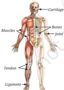
- Bone — hard and rigid because its matrix holds calcium and phosphorus compounds; gives strength, support and protection.
- Cartilage — soft and flexible with a jelly-like matrix; cushions the ends of bones and absorbs shock.
- Tendon — connects muscle to bone; when a muscle contracts, the tendon transmits the force and produces movement.
- Ligament — connects bone to bone; provides stability, limits movement and prevents dislocation.
Muscle and Nerve Tissues: Voluntary, Involuntary and the Neuron
Movements under conscious control — running, writing, lifting — are voluntary movements, produced by skeletal muscles attached to the skeleton. Movements that happen automatically, like food moving through the intestine or the heart beating, are involuntary movements (NCERT, p. 10).
| Muscle | Control | Location | Cell shape | Nuclei | Striations | Special feature |
|---|---|---|---|---|---|---|
| Skeletal | Voluntary | Attached to skeleton | Long, cylindrical, unbranched fibres | Many (multinucleate) | Present | Moves the body by pulling on bones |
| Smooth | Involuntary | Stomach, intestines | Spindle-shaped | Single | Absent | Slow, continuous movement like digestion |
| Cardiac | Involuntary | Only in the heart | Cylindrical, branched | Single | Faint | Works tirelessly and rhythmically without fatigue |
Cardiac muscle never tires because its cells carry a high number of mitochondria and enjoy an abundant blood supply — both needed to keep producing energy for every beat (NCERT, p. 17).
Nervous tissue: the control network
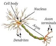
Nervous tissue forms the body’s control and coordination network. The brain is the control centre, coordinating activities, memory and responses; muscles cannot act on their own and receive instructions from it (NCERT, p. 11). During exercise, for example, the brain signals the heart to beat faster to meet the extra oxygen demand.
The nerve cell, the neuron, is specialised to receive, process and transmit messages. It has three parts (NCERT, p. 11):
- Cell body — contains the nucleus and controls the cell’s activities.
- Dendrites — receive signals from other neurons.
- Axon — a long fibre that carries messages away; it ends at axon terminals, which pass the message on to other cells.
The Musculoskeletal System and the Four Types of Joints
The musculoskeletal system — bones, muscles, joints, cartilage, tendons and ligaments — lets us stand upright, move, maintain posture and protect delicate organs. It works under nervous control: muscles pull on bones through tendons, and the tendon transmits the muscle’s force to the bone, producing movement at a joint (NCERT, p. 11).
The adult human skeleton makes up about 12–15 per cent of body weight, though the value varies with age, gender and body composition (NCERT, p. 11).
Skeletal system: spine and rib cage
The skeletal system — skull, vertebral column and rib cage — provides strength and protects internal organs (NCERT, p. 14).
- Vertebral column (backbone) — a flexible column of small bones called vertebrae; it supports the body and keeps us upright. Between vertebrae sit cartilage discs that cushion and allow bending and twisting without injuring the spinal cord.
- Rib cage — formed by 12 pairs of ribs, attached to the spine at the back and the sternum (breast bone) at the front through flexible cartilage. That flexibility lets the rib cage expand and contract during breathing; injured ribs make breathing painful.
Joints compared
A joint is a junction between two or more bones. Joints allow movement, but they do not move bones themselves — muscles provide the pull (NCERT, p. 13).
| Joint | Where | Structure | Movement it allows |
|---|---|---|---|
| Ball and socket | Shoulder | Rounded top of the upper arm bone fits into a shallow hollow of the shoulder bone | Forward, backward, sideways and circular |
| Hinge | Elbow, knee | Bends like a door hinge; the kneecap protects the knee | Bending and straightening in one direction only |
| Pivot | Neck | Skull connected to the backbone | Head turns side to side like a doorknob |
| Fixed | Skull | Flat bones joined tightly | No movement; protects the brain, eyes and ears |
That is why classical and folk dance poses vary so much — circular shoulder and hip movement needs ball-and-socket joints, deep knee bends need hinge joints, and head turns need a pivot joint (NCERT, p. 13).
Totipotency: One Cell Grows a Whole Plant
In 1958, F. C. Steward showed that single vascular phloem cells of carrot can regenerate whole plants — the first demonstration of this ability (NCERT, p. 15).
- Steward grew the phloem cells in a nutrient medium containing simple sugars and hormones.
- The cells divided to form a mass of cells — they first dedifferentiated, regaining the ability to divide.
- Grown with nutrients and growth chemicals, the mass redifferentiated into roots, shoot and a complete plant.
- This ability of mature plant cells to undifferentiate, divide and redifferentiate into a new plant is totipotency — the same power a zygote has (NCERT, p. 15).
Steward’s culture conditions also revealed what growth needs: a liquid nutrient medium with light and air gave a 20 per cent increase in fresh weight, while solid medium without air, or no light, reduced growth. In other words, both light and air are required (NCERT, p. 15).
The practical payoff is tissue culture. Sipra Guha Mukherjee and S. C. Maheshwari developed a complete plant through anther culture on an artificial nutrient medium, a breakthrough that supported crop improvement (NCERT, p. 7).
Crown gall: a disease turned into a tool
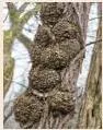
In crown gall disease, tumour-like swellings develop on stems through rapid, uncontrolled cell division, caused by the bacterium Agrobacterium tumefaciens (NCERT, p. 16).
- Instead of only treating the disease, scientists studied how the bacterium transfers its genetic material into plant cells.
- That knowledge turned Agrobacterium into a tool for introducing useful genes into plants.
- Applications today: production of valuable phytochemicals, improved crops and disease-resistant varieties.
Key Numbers and Relationships in Tissues in Action
This chapter carries no formulas to memorise, but questions in tests do reward its measurable relationships (NCERT, pp. 3, 11–12, 17):
- Annual rings = age — the number of annual rings equals the tree’s age in years.
- Ring width = growth conditions — wide rings mark favourable years, narrow rings unfavourable years.
- Skeleton ≈ 12–15% of adult body weight.
- Muscle mass ≈ 40–50% in adult males, 30–40% in adult females (bone percentage is similar across adults).
- RBCs live about 4 months and are replaced regularly.
- 12 pairs of ribs form the rib cage.
Worked Example 1: Estimating Bone and Muscle Weight from Body Weight
This follows Activity 3.4’s method — multiply total body weight by the bone and muscle percentages (NCERT, p. 12). Use midpoints of the given ranges.
Step 1: Take a body weight, say 45 kg.
Step 2: Bone is about 12–15% of body weight; use the midpoint 13%.
\[ \text{Bone mass} = 45 \times 13\% = 5.85\ \text{kg} \]
Step 3: For a female student, muscle is about 30–40%; use the midpoint 35%.
\[ \text{Muscle mass} = 45 \times 35\% = 15.75\ \text{kg} \]
Step 4: Combine the estimates.
\[ 5.85 + 15.75 = 21.6\ \text{kg} \approx 48\%\ \text{of body weight} \]
Final answer: For a 45 kg student, bones ≈ 5.85 kg and muscles ≈ 15.75 kg, together about 48% of body weight.
Try the same method on a 52 kg boy using the male muscle midpoint of 45%: bone = \( 52 \times 13\% = 6.76\ \text{kg} \) and muscle = \( 52 \times 45\% = 23.4\ \text{kg} \). State your assumed percentage before calculating — these ranges vary with age, gender, ethnicity and body composition, so every estimate is approximate.
Worked Example 2: Reading the Teak Tree Growth Data
The chapter’s data table (NCERT, p. 17) records the age, DBH (diameter at breast height) and annual-ring count of a teak tree:
| Age of the teak tree (years) | DBH of tree (cm) | Number of annual rings formed |
|---|---|---|
| 5 | 4 | 5 |
| 10 | 8 | 10 |
| 20 | 24 | 20 |
| 25 | 28 | 25 |
| 30 | 32 | 30 |
| 40 | 40 | 40 |
Step 1: Compare the ring column with age.
In every row the number of annual rings equals the age in years — rings directly record age.
Step 2: Find the total diameter gain from age 5 to age 40.
\[ 40 – 4 = 36\ \text{cm over } 35\ \text{years} \Rightarrow \frac{36}{35} \approx 1.03\ \text{cm/year on average} \]
Step 3: Compare growth per decade.
\[ \text{Age }5\text{–}10:\ \frac{8-4}{5} = 0.8\ \text{cm/yr}; \qquad \text{Age }10\text{–}20:\ \frac{24-8}{10} = 1.6\ \text{cm/yr} \]
\[ \text{Age }20\text{–}30:\ \frac{32-24}{10} = 0.8\ \text{cm/yr}; \qquad \text{Age }30\text{–}40:\ \frac{40-32}{10} = 0.8\ \text{cm/yr} \]
Step 4: Interpret.
Diameter rises with age, fastest between 10 and 20 years; wide rings mark good growth years.
Step 5: Name the tissue responsible for girth — the lateral meristem, located as a ring of dividing cells in the stem.
Final answer: Rings reveal age, DBH increases with age but not at a constant rate, and the lateral meristem (a ring in the stem) adds girth.
Common Mistakes Students Make in Tissues in Action
Six errors appear again and again in tests. Each row pairs the mistake with the rule that fixes it and a quick self-check.
| Mistake | Correct rule | How to check your answer |
|---|---|---|
| “Xylem transports food.” | Xylem transports water and minerals; phloem transports food. | Ask where it starts: roots → water up; leaves → food out. |
| “Sclerenchyma is living tissue that stores food.” | Sclerenchyma is mostly dead, with thick lignified walls for strength; storage is parenchyma’s job. | Coconut husk is hard because its sclerenchyma cells are dead and lignified. |
| “Meristem exists only at root and shoot tips.” | Apical meristem is at the tips; lateral meristem runs as a ring in the stem; intercalary meristem sits above the node. | Grass regrows after mowing — that is intercalary meristem, not apical. |
| Tendon and ligament swapped. | Tendon connects muscle to bone; ligament connects bone to bone and prevents dislocation. | T = Tough muscle attachment; L = Links bone to bone. |
| “Cardiac muscle tires quickly.” | Cardiac muscle works without fatigue because its cells have many mitochondria and an abundant blood supply. | Your heart has beaten continuously since birth. |
| “Cuticle is the epidermis.” | The epidermis is the cell layer; the cuticle is the waxy cutin coating on it that reduces water loss. | Cuticle is a waterproof coating, not a layer of cells. |
Exam Notes: What This Chapter’s Questions Reward
These observations come from the chapter’s own Revise–Reflect–Refine section (NCERT, pp. 17–18); they show the question styles the chapter trains you for, not predictions.
- Assertion–reason pairs — the chapter tests whether the reason actually explains the assertion. Its own example: cardiac muscle works without fatigue BECAUSE its cells have many mitochondria and an abundant blood supply — both statements are true and the reason explains the assertion. Stating both halves together earns the mark.
- Component-naming scores — a question on xylem expects “tracheids, vessels, xylem parenchyma, xylem fibres”, not just the word xylem.
- Living-vs-dead classification repeats — xylem parenchyma is living; tracheids, vessels and fibres are primarily sclerenchymatous; phloem is mostly living.
- Data and graph questions — the teak table (p. 17) links three ideas: rings equal age, diameter grows with age, and the lateral meristem adds girth. Reading the trend matters as much as plotting it.
- Joint questions reward the movement type — ball-and-socket = many directions, hinge = one plane, pivot = side to side, fixed = none.
- Single-answer multiple-choice questions on cell features — repeated division is possible because meristematic cells have thin walls, dense cytoplasm and a large prominent nucleus.
- Why linings are thin — epithelial linings are one or a few cells thick so materials can diffuse across quickly.
The same graph-reading skill appears in the motion chapter notes — the habit of interpreting a trend from plotted data transfers directly.
Revision Summary: Tissues in Action Class 9 Notes in One Sheet
The whole chapter in one breath (compare the textbook’s At a Glance, NCERT p. 16):
- Tissue = a group of similar cells working together for one function; tissues → organs → organ systems → organism.
- Plant tissues: meristematic (dividing) vs permanent (differentiated).
- Meristems: apical (length), lateral (girth), intercalary (regrowth) — remember A-L-I.
- Simple permanent tissues: parenchyma (living, storage), collenchyma (living, flexibility), sclerenchyma (mostly dead, strength).
- Complex permanent tissues: xylem (water and minerals up; only parenchyma living) and phloem (food from leaves; mostly living).
- Protective layer: epidermis with cuticle; root hair absorbs, stomata exchange gases.
- Animal tissues (four): epithelial (covering and lining), connective (blood, bone, cartilage, tendon, ligament), muscular (skeletal, smooth, cardiac), nervous (neurons).
- Musculoskeletal system: bones + muscles + joints + cartilage + tendons + ligaments, under nervous control; skeleton ≈ 12–15% of body weight.
- Joints: ball-and-socket (shoulder), hinge (elbow, knee), pivot (neck), fixed (skull).
- Totipotency: one mature plant cell can dedifferentiate and redifferentiate into a whole plant (Steward, 1958); the basis of tissue culture and Agrobacterium-based genetic engineering.
| Term | Meaning | Example |
|---|---|---|
| Differentiation | Meristematic cells losing the ability to divide and becoming specialised | A meristematic cell becoming a xylem vessel |
| Dedifferentiation | Mature cells regaining the ability to divide | Carrot phloem cells in Steward’s culture |
| Totipotency | One cell regenerating a whole organism | Carrot cell → complete plant |
| Meristem | Actively dividing plant tissue | Apical, lateral, intercalary |
| Sieve tube | Long tubular phloem cells joined end-to-end by perforated walls | Food transport in phloem |
| Matrix | Non-cellular substance between connective tissue cells | Jelly-like in blood, rigid in bone |
For the full set of chapter pages, browse the Class 9 Science notes; for other subjects and classes, see all Class 9 notes or the complete CBSE notes collection.
FAQs on Tissues in Action
What is the difference between meristematic and permanent tissues?
Meristematic tissues divide continuously — their cells are small and thin-walled, with dense cytoplasm, a large prominent nucleus and no vacuoles. Permanent tissues have lost the ability to divide; through differentiation their cells become specialised for jobs like support, transport or storage (NCERT, pp. 2–4).
Why does grass grow back after being cut while a cut tree branch does not?
Grasses carry intercalary meristem at the base of the internode, just above the node, so after mowing or grazing the remaining tissue regenerates the plant. When a young stem’s tip is cut, the stem stops growing in length; new branches arise from the nodes instead (NCERT, p. 4).
Which plant tissue transports water and which transports food?
Xylem transports water and minerals from the roots to the rest of the plant; phloem transports food from the leaves to other parts. Only xylem parenchyma is living in xylem, while phloem is mostly living (NCERT, p. 6).
Why can cardiac muscle keep beating without getting tired?
Cardiac muscle cells have many mitochondria and an abundant blood supply, so they keep producing the energy needed for every beat. That is why the heart works tirelessly and rhythmically throughout life (NCERT, pp. 10, 17).
What is the difference between a tendon and a ligament?
A tendon connects muscle to bone and transmits the muscle’s pulling force to produce movement; a ligament connects bone to bone, providing stability, limiting movement and preventing dislocation (NCERT, p. 10).
Which type of joint lets your shoulder move in a circle but only lets your elbow bend one way?
The shoulder is a ball-and-socket joint — the rounded top of the upper arm bone fits into a shallow hollow of the shoulder bone — so it moves forward, backward, sideways and in a circle. The elbow is a hinge joint that bends and straightens in one direction only, like a door hinge (NCERT, p. 13).
Reference: NCERT Class 9 Science textbook, chapter ‘Tissues in Action’.
Explore Class 9 Science Notes
- Class 9 Science Notes
- Class 9 CBSE Notes
- CBSE Notes for Classes 1 to 12
- Previous: Cell: The Building Block of Life
- Next: Describing Motion Around Us
Related chapters:
- Exploration: Entering the World of Secondary Science notes
- Exploring Mixtures and their Separation notes
- How Forces Affect Motion notes
Official source: download the NCERT textbook free from ncert.nic.in.