Cell the Building Block of Life Class 9 notes — the whole chapter condensed for revision. These notes give you every definition, organelle job, the osmosis rules, cell division summaries, and exam pointers from the NCERT textbook (p. 1–19), in original words with page references so you can verify anything you are unsure about.
This page is written for a Class 9 student revising before a test. Read it straight through, or jump straight to the section you need — the table of contents below links to each topic, and the definitions table and revision summary at the end work as a night-before recap.
For the full official text and figures, open the NCERT Class 9 Science textbook PDF for this chapter.
Chapter 2 at a Glance: From Hot Springs to Cell Theory
Scientists believe life originated in water. The hot springs of Puga Valley in Ladakh stay near boiling point even in a cold climate, and they are home to heat-loving bacteria called thermophiles, which are unicellular — one cell performs every life function (NCERT, p. 1). From bacteria to humans, every living organism is built from cells.
The chapter’s key hierarchy: a group of similar cells forms a tissue, tissues form an organ, and organs work together in organ systems — for example, nasal pores, nasal cavity, trachea and lungs together form the respiratory system (NCERT, p. 1). Yet the cell remains the fundamental unit of structure and function.
The chapter builds towards the classical Cell Theory (NCERT, p. 16), which rests on three statements:
- All living organisms are made up of one or more cells.
- The cell is the basic unit of structure and function in living beings.
- All cells arise from pre-existing cells.
The history: Schleiden (1838) reported all plants are made of cells, Schwann (1839) found the same for animals, and Virchow (1855) added that new cells form only from pre-existing cells.
How to Study Cells: Resolution, Magnification and Microscopes
Before studying cells, you need the tools to see them. The limit of resolution of the human eye is 0.1 mm — two objects closer than that appear as one point when viewed from about 25 cm, the near point of the eye (NCERT, p. 2). A cell is usually smaller than this, so a microscope is essential.
A convex lens or a combination of lenses gives magnification — it makes the object appear larger. Three features of a microscope have been improved over the years: resolution (clarity), contrast (difference in brightness between parts of an object) and magnification (NCERT, p. 3).
Robert Hooke was the first person to observe a cell in 1665. Using a self-designed microscope capable of about 200–300X magnification, he examined a thin slice of cork and saw small box-like compartments, which he named cells (NCERT, p. 2).
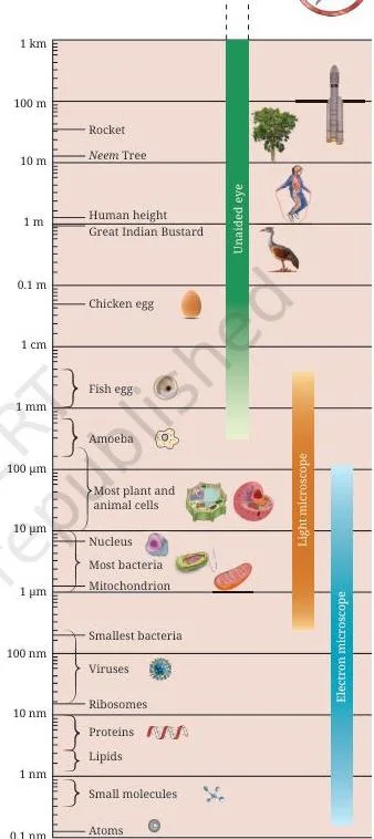
School light microscopes use objective lenses such as 10X and 40X for better magnification and resolution under visible light. Electron microscopes go further: they use a beam of electrons instead of light and reveal cell structure at the nanometre scale — one nanometre is one-billionth of a metre (NCERT, p. 3). The image below shows what such a microscope reveals.
These cell the building block of life class 9 notes now move inside the cell — starting with the boundary that defines it.
Cell Membrane and Osmosis: The Gatekeeper of the Cell
The cell membrane (also called the plasma membrane) is a thin boundary that surrounds the cell and protects its contents (NCERT, p. 4). It defines the individuality of the cell and is selectively permeable — it allows some substances through while blocking others.
Analogy to remember: think of the cell membrane as a bouncer at a club gate. The bouncer checks every visitor — some are allowed in, some are turned away, and the club’s contents stay protected. The membrane does the same for the cell: it lets oxygen, water and nutrients in, keeps waste out, and stops useful molecules from leaking away.
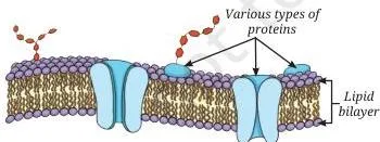
The fluid-mosaic model explains the membrane’s structure (NCERT, p. 5):
- It is a lipid bilayer — two layers of fat molecules with water-attracting heads outward and water-repelling tails inward.
- Proteins are embedded in the bilayer and act like gatekeepers, helping substances pass through.
- The molecules can move sideways, flip and rotate — so the membrane is fluid.
- The molecules are arranged like tiles in a mosaic — hence the mosaic part of the name.
Two movement processes matter here (NCERT, p. 5):
- Diffusion — the net movement of particles from higher to lower concentration. It occurs even without a membrane.
- Osmosis — the diffusion of water across a selectively permeable membrane. Water moves from a more dilute solution to a more concentrated one until the concentrations equalise. In plants, water from the soil enters root cells by osmosis.
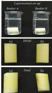
The potato experiment (Activity 2.2, NCERT, p. 4–5) proves osmosis: a potato piece in plain water swells, while a piece in 20% salt or sugar solution shrinks — because the membrane lets water pass but not the sugar or salt molecules.
Real-life application: hospitals give patients isotonic saline drips — salt solution with the same solute concentration as blood. Because the drip is isotonic, red blood cells neither gain water and swell nor lose water and shrink. A wrong concentration would damage the cells.
| Solution type | Solute concentration vs cell interior | Water movement | Cell outcome |
|---|---|---|---|
| Isotonic | Same (equal) | No net movement | Cell stays the same size |
| Hypotonic | Lower outside | Water enters the cell | Cell swells |
| Hypertonic | Higher outside | Water leaves the cell | Cell shrinks |
Cell Wall: Why Plant Cells Stay Firm
Plants are fixed in one place, so they need a rigid structure to withstand wind and rain. Hence plant cells have an additional covering outside the cell membrane called the cell wall (NCERT, p. 6). It keeps leaves and flowers firm and helps plants stay upright.
The cell wall is permeable — water and some dissolved minerals pass through it. This is the classic contrast: the wall is permeable, while the membrane is selectively permeable. Together they let plant roots absorb water and nutrients from the soil.
The wall is mainly made of cellulose, a carbohydrate formed by many glucose units linked together. Cellulose in our diet acts as roughage, helping digestion. Fungi and bacteria also have cell walls.
In a concentrated sugar solution, a Rhoeo or onion cell loses water by osmosis but does not shrink — the rigid wall keeps the outer outline while the inner contents shrink and the membrane pulls away from the wall. Animal cheek cells have no wall, so they shrink considerably. This flexibility lets animal cells change shape easily, supporting movement.
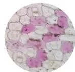
Notice the shapes in the figure: the onion cells are box-shaped and regularly arranged, while the cheek cells are irregular. The reason is exactly the cell wall — plant cells have it, animal cells do not.
Prokaryotic and Eukaryotic Cells: Two Blueprints of Life
Most cells have three basic parts (NCERT, p. 7): a selectively permeable plasma membrane, a semi-fluid jelly-like cytoplasm, and a prominent nucleus. But the presence of a true nucleus divides all cells into two blueprints.
- Prokaryotic cells (pro = primitive, karyon = nucleus) lack a well-defined nucleus and membrane-bound organelles. Most cellular activities happen directly in the cytoplasm. Their DNA is a single circular molecule in a region called the nucleoid (NCERT, p. 9).
- Eukaryotic cells (eu = true, karyon = nucleus) have a well-defined nucleus and several membrane-bound organelles (NCERT, p. 7–8).
| Characteristic | Prokaryotic cell | Eukaryotic cell |
|---|---|---|
| Diameter of a typical cell | 1–10 μm | 10–100 μm |
| Number of cells in an organism | Usually unicellular | Unicellular or multicellular |
| Membrane-bound nucleus | Absent | Present |
| Membrane-bound organelles | Absent | Present |
This is the completed revision version of NCERT Table 2.2 (p. 8). Two extra details from the Ready to Go Beyond box (p. 8): the cytoskeleton is a network of fine fibres that provides structural support, maintains cell shape, and enables movement and internal transport; and cell inclusions are stored materials such as starch, calcium oxalate crystals and silica crystals.
Also note that viruses, viroids and prions are acellular (no cells) — viruses have genetic material with a protein coat, viroids lack the protein coat, and prions are misfolded proteins without genetic material.
All the Organelles and Their Jobs: The Cell at Work
Eukaryotic cells carry out life processes in different organelles independently at the same time — the cell works like a tiny living factory, with each part doing a specific job (NCERT, p. 9). Here is the full line-up, in the order a teacher would build it.
Nucleus — the house of coded instructions (p. 9)
- Double-layered nuclear membrane with pores that allow transfer of material between nucleus and cytoplasm.
- Nucleolus — dense round body where ribosomal subunits are made; these exit to the cytoplasm and assemble into ribosomes.
- Chromatin — an entangled mass of thread-like structures seen in a non-dividing cell.
- When the cell is about to divide, chromatin organises into rod-shaped chromosomes.
- Chromosomes = DNA + proteins; the functional segments of DNA are called genes.
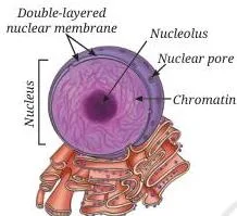
The chain worth memorising from this figure: cell → nucleus → chromosome → DNA → gene.
Ribosomes — the protein factories (p. 10)
Tiny structures, either free in the cytoplasm or attached to the endoplasmic reticulum. They are the sites of protein synthesis.
Endoplasmic reticulum (ER) — the manufacturing factory (p. 10)
- A large organelle that spreads like a network through the cytoplasm and is continuous with the outer membrane of the nuclear envelope.
- RER (rough, with ribosomes attached) — protein synthesis and secretion, for example in pancreatic gland cells.
- SER (smooth, no ribosomes) — synthesis and storage of fats and hormones.
Golgi apparatus — packaging and shipping centre (p. 10)
Stacks of flattened, sac-like structures, functionally linked to the ER and the cell membrane. It modifies, sorts and packages proteins and lipids into vesicles for transport, secretion or lysosome formation. It was first observed in 1898 by the Italian scientist Camillo Golgi in the nerve cells of a barn owl.
Analogy to remember — the protein dispatch route: the ribosome is the kitchen where the protein is cooked, the ER is the assembly line that folds and refines it, the Golgi apparatus is the packing desk that boxes it into vesicles, and the vesicle is the delivery van that carries the package to the cell membrane for export.
NCERT’s exercise asks exactly this path, so learn it as one route.
Lysosomes — the clean-up system (p. 10–11)
Single membrane-bound sacs filled with enzymes that break down unwanted proteins, carbohydrates, fats and even damaged parts of the cell. The breakdown products are released into the cytoplasm for reuse. Human sperm cells contain lysosomal enzymes that help break down the outer layer of the egg during fertilisation.
Mitochondria — the powerhouse of the cell (p. 11)
- Surrounded by two membranes: the outer membrane is smooth and porous; the inner membrane is folded into finger-like projections called cristae, which increase the surface area for chemical reactions.
- Glucose and other molecules are broken down in cellular respiration.
- The released energy is stored as ATP (Adenosine Triphosphate), the energy currency used for most cellular activities.
Plastids — centre for food synthesis in plant cells (p. 11–12)
- Chloroplasts — contain the green pigment chlorophyll, which absorbs sunlight for photosynthesis. They are double-membrane-bound; inside is a semi-fluid stroma with disc-shaped membrane structures containing chlorophyll.
- Chromoplasts — contain yellow, orange or red pigments that give flowers and fruits their bright colours and attract pollinators.
- Leucoplasts — colourless plastids that store starch, oils or proteins, for example in potato and taro cells.
Vacuoles — organelles for storage and support (p. 12)
A mature plant cell usually has one large central vacuole surrounded by a single selectively permeable membrane, filled with watery cell sap. It stores water, minerals, sugars and waste, and maintains pressure inside the cell that keeps the plant firm — when water is lost, the plant wilts. Animal vacuoles are small and temporary.
| Feature | Mitochondria | Chloroplasts |
|---|---|---|
| Membrane | Double membrane; inner folded into cristae | Double membrane; inner disc-shaped structures in stroma |
| Own DNA | Yes | Yes |
| Own ribosomes | Yes | Yes |
| What it produces | ATP through cellular respiration | Sugars through photosynthesis |
| Where found | Almost all eukaryotic cells | Photosynthetic plant cells |
These shared features — own DNA and ribosomes — suggest both organelles share an evolutionary history with bacteria (NCERT, p. 11). Memory device: the MP3 rule — Mito + Plastids have their o3wn DNA and ribosomes. If an exam question asks which organelles contain DNA (other than the nucleus), the answer is mitochondria and plastids.
Two curiosity facts worth a line each: mature human RBCs lose their nucleus to make room for haemoglobin, so they cannot divide and survive about 120 days (p. 9); and in 2010, J. Craig Venter’s team inserted chemically synthesised DNA of Mycoplasma mycoides into a bacterium whose own DNA had been removed — the cell grew and divided following the synthetic DNA’s instructions, proving DNA controls the structure and activities of a cell (p. 13).
Definitions: The Terms That Earn Marks
Reproduce these one-line definitions in one-mark answers. The table groups the chapter’s tested vocabulary by theme.
| Term | Meaning | Example |
|---|---|---|
| Cell boundary and movement | ||
| Cell | The basic structural and functional unit of a living organism | Nerve cell, onion cell |
| Plasma membrane | Thin selectively permeable boundary that defines and protects the cell | Boundary of every cell |
| Selectively permeable | Allows some substances to pass through while blocking others | Membrane lets water pass but not sugar |
| Diffusion | Net movement of particles from higher to lower concentration, no membrane needed | Oxygen moving into cells |
| Osmosis | Diffusion of water across a selectively permeable membrane | Water entering root cells from the soil |
| Isotonic solution | Solute concentration outside equals that inside the cell | Saline drip in hospitals |
| Hypotonic solution | Solute concentration outside is lower than inside | Pure water around a cell |
| Hypertonic solution | Solute concentration outside is higher than inside | 20% salt solution around a potato piece |
| Cell wall | Rigid permeable outer layer of plant, fungal and bacterial cells | Onion peel cell wall |
| Cellulose | Carbohydrate made of many glucose units; main material of the plant cell wall | Roughage in our diet |
| Interior and genetic material | ||
| Cytoplasm | Semi-fluid, jelly-like substance inside the cell | Medium holding organelles |
| Organelle | Sub-cellular component in the cytoplasm with a specific job | Mitochondrion, ribosome |
| Nucleus | Organelle with the cell’s genetic material; house of coded instructions | Controls cell activities |
| Nucleolus | Dense round body in the nucleus where ribosomal subunits are made | Inside the nucleus |
| Chromatin | Thread-like mass of DNA and proteins in a non-dividing cell | Entangled threads in the nucleus |
| Chromosome | Rod-shaped structure formed when chromatin organises before cell division | Visible only when cell is about to divide |
| Gene | Functional segment of DNA | Gene for eye colour |
| DNA | Molecule carrying genetic information; makes up chromosomes with proteins | Deoxyribonucleic acid |
| Nucleoid | Region in a prokaryotic cell containing its circular DNA | In bacterial cells |
| Cytoskeleton | Network of fine fibres providing support, shape, movement and transport | In eukaryotic cells |
| Cell inclusions | Stored materials in the cytoplasm | Starch, calcium oxalate crystals |
| Organelles | ||
| Ribosome | Tiny structure that is the site of protein synthesis | Free or on the ER |
| RER | Rough ER with ribosomes attached; makes and secretes proteins | In pancreatic gland cells |
| SER | Smooth ER without ribosomes; makes and stores fats and hormones | In cells making lipids |
| Golgi apparatus | Stacks of flattened sacs that modify, sort and package proteins and lipids | Cell’s post office |
| Lysosome | Sac of enzymes that breaks down unwanted materials and damaged organelles | Cell’s clean-up system |
| Mitochondria | Organelle where glucose is broken down to release energy as ATP | Powerhouse of the cell |
| Cristae | Finger-like folds of the inner mitochondrial membrane that increase surface area | Inside mitochondria |
| ATP | Adenosine Triphosphate; molecule that stores and supplies energy | Energy currency of the cell |
| Plastid | Plant organelle for food synthesis and storage | Chloroplast |
| Chloroplast | Green plastid containing chlorophyll; site of photosynthesis | In leaf cells |
| Chromoplast | Plastid with yellow, orange or red pigments | In flower petals and fruits |
| Leucoplast | Colourless plastid that stores starch, oils or proteins | In potato and taro cells |
| Vacuole | Membrane-bound sac storing water, minerals, sugars and waste | Large central vacuole in plant cells |
| Cell sap | Watery fluid inside the vacuole | Keeps plant cells firm |
| Life and division | ||
| Unicellular | Organism made of a single cell | Bacteria, yeast |
| Multicellular | Organism made of many cells working together | Plants, fish, humans |
| Prokaryotic | Cell without a well-defined nucleus or membrane-bound organelles | Bacterial cell |
| Eukaryotic | Cell with a well-defined nucleus and membrane-bound organelles | Plant and animal cells |
| Mitosis | Cell division producing two genetically identical daughter cells | Growth, repair, asexual reproduction |
| Meiosis | Two-step division producing four gametes with half the chromosome number | Sperm and egg formation |
| Gamete | Sex cell produced by meiosis | Sperm, egg |
| Contact inhibition | Cell division stops when cells touch neighbouring cells | Normal animal cells |
| Tumour | Mass formed by uncontrolled cell division | Cancerous growth |
| Programmed cell death | Genetically controlled death of cells to maintain balance | Removing cells between developing fingers |
| Totipotency | A plant cell’s ability to develop into a complete plant | Basis of plant tissue culture |
| Cell theory | All organisms are made of cells; the cell is the basic unit; all cells come from pre-existing cells | Schleiden, Schwann, Virchow |
Microscope Maths: The Formulas and Unit Conversions
The chapter’s quantitative tools come from Activity 2.1 (NCERT, p. 3). Know these three relationships and the conversions.
Formula 1 — estimating cell size:
\[ \text{Estimated size of a cell} = \frac{\text{Diameter of visible field (in }\mu\text{m)}}{\text{Number of cells along the diameter}} \]
Formula 2 — total magnification:
\[ \text{Total magnification} = \text{Eyepiece magnification} \times \text{Objective lens magnification} \]
Unit conversions you must apply before dividing:
- \( 1\ \text{mm} = 1000\ \mu\text{m} \)
- \( 1\ \text{nm} = 0.000001\ \text{mm} \)
- \( 1\ \text{nm} = \) one-billionth of a metre
The symbol X means “times” — a magnification of 450X means the image is 450 times larger than the real object.
Worked Examples: Estimating Cell Size and Magnification
Mirror this exact method — convert first, then divide or multiply, and write the unit in every line.
Example 1: Estimating the size of an onion cell
Method: estimated size = field diameter ÷ number of cells across the diameter.
Step 1: A student measures the visible field as 4 mm and counts 40 onion cells along the diameter.
Convert the field diameter from mm to μm:
\[ 4\ \text{mm} \times 1000 = 4000\ \mu\text{m} \]
Step 2: Divide the field diameter by the number of cells:
\[ \text{Estimated size} = \frac{4000\ \mu\text{m}}{40} = 100\ \mu\text{m per cell} \]
Final answer: one onion cell is about 100 μm long.
Example 2: Finding total magnification and the apparent size of a cell
Method: multiply the eyepiece and objective powers; then multiply the real length by the magnification.
Step 1: A microscope has an eyepiece of 10X and an objective of 45X.
Total magnification is:
\[ 10 \times 45 = 450\text{X} \]
Step 2: A cell of real length 40 μm is viewed at 450X.
Its apparent length is:
\[ 40\ \mu\text{m} \times 450 = 18000\ \mu\text{m} = 18\ \text{mm} \]
Final answer: the cell appears 18 mm across — large enough to see with the naked eye.
Example 3: Reverse calculation — real size from a labelled drawing
Method: real size = drawn size ÷ magnification.
Convert the drawing width to μm first.
Step 1: A chloroplast is drawn 6 mm wide and the drawing is labelled 300X.
Convert 6 mm to μm:
\[ 6\ \text{mm} \times 1000 = 6000\ \mu\text{m} \]
Step 2: Divide the drawn width by the magnification:
\[ \text{Real width} = \frac{6000\ \mu\text{m}}{300} = 20\ \mu\text{m} \]
Final answer: the real chloroplast is about 20 μm wide.
Common Mistakes: Where Students Lose Marks
Each error below is paired with the correction and the reason. The first one is worth special attention.
| Student writes… | Correct is… | Reason |
|---|---|---|
| “All cells have a nucleus.” | Prokaryotic cells have only a nucleoid, and mature human RBCs have no nucleus at all. | A bacterium’s DNA sits in a nucleoid without a membrane (p. 7, 9); RBCs lose the nucleus to make room for haemoglobin (p. 9). This is a misconception autopsy — the word “nucleus” is not the same as “genetic material.” |
| “Diffusion and osmosis are the same.” | Osmosis is the diffusion of water across a selectively permeable membrane; diffusion needs no membrane and can involve any particle. | The membrane is the deciding feature — diffusion happens even without one (p. 5). |
| “The cell wall is selectively permeable.” | The cell wall is permeable; selective permeability belongs to the cell membrane. | Water and dissolved minerals pass freely through the wall, but the membrane decides what enters the cell (p. 4, 6). |
| “Chromosomes are visible in a resting cell.” | A non-dividing cell shows chromatin; chromosomes appear only when the cell is about to divide. | Chromatin organises into rod-shaped chromosomes just before division (p. 9). |
| “Meiosis produces two cells.” | Meiosis produces four daughter cells, each with half the chromosome number. | The parent cell divides twice, so 1 → 2 → 4 (p. 15). |
| “Errors in meiosis cause tumours.” | Errors in mitosis cause uncontrolled division and tumours; errors in meiosis cause genetic disorders. | Body-cell division goes wrong in cancer; sex-cell division errors affect offspring (p. 15–16). |
Exam Notes: How This Chapter Gets Asked
These patterns come from the chapter’s own Revise–Reflect–Refine exercise set (NCERT, p. 17–19). Learn the pattern, and the question style stops being a surprise. All Class 9 Science revision resources are grouped on the Class 9 Science notes page.
- Pattern 1 — difference questions with the clue in brackets. Cell membrane vs cell wall (permeability), RER vs SER (structure), chloroplast vs chromoplast (pigments). The mark-earning step: start each point with the clue word — “Cell membrane is selectively permeable, whereas cell wall is permeable.”
- Pattern 2 — diagram-based identification. A labelled cell diagram is given and parts (a) to (g) must be matched with functions such as controlling activities, site of respiration, storage plus rigidity, separation from surroundings, and packing ER products (p. 18). The skill: learn each organelle by its function, not its drawing.
- Pattern 3 — predict-the-outcome of osmosis. Two similar cells or carrots placed in plain water vs salt solution (p. 18–19). The mark-earning step: state the direction of water movement first, then the result — “water moves out of the cell into the salt solution, so the cell shrinks.”
- Pattern 4 — single-choice pairs. Organelle matched with function (ribosome — protein synthesis; SER — lipid and cellulose synthesis; lysosome — digestion) and DNA-containing pairs (mitochondria + nucleus). One wrong match in the option list makes it a quick elimination question.
NCERT’s own exercise set is the best predictor of the question style for this chapter — mastering those 16 questions covers the chapter’s reasoning demands.
Revision Summary: The Cell in 10 Points
Ten bullets rewritten from NCERT’s At a Glance list (p. 17) — your night-before recap.
- The cell is the basic structural and functional unit of all living organisms.
- Prokaryotic cells have a nucleoid and no membrane-bound organelles.
- Eukaryotic cells are larger, with a well-defined nucleus and membrane-bound organelles.
- All cells have a cell membrane; plants, fungi and bacteria have an additional cell wall.
- The nucleus holds chromosomes made of DNA and proteins; genes are functional segments of DNA.
- Cytoplasm fills every cell and carries organelles, each with a specific job.
- Key organelles: nucleus, endoplasmic reticulum, mitochondria, Golgi apparatus, ribosomes and lysosomes.
- Plant cells add plastids — chloroplasts, chromoplasts and leucoplasts.
- Mitosis gives two identical daughter cells; meiosis gives four cells with half the chromosomes.
- Normal cells grow, function and die in a controlled way; cancer cells divide uncontrollably and form tumours.
Memorise the chain: cell → nucleus → chromosome → DNA → gene. For the broader picture of how cells build tissues and organs, see the Class 9 Tissues notes.
Figure Walkthrough: The Diagrams Worth a Second Look
Examiners reuse the chapter’s diagrams because each one teaches one idea. Here is what to notice in each.
- Fig. 2.1 and 2.2 (p. 2): object sizes from unaided to aided eye, and the structure of a light microscope — know the eyepiece, objective lens and stage.
- Fig. 2.4 (p. 4): Colocasia leaf stomata under an electron microscope — what nanometre-scale detail looks like.
- Fig. 2.6 (p. 5): a cell in isotonic, hypotonic and hypertonic solutions — read it as water movement arrows: none, inward, outward.
- Fig. 2.7 (p. 5): membrane lipid bilayer with embedded proteins — the fluid-mosaic model in one picture.
- Fig. 2.8 (p. 6): onion peel cells box-shaped vs cheek cells irregular — the cell wall makes the difference.
- Fig. 2.10 (p. 7): bacterial, plant and animal cells side by side — the basis of the prokaryote vs eukaryote comparison.
- Fig. 2.12 (p. 9): cell → nucleus → chromosome → DNA — learn the packaging order.
- Fig. 2.13 (p. 10): ER–Golgi pathway for protein processing and secretion — the dispatch route.
- Fig. 2.14 (p. 11): mitochondrion with outer membrane and folded inner membrane (cristae) — surface area is the point.
- Fig. 2.15 (p. 11): chloroplast showing stroma and disc-shaped membrane structures.
- Fig. 2.17 (p. 13): onion root tip cells at different stages of division — why root tips are used.
- Fig. 2.18 and 2.19 (p. 14–15): mitosis producing two identical cells vs meiosis producing four gametes — count the daughter cells.
For each diagram, an examiner can ask one of three things: name the part, match the function, or predict what happens next. Practise covering the labels and stating the function from memory.
Activities Insight: What Each NCERT Activity Proves
These five activities show up in “what does this experiment show?” questions and practical vivas.
- Activity 2.1 (p. 3): measure the diameter of the visible field in mm, convert to μm, divide by the number of cells along the diameter — you get the real size of one cell. This links directly to the microscope-maths section above.
- Activity 2.2 (p. 4): potato pieces weighed before and after soaking — the piece in plain water gains weight and swells, the piece in 20% salt or sugar solution loses weight and shrinks. Direct evidence of osmosis.
- Activity 2.3 (p. 6): onion or Rhoeo peel cells are box-shaped and regular; cheek cells are irregular because animal cells lack a wall. In 20% sugar solution the plant cell’s contents shrink while the wall keeps the outline — plasmolysis.
- Activity 2.4 (p. 7): comparing bacterial, plant and animal cell diagrams fills in the prokaryote vs eukaryote comparison table.
- Activity 2.5 (p. 13): onion root tips are squashed and stained with aceto-carmine; cells at different division stages appear because root tip cells divide continuously — that is why root tips are chosen for observing cell division.
Frequently Asked Questions
Why is the cell called the structural and functional unit of life?
Because every living organism is made of cells, and every function of life happens inside them. Cells group into tissues, tissues into organs, and organs into organ systems, yet the cell remains the fundamental unit of structure and function (NCERT, p. 1, 16).
Why do plant cells not shrink in a concentrated sugar solution while animal cells do?
Because the rigid cell wall maintains the cell’s shape. The inner contents shrink and the membrane pulls away from the wall, but the outer outline stays fixed; cheek cells have no wall, so they shrink considerably (NCERT, p. 6).
What is the difference between diffusion and osmosis?
Diffusion is the net movement of any particles from higher to lower concentration and needs no membrane. Osmosis is specifically the diffusion of water across a selectively permeable membrane (NCERT, p. 5).
What is the difference between chromatin and chromosomes?
They are the same genetic material in different forms. In a non-dividing cell, DNA and proteins appear as thread-like chromatin; when the cell is about to divide, chromatin organises into rod-shaped chromosomes (NCERT, p. 9).
Why are mitochondria called the powerhouse of the cell?
Because cellular respiration breaks down glucose in the mitochondria and releases energy stored as ATP. The folded inner membrane, with cristae, increases the surface area for these energy-producing reactions (NCERT, p. 11).
Do bacterial cells have a nucleus?
No. Bacteria are prokaryotic — they lack a well-defined, membrane-bound nucleus. Their DNA is a single circular molecule in a region called the nucleoid (NCERT, p. 7, 9).
Reference: NCERT Class 9 Science textbook, chapter Cell: The Building Block of Life.
Explore Class 9 Science Notes
- Class 9 Science Notes
- Class 9 CBSE Notes
- CBSE Notes for Classes 1 to 12
- Previous: Exploration: Entering the World of Secondary Science
- Next: Tissues in Action
Related chapters:
- Describing Motion Around Us notes
- Exploring Mixtures and their Separation notes
- How Forces Affect Motion notes