
These biomolecules class 12 notes cover the complete CBSE Unit 10 — carbohydrates, proteins, enzymes, vitamins, nucleic acids and hormones — in the teaching order a teacher would follow. Every definition, table and reaction here is grounded in the rationalised NCERT Class 12 Chemistry Part II textbook.
Revise in one pass: skim the overview, drill the disaccharide, vitamin and DNA-versus-RNA tables, then attempt the two solved examples before checking the common-mistakes table. The exam notes near the end tell you which details earn marks in board answers.
For the rest of the syllabus, pair this page with the Class 12 Chemistry notes hub and the Class 12 notes index.
Reference: NCERT Class 12 Chemistry textbook, chapter Biomolecules.
Biomolecules Class 12 Notes: The Six Families to Master
Biochemistry is the study of what goes on chemically inside a living system. This unit surveys the six families of biomolecules that make up the molecular logic of life. Each family below is developed in the same four steps: definition → classification → structure → function.
- Carbohydrates — optically active polyhydroxy aldehydes or ketones; the body’s main energy food.
- Proteins — polymers of alpha-amino acids joined by peptide bonds; the basis of body structure and function.
- Enzymes — biocatalysts, almost all globular proteins, that speed up reactions under mild conditions.
- Vitamins — essential organic food factors required in small amounts.
- Nucleic acids — DNA and RNA; carry heredity and direct protein synthesis.
- Hormones — intercellular messengers produced by endocrine glands.
Carbohydrates: Definition and the Three-Way Classification
Carbohydrates are optically active polyhydroxy aldehydes or ketones, or compounds that give such units on hydrolysis (NCERT, p. 281). Sweet ones are called sugars; the common home sugar is sucrose and the sugar in milk is lactose.
The old name “hydrate of carbon” came from the general formula \( \mathrm{C}_x(\mathrm{H}_2\mathrm{O})_y \), but the formula is only a hint, not the definition.
The formula trap: acetic acid and rhamnose
Two examples break the hydrate formula as a definition. Acetic acid \( \mathrm{CH}_3\mathrm{COOH} \) fits \( \mathrm{C}_2(\mathrm{H}_2\mathrm{O})_2 \), yet it is not a carbohydrate — it has no polyhydroxy aldehyde or ketone structure. Rhamnose \( \mathrm{C}_6\mathrm{H}_{12}\mathrm{O}_5 \) is a genuine carbohydrate but does not fit the formula at all.
The lesson: always test the chemical definition, never just the empirical formula. This is the chapter’s favourite trick question.
On the basis of behaviour on hydrolysis, carbohydrates fall into three groups (NCERT, p. 282):
- Monosaccharides — cannot be hydrolysed further (glucose, fructose, ribose). About 20 occur in nature.
- Oligosaccharides — yield two to ten monosaccharide units on hydrolysis (disaccharides, trisaccharides, and so on).
- Polysaccharides — yield a large number of monosaccharide units (starch, cellulose, glycogen, gums); not sweet, hence called non-sugars.
A monosaccharide with an aldehyde group is an aldose; one with a keto group is a ketose. The carbon count also enters the name, as Table 10.1 shows.
| Carbon atoms | General term | Aldehyde form | Ketone form |
|---|---|---|---|
| 3 | Triose | Aldotriose | Ketotriose |
| 4 | Tetrose | Aldotetrose | Ketotetrose |
| 5 | Pentose | Aldopentose | Ketopentose |
| 6 | Hexose | Aldohexose | Ketohexose |
| 7 | Heptose | Aldoheptose | Ketoheptose |
All monosaccharides, whether aldose or ketose, are reducing sugars — they reduce Fehling’s solution and Tollens’ reagent. The full reducing versus non-reducing picture comes with disaccharides.
Glucose: Preparation and Evidence for the Open-Chain Structure
Glucose occurs free in sweet fruits and honey, and in large amounts in ripe grapes. Its formula is \( \mathrm{C}_6\mathrm{H}_{12}\mathrm{O}_6 \); it is an aldohexose also called dextrose, and it is the monomer of starch and cellulose (NCERT, p. 283). Two commercial preparations matter for exams:
- From sucrose: boiling cane sugar with dilute HCl or \( \mathrm{H}_2\mathrm{SO}_4 \) in alcoholic solution gives equal amounts of glucose and fructose (NCERT, p. 282).
- From starch: hydrolysis of starch with dilute \( \mathrm{H}_2\mathrm{SO}_4 \) at 393 K under 2–3 atm pressure gives glucose.
The open-chain structure was fixed by six pieces of evidence. The product name in each case is the exam-winning detail:
| Reagent / condition | Product | What it proves |
|---|---|---|
| Molecular formula determination | \( \mathrm{C}_6\mathrm{H}_{12}\mathrm{O}_6 \) | Composition of glucose |
| HI, heat | n-Hexane | All six carbons linked in a straight chain |
| Hydroxylamine / HCN | Oxime / cyanohydrin | Presence of a carbonyl group |
| Bromine water (mild oxidant) | Gluconic acid | Carbonyl is an aldehyde, not a ketone |
| Acetic anhydride | Glucose pentaacetate | Presence of five –OH groups |
| Nitric acid | Saccharic acid (dicarboxylic acid) | Presence of a primary –OH group |
The name is D-(+)-glucose. ‘D’ is a configuration: the molecule is correlated to D-(+)-glyceraldehyde by comparing the lowest asymmetric carbon, whose –OH lies on the right. ‘(+)’ is optical rotation: the molecule turns plane-polarised light to the right. ‘D’ and ‘L’ have no relation to optical activity — D-fructose, for example, is laevorotatory (NCERT, p. 284).
Cyclic Glucose and Fructose: Anomers, Pyranose and Furanose
The open-chain structure could not explain three facts about glucose (NCERT, p. 285):
- Glucose does not give Schiff’s test and forms no NaHSO\(_3\) addition product, despite having an aldehyde group.
- The pentaacetate of glucose does not react with hydroxylamine — no free –CHO group.
- Glucose exists as two crystalline forms: alpha (m.p. 419 K) and beta (m.p. 423 K).
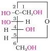
Explanation: one –OH group at C5 adds across the –CHO, forming a cyclic hemiacetal. The ring has six members, so glucose gets the pyranose name, in analogy with the parent compound pyran. The figure below shows this ring formation.
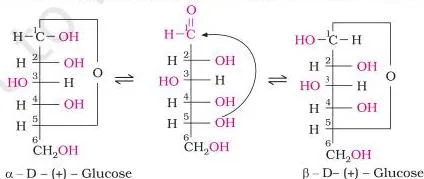
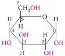
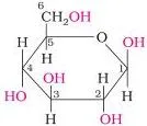
The anomeric carbon is C1 — the aldehyde carbon before cyclisation. The alpha and beta forms differ only in the –OH configuration at C1, so they are called anomers, and they exist in equilibrium with the open chain. The accurate way to draw these rings is the Haworth structure.
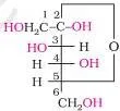
Fructose is a ketohexose with the same formula as glucose, obtained with glucose on hydrolysis of sucrose. Its ketone group sits at C2; the –OH at C5 adds to it, forming a five-membered ring called furanose, named after furan. Fructose belongs to the D-series but is laevorotatory, so it is written D-(-)-fructose; its anomers differ at C2 (NCERT, p. 286).
Disaccharides: Glycosidic Linkages and Reducing Behaviour
A glycosidic linkage is an oxide linkage formed by the loss of a water molecule between two monosaccharide units (NCERT, p. 287). If the reducing groups — aldehydic or ketonic — are bonded in the linkage, the sugar is non-reducing; if they remain free, or can become free, it is reducing.
| Disaccharide | Units | Linkage carbons | Reducing or non-reducing |
|---|---|---|---|
| Sucrose | alpha-D-glucose + beta-D-fructose | C1 of glucose to C2 of fructose | Non-reducing |
| Maltose | Two alpha-D-glucose units | C1 of glucose I to C4 of glucose II | Reducing (free aldehyde possible at C1 of second unit) |
| Lactose | beta-D-galactose + beta-D-glucose | C1 of galactose to C4 of glucose | Reducing (free aldehyde at C1 of glucose); milk sugar |
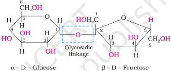
Invert sugar: sucrose is dextrorotatory, but hydrolysis gives glucose \( (+52.5^{\circ}) \) and fructose \( (-92.4^{\circ}) \). The laevorotation of fructose outweighs the dextrorotation of glucose, so the mixture turns laevorotatory — the sign of rotation inverts, hence the name (NCERT, p. 287).
The table below links reducing behaviour to the two classic reagent tests. “Reduces” means the sugar reduces the reagent, giving the characteristic colour change or precipitate.
| Sugar | Family | Fehling’s solution | Tollens’ reagent |
|---|---|---|---|
| Glucose | Monosaccharide (aldose) | Reduces | Reduces |
| Fructose | Monosaccharide (ketose) | Reduces | Reduces |
| Sucrose | Disaccharide | Does not reduce | Does not reduce |
| Maltose | Disaccharide | Reduces | Reduces |
| Lactose | Disaccharide | Reduces | Reduces |
Why this pattern? All monosaccharides reduce both reagents. Sucrose is the odd one out because both carbonyl groups are trapped in the glycosidic linkage; maltose and lactose can regenerate a free aldehyde at C1 of the second unit.
Polysaccharides: Starch, Cellulose and Glycogen Compared
Polysaccharides hold a large number of monosaccharide units joined by glycosidic linkages and act mainly as food storage or structural material (NCERT, p. 288).
| Polymer | Monomer | Linkage | Branching | Role and source |
|---|---|---|---|---|
| Starch | alpha-D-glucose | C1–C4 chains | Amylose (15–20%): unbranched; amylopectin (80–85%): branched via C1–C6 | Main storage polysaccharide of plants; cereals, roots, tubers |
| Cellulose | beta-D-glucose | C1–C4 | Straight chain, no branching | Cell walls of plants; most abundant organic substance in the plant kingdom |
| Glycogen | alpha-D-glucose | C1–C4 chains | More highly branched than amylopectin | Animal starch; liver, muscles, brain; also in yeast and fungi |
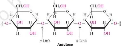
The alpha versus beta difference has a real consequence: starch (alpha) is our most important dietary source, while cellulose (beta) works as structural material in wood and cotton. Glycogen is the animal reserve — when the body needs glucose, enzymes break glycogen down to glucose.
Real-life application — carbo-loading: because muscles store glycogen as a quick glucose reserve, endurance athletes “carbohydrate-load” before long events to pack their liver and muscles with glycogen. During the event, enzymes release glucose from glycogen on demand — the textbook fact is the athlete’s strategy.
Importance of carbohydrates (NCERT, p. 289):
- Storage molecules — starch in plants, glycogen in animals.
- Cellulose builds the cell wall of plants and bacteria.
- Wood (furniture) and cotton fibre (clothing) are cellulose; it feeds the textiles, paper, lacquer and brewery industries.
- Honey has long been used in Ayurveda as an instant source of energy.
Key Definitions: Carbohydrates to Hormones in One Table
Here is the complete glossary for the chapter, in words you can use directly in short-answer definitions.
| Term | Meaning | Example |
|---|---|---|
| Monosaccharide | A carbohydrate that cannot be hydrolysed further into a simpler polyhydroxy aldehyde or ketone unit | Glucose, fructose, ribose |
| Oligosaccharide | Yields two to ten monosaccharide units on hydrolysis | Sucrose (a disaccharide) |
| Polysaccharide | Yields a large number of monosaccharide units on hydrolysis; not sweet | Starch, cellulose, glycogen |
| Glycosidic linkage | Oxide linkage formed between two monosaccharide units with loss of water | Sucrose: C1–C2 linkage |
| Reducing sugar | Sugar that reduces Fehling’s solution and Tollens’ reagent | Glucose, maltose, lactose |
| Peptide bond | Amide (\( -\mathrm{CO}-\mathrm{NH}- \)) formed between \( -\mathrm{COOH} \) and \( -\mathrm{NH}_2 \) with loss of water | Glycylalanine |
| Zwitter ion | Dipolar, neutral form of an amino acid carrying both positive and negative charges | Glycine in water |
| Denaturation | Loss of biological activity when a protein’s secondary and tertiary structures are destroyed | Boiled egg white |
| Enzyme | Biocatalyst, almost always a globular protein, specific to a substrate and a reaction | Maltase |
| Vitamin | Organic compound required in small amounts in the diet for normal growth and health | Vitamin C |
| Nucleoside | Base attached to the 1′ position of a sugar | Adenosine |
| Nucleotide | Nucleoside linked to phosphoric acid at the 5′ position | AMP |
| Hormone | Intercellular messenger produced by endocrine glands and carried by blood | Insulin |
Amino Acids: Essential or Non-Essential, and the Zwitter Ion
Proteins are polymers of alpha-amino acids. The general skeleton is \( \mathrm{R}-\mathrm{CH}(\mathrm{NH}_2)-\mathrm{COOH} \), where R is the side chain. Depending on the position of \( -\mathrm{NH}_2 \) relative to \( -\mathrm{COOH} \), amino acids are classified as alpha, beta, gamma and so on — but only alpha-amino acids are obtained on hydrolysis of proteins (NCERT, p. 290).
Amino acids are classified as acidic, basic or neutral by counting groups (NCERT, p. 291):
| If the molecule has… | Classification | Example side chain R |
|---|---|---|
| Equal \( -\mathrm{NH}_2 \) and \( -\mathrm{COOH} \) | Neutral | Alanine (\( -\mathrm{CH}_3 \)) |
| More \( -\mathrm{COOH} \) than \( -\mathrm{NH}_2 \) | Acidic | Aspartic acid (\( -\mathrm{CH}_2\mathrm{COOH} \)) |
| More \( -\mathrm{NH}_2 \) than \( -\mathrm{COOH} \) | Basic | Lysine (\( -(\mathrm{CH}_2)_4-\mathrm{NH}_2 \)) |
Essential amino acids cannot be synthesised in the body and must come from the diet — there are ten of them, marked with an asterisk in Table 10.2 (valine, leucine, isoleucine, lysine, and others). Non-essential amino acids can be synthesised in the body.
Amino acids are colourless, crystalline, water-soluble solids with high melting points — they behave like salts, not like simple amines or acids. In water the \( -\mathrm{COOH} \) group loses a proton and the \( -\mathrm{NH}_2 \) group gains one, giving a zwitter ion: a neutral, dipolar ion carrying both charges.
Because of this form, amino acids show amphoteric behaviour, reacting with both acids and bases.
Except glycine, all naturally occurring alpha-amino acids are optically active because the alpha-carbon is asymmetric. Most natural amino acids have the L-configuration, drawn with \( -\mathrm{NH}_2 \) on the left. This is the same functional-group logic you met in the Amines chapter.
Trivial names often tell a story: glycine comes from the Greek glykos (sweet), and tyrosine was first obtained from cheese (tyros).
Peptide Bonds and Protein Structure at Four Levels
A peptide bond is an amide linkage \( -\mathrm{CO}-\mathrm{NH}- \) formed between the \( -\mathrm{COOH} \) group of one amino acid and the \( -\mathrm{NH}_2 \) group of another, with elimination of a water molecule. Combining glycine with alanine gives the dipeptide glycylalanine (NCERT, p. 292).
- Dipeptide (2 amino acids), tripeptide (3), tetrapeptide (4), pentapeptide (5), hexapeptide (6).
- More than ten residues: polypeptide.
- A polypeptide with more than 100 residues and molecular mass above 10,000 u is called a protein. Insulin (51 residues) counts because it has a well-defined protein conformation.
| Feature | Fibrous proteins | Globular proteins |
|---|---|---|
| Shape | Polypeptide chains run parallel, forming fibres | Chains coil around into a spherical shape |
| Holding forces | Hydrogen and disulphide bonds between chains | Folding of the chain itself |
| Solubility | Generally insoluble in water | Usually soluble in water |
| Examples | Keratin (hair, wool, silk), myosin (muscles) | Insulin, albumins |
Protein structure is studied at four levels, each more complex than the last (NCERT, p. 293):
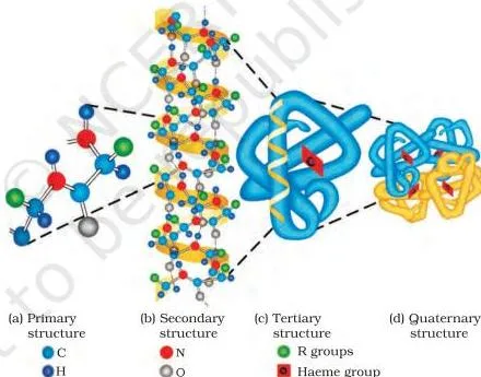
- Primary: the specific sequence of amino acids in the polypeptide chain. Changing the sequence creates a different protein.
- Secondary: regular folding of the backbone into an alpha-helix or beta-pleated sheet, stabilised by hydrogen bonds between the \( -\mathrm{NH} \) and carbonyl (C=O) groups of the peptide bond.
- Tertiary: overall folding of the chain, giving fibrous or globular shapes.
- Quaternary: spatial arrangement of two or more polypeptide subunits.
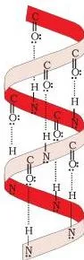
The alpha-helix figure shows the chain twisted into a right-handed screw; every \( -\mathrm{NH} \) group of one residue is hydrogen-bonded to the C=O of an adjacent turn. The four-level diagram below shows how primary structure folds into secondary, tertiary and quaternary levels — each coloured ball is an amino acid.
The main forces that stabilise secondary and tertiary structures are hydrogen bonds, disulphide linkages, van der Waals forces and electrostatic forces of attraction.
Denaturation: secondary and tertiary gone, primary intact
A protein in its native form has a unique three-dimensional structure and biological activity. When temperature or pH changes disturb its hydrogen bonds, globules unfold, helices uncoil and the protein loses its activity — this is denaturation. Secondary and tertiary structures are destroyed while the primary sequence stays intact.
Boiling coagulates egg white; milk curdles because bacteria form lactic acid (NCERT, p. 294).
Worked Examples: Hydrolysis Products and Reducing Behaviour
Two application problems, solved step by step, to fix the chapter’s most examinable logic.
Example A: Identify a reducing disaccharide from its hydrolysis
Step 1: Note the clues.
X has formula \( \mathrm{C}_{12}\mathrm{H}_{22}\mathrm{O}_{11} \), reduces Fehling’s solution, and gives only glucose on hydrolysis.
Reducing behaviour means a free aldehyde can form at C1; “only glucose” means both units are glucose.
Step 2: Identify X.
Sucrose would give glucose + fructose, and lactose gives galactose + glucose.
Only maltose gives glucose alone.
Step 3: Give the linkage.
In maltose, C1 of one alpha-D-glucose links to C4 of the second glucose unit — a C1–C4 glycosidic linkage.
Step 4: Write the hydrolysis and the mass balance for 342 g of maltose:
\[ \mathrm{C}_{12}\mathrm{H}_{22}\mathrm{O}_{11} + \mathrm{H}_{2}\mathrm{O} \rightarrow 2\mathrm{C}_{6}\mathrm{H}_{12}\mathrm{O}_{6} \]
\[ 342\ \text{g maltose} + 18\ \text{g water} = 360\ \text{g glucose} \]
Final answer: X is maltose (C1–C4 linkage). 342 g of maltose plus 18 g of water gives 360 g of glucose; mass is conserved.
Example B: Classify amino acid side chains
- Step 1: State the decision rule — count \( -\mathrm{COOH} \) groups against \( -\mathrm{NH}_2 \) groups in the whole molecule.
- Step 2: R = \( -\mathrm{CH}_3 \) (alanine): one \( -\mathrm{COOH} \) and one \( -\mathrm{NH}_2 \) → neutral.
- Step 3: R = \( -\mathrm{CH}_2\mathrm{COOH} \) (aspartic acid): an extra \( -\mathrm{COOH} \) makes the count acidic.
- Step 4: R = \( -(\mathrm{CH}_2)_4-\mathrm{NH}_2 \) (lysine): an extra \( -\mathrm{NH}_2 \) makes the count basic.
- Step 5: Zwitter ion behaviour — in neutral water, \( -\mathrm{COOH} \) loses \( \mathrm{H}^{+} \) and \( -\mathrm{NH}_2 \) gains it, so the molecule becomes dipolar and neutral overall, whatever the side chain type.
Final answer: \( -\mathrm{CH}_3 \) neutral; \( -\mathrm{CH}_2\mathrm{COOH} \) acidic; \( -(\mathrm{CH}_2)_4-\mathrm{NH}_2 \) basic. All three form zwitter ions in water.
Enzymes: Biocatalysts That Lower Activation Energy
Enzymes are biocatalysts, and almost all of them are globular proteins. They work under the mild conditions of the body and are highly specific — for a particular reaction and a particular substrate (NCERT, p. 295).
- Named after the substrate: maltase catalyses the hydrolysis of maltose into glucose.
- Named after the reaction type: oxidoreductase enzymes catalyse oxidation of one substrate with simultaneous reduction of another.
- The ending -ase marks an enzyme.
\[ \mathrm{C}_{12}\mathrm{H}_{22}\mathrm{O}_{11} \xrightarrow{\text{Maltase}} 2\mathrm{C}_{6}\mathrm{H}_{12}\mathrm{O}_{6} \]
Enzymes, like chemical catalysts, reduce the magnitude of activation energy. Acid hydrolysis of sucrose needs \( 6.22\ \text{kJ mol}^{-1} \); the enzyme sucrase brings it down to \( 2.15\ \text{kJ mol}^{-1} \). That comparison is the quantitative point to quote.
Vitamins: Sources and Deficiency Diseases in One Table
Vitamins are organic compounds required in small amounts in the diet to perform specific biological functions for normal growth and health (NCERT, p. 295). The name came from vital + amine; the ‘e’ was dropped when later work showed most vitamins lack amino groups.
Most cannot be synthesised in the body, so they are essential food factors — and excess of vitamins is harmful.
- Fat-soluble: vitamins A, D, E and K — soluble in fats and oils, stored in the liver and adipose (fat-storing) tissue.
- Water-soluble: B-group vitamins and vitamin C — readily excreted in urine, cannot be stored (except \( \mathrm{B}_{12} \)), so they must be supplied regularly.
Memory device: think of the phrase “KADE is kept in the liver” — the four fat-soluble vitamins (K, A, D, E) are the ones stored in the liver and fat tissue; B and C are washed out in urine.
| Vitamin | Representative sources | Deficiency disease |
|---|---|---|
| A | Fish liver oil, carrots, butter, milk | Xerophthalmia (hardening of cornea), night blindness |
| B\(_1\) (Thiamine) | Yeast, milk, green vegetables, cereals | Beri beri (loss of appetite, retarded growth) |
| B\(_2\) (Riboflavin) | Milk, egg white, liver, kidney | Cheilosis (fissuring at corners of mouth and lips), digestive disorders |
| B\(_6\) (Pyridoxine) | Yeast, milk, egg yolk, cereals, grams | Convulsions |
| B\(_{12}\) | Meat, fish, egg, curd | Pernicious anaemia (RBC deficient in haemoglobin) |
| C (Ascorbic acid) | Citrus fruits, amla, green leafy vegetables | Scurvy (bleeding gums) |
| D | Exposure to sunlight, fish, egg yolk | Rickets (bone deformities in children), osteomalacia (soft bones in adults) |
| E | Vegetable oils (wheat germ oil, sunflower oil) | Increased fragility of RBCs, muscular weakness |
| K | Green leafy vegetables | Increased blood clotting time |
Nucleic Acids: DNA versus RNA
Complete hydrolysis of DNA or RNA yields a pentose sugar, phosphoric acid and nitrogen-containing bases. DNA has beta-D-2-deoxyribose; RNA has beta-D-ribose. DNA carries adenine (A), guanine (G), cytosine (C) and thymine (T); RNA has uracil (U) in place of thymine (NCERT, p. 297).
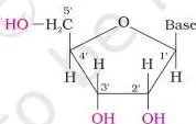
A nucleoside is a base attached to the 1′ position of the sugar. Sugar carbons are numbered 1′, 2′, 3′ and so on to distinguish them from base atoms. When the nucleoside is linked to phosphoric acid at the 5′ position, it becomes a nucleotide (NCERT, p. 298). The figure below shows the base-sugar unit of a nucleoside.
Nucleotides are joined together by a phosphodiester linkage between the 5′ carbon of one sugar and the 3′ carbon of the next. The dinucleotide figure below shows exactly this connection.
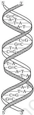
Watson and Crick proposed a double-strand helix for DNA: two chains wound about each other, held by hydrogen bonds between specific base pairs — A with T, C with G. The strands are therefore complementary, which explains how identical copies of parental DNA pass to daughter cells.
RNA, in its secondary structure, is a single-stranded helix that sometimes folds back on itself (NCERT, p. 299).
- DNA is the chemical basis of heredity; it holds the coded message for protein synthesis and self-duplicates during cell division.
- RNA carries out the message. There are three types: messenger RNA (m-RNA), ribosomal RNA (r-RNA) and transfer RNA (t-RNA).
Because a person’s DNA base sequence is unique and cannot be altered by surgery or treatment, DNA fingerprinting is used (NCERT, p. 300):
- in forensic laboratories, to identify criminals;
- to determine the paternity of an individual;
- to identify dead bodies in accidents by comparing the DNA of parents or children;
- to identify racial groups and rewrite biological evolution.
| Feature | DNA | RNA |
|---|---|---|
| Sugar | beta-D-2-deoxyribose | beta-D-ribose |
| Bases | Adenine, guanine, cytosine, thymine | Adenine, guanine, cytosine, uracil |
| Strands | Double helix; two complementary strands | Single strand that can fold back on itself |
| Function | Chemical basis of heredity; stores the coded message for proteins | Carries out protein synthesis (m-RNA, r-RNA, t-RNA) |
Hormones: Steroid, Peptide and Amino Acid Messengers
Hormones act as intercellular messengers. They are produced by endocrine glands, poured directly into the bloodstream, and transported to the site of action (NCERT, p. 300).
| Chemical class | Examples | Key function |
|---|---|---|
| Steroids | Estrogens, androgens, glucocorticoids, mineralocorticoids | Secondary sex characters; carbohydrate metabolism; water and salt balance |
| Polypeptides | Insulin, glucagon, endorphins | Regulation of blood glucose — insulin lowers it, glucagon raises it |
| Amino acid derivatives | Epinephrine, norepinephrine, thyroxine | Response to external stimuli; control of metabolism |
- Thyroxine is an iodinated derivative of tyrosine. Low levels cause hypothyroidism (lethargy, obesity); high levels cause hyperthyroidism. Low dietary iodine enlarges the thyroid, which is why commercial table salt is “iodised” with sodium iodide.
- Glucocorticoids control carbohydrate metabolism and modulate inflammatory and stress reactions; mineralocorticoids control the excretion of water and salt by the kidney.
- Failure of the adrenal cortex can cause Addison’s disease — hypoglycemia, weakness, increased susceptibility to stress — which is fatal unless treated with glucocorticoids and mineralocorticoids.
- Gonadal hormones: testosterone (male secondary characters), estradiol (female characters and the menstrual cycle), and progesterone (prepares the uterus for implantation of the fertilised egg).
Common Mistakes Students Make in Biomolecules
These seven errors are the classic marks-losing slips. Each row shows the wrong idea and the rule that fixes it.
| Students write… | Correct rule | How to check your answer |
|---|---|---|
| “Any compound fitting \( \mathrm{C}_x(\mathrm{H}_2\mathrm{O})_y \) is a carbohydrate” | It must be a polyhydroxy aldehyde or ketone, or give such units on hydrolysis. Acetic acid fits the formula but is not one. | Look for multiple –OH plus –CHO or C=O. Rhamnose is a sugar without the formula. |
| “D means dextrorotatory” | D/L is configuration relative to glyceraldehyde; (+) / (−) is optical rotation. | Glucose is D(+), fructose is D(−) — configuration and rotation are independent. |
| “Sucrose is reducing because fructose has a ketone” | Both reducing groups are bonded in the glycosidic linkage (C1–C2), so no free carbonyl remains. | Sucrose does not reduce Fehling’s or Tollens’; maltose and lactose do. |
| “Glycogen is an unbranched starch” | Glycogen is more highly branched than amylopectin. | Glycogen branches by C1–C6 linkages and does so more often. |
| “Denaturation destroys the primary structure” | Only secondary and tertiary structures are destroyed; the amino acid sequence survives. | Boiled egg white is still protein — coagulation is denaturation, not hydrolysis. |
| “Enzymes raise activation energy” | Enzymes lower activation energy — from 6.22 to 2.15 kJ mol\(^{-1}\) for sucrose hydrolysis. | Compare the two numbers: a lower activation energy means a faster reaction. |
| “A nucleotide is base + sugar” | That is a nucleoside. A nucleotide is a nucleoside plus phosphate at the 5′ position. | Count three parts in a nucleotide: base + sugar + phosphate. |
Exam Notes: What Earns the Marks
These points come from reading the chapter’s end-of-unit exercises (10.1–10.25) with an examiner’s eye. They are observations of how the content is examined, not predictions.
| NCERT exercise | Topic | The detail that earns the mark |
|---|---|---|
| 10.9, 10.10 | Glucose reactions | Name the products — n-hexane, gluconic acid, saccharic acid. Quote the three open-chain failures: no Schiff’s test, no NaHSO\(_3\) adduct, two crystalline forms. |
| 10.2, 10.5, 10.7 | Reducing sugars, glycosidic linkage | Define the linkage as an oxide linkage through oxygen. State hydrolysis products: sucrose → glucose + fructose; lactose → galactose + glucose. |
| 10.6, 10.8 | Starch, glycogen, cellulose | Glycogen is more branched than amylopectin and stored in liver, muscle, brain. Cellulose is straight-chain beta-D-glucose with C1–C4 links. |
| 10.11–10.16 | Amino acids, peptides, proteins | Zwitter ion answers must say “dipolar” and “amphoteric”. The alpha-helix credit point is hydrogen bonding between the –NH of one residue and the C=O of an adjacent turn. |
| 10.17, 10.18 | Enzymes, denaturation | Enzymes reduce activation energy; denaturation destroys only secondary and tertiary structure. |
| 10.19, 10.20 | Vitamins | Deficiency-disease recall from Table 10.3 — A: night blindness/xerophthalmia, D: rickets, K: clotting time. Give sources for A and C. |
| 10.21–10.25 | Nucleic acids | Nucleoside vs nucleotide difference; A–T and C–G pairing; DNA vs RNA in separate points — sugar, bases, strands, function. |
Definition questions (glycosidic linkage, peptide bond, denaturation, nucleoside, nucleotide) are short-answer staples: write the definition, then one structural detail. For linkage questions, always state the carbons — sucrose C1–C2; maltose and lactose C1–C4.
Key Reactions of Biomolecules to Memorise
The reaction list every answer needs. Conditions matter — dilute H\(_2\)SO\(_4\) at 393 K under 2–3 atm for starch, H\(^+\) for sucrose. Verify each equation against the official rationalised NCERT Chemistry Part II text (Unit 10) at ncert.nic.in.
| Reaction | Reagents and conditions | Product(s) |
|---|---|---|
| Sucrose hydrolysis | Dilute acid, H\(^+\) | \( \mathrm{C}_{12}\mathrm{H}_{22}\mathrm{O}_{11} + \mathrm{H}_{2}\mathrm{O} \xrightarrow{\mathrm{H}^{+}} \mathrm{C}_{6}\mathrm{H}_{12}\mathrm{O}_{6} + \mathrm{C}_{6}\mathrm{H}_{12}\mathrm{O}_{6} \) (glucose + fructose) |
| Starch hydrolysis | Dilute \( \mathrm{H}_{2}\mathrm{SO}_{4} \), 393 K, 2–3 atm | \( (\mathrm{C}_{6}\mathrm{H}_{10}\mathrm{O}_{5})_{n} + n\mathrm{H}_{2}\mathrm{O} \xrightarrow[393\ \mathrm{K},\ 2\text{-}3\ \text{atm}]{\mathrm{H}^{+}} n\mathrm{C}_{6}\mathrm{H}_{12}\mathrm{O}_{6} \) (glucose) |
| Glucose + HI | HI, heat | n-Hexane — proves the straight six-carbon chain |
| Glucose + bromine water | Mild oxidation | Gluconic acid — proves the aldehyde group |
| Glucose + HNO\(_3\) | Nitric acid | Saccharic acid — proves the primary –OH group |
| Maltose hydrolysis | Maltase enzyme | 2 glucose |
| Glycine + alanine | Condensation, loss of water | Glycylalanine — peptide bond formed |
Biomolecules Revision Summary in One Table
The whole chapter on one screen — family, building block, and the single fact most likely to be asked.
| Family | Building block | One exam-critical point |
|---|---|---|
| Carbohydrates | Monosaccharides joined by glycosidic linkages | Glucose is pyranose (six-membered), fructose is furanose (five-membered); sucrose is non-reducing |
| Proteins | alpha-Amino acids joined by peptide bonds | Four structure levels; denaturation destroys secondary and tertiary structure only |
| Enzymes | Globular proteins | Lower activation energy (sucrose: 6.22 → 2.15 kJ mol\(^{-1}\)) |
| Vitamins | Organic food factors | Fat-soluble A, D, E, K stored in the liver; water-soluble B and C excreted |
| Nucleic acids | Nucleotides (base + sugar + phosphate) | DNA double helix with A–T and C–G; RNA single strand with U |
| Hormones | Steroids, polypeptides, amino acid derivatives | Intercellular messengers carried by blood |
If you remember only one fact per family, this table is it — everything above is the proof and the drill. For every chapter in the syllabus, browse the complete set of CBSE notes.
Frequently Asked Questions
Why is sucrose a non-reducing sugar even though glucose and fructose are reducing sugars?
Because in sucrose the reducing groups of both units are used up in the glycosidic linkage — C1 of alpha-D-glucose bonds to C2 of beta-D-fructose. No free aldehyde or ketone group remains to reduce Fehling’s solution or Tollens’ reagent.
What is the difference between a nucleoside and a nucleotide?
A nucleoside is a base attached to the 1′ position of a pentose sugar. A nucleotide is a nucleoside linked to phosphoric acid at the 5′ position. Nucleic acids are polynucleotides joined by phosphodiester linkages.
Why can vitamin C not be stored in the body while vitamin A can?
Vitamin C is water-soluble, so it is readily excreted in urine and must be supplied regularly. Vitamin A is fat-soluble and is stored in the liver and adipose tissue, so the body keeps a reserve.
How is glycogen different from starch in structure and function?
Both are polymers of glucose. Glycogen is the animal storage form found in liver, muscle and brain, and its structure resembles amylopectin but is more highly branched. Starch is the plant storage form: unbranched amylose (15–20%) plus branched amylopectin (80–85%).
Why does glucose not give Schiff’s test despite having an aldehyde group?
Because in solution glucose exists mainly as the cyclic hemiacetal, in which the aldehyde carbon is tied up in the ring. The open-chain form is present only in a tiny equilibrium amount, too small to react with Schiff’s reagent or NaHSO\(_3\).
What is the difference between D-glucose and L-glucose?
They are mirror-image stereoisomers. D and L describe configuration relative to glyceraldehyde, judged by the lowest asymmetric carbon — not the direction of rotation. D-glucose is dextrorotatory (+), but D-fructose is laevorotatory (−), which shows the two labels are independent.
Reference: NCERT Class 12 Chemistry textbook, chapter Biomolecules.
Explore Class 12 Chemistry Notes
Class 12 Chemistry on LearnCBSE:
Related chapters: