
Human reproduction class 12 notes — the complete Chapter 2 of the NCERT Biology textbook compressed into one revision page. These notes follow the CBSE 2026-27 session and the rationalised NCERT syllabus, and many sections carry their textbook page reference so you can check the source when you need more detail.
What you get here: the male and female reproductive systems, gametogenesis (with the full spermatogenesis-versus-oogenesis comparison), the menstrual cycle, fertilisation and implantation, pregnancy and embryonic development, parturition and lactation — plus the hormone controls and every key number that connects them.
How to use this page: read the reproductive journey flow first to build the mental map, then work through the systems and processes in section order. Finish with the data bank of key numbers, the definitions table and the one-page revision summary. If you want to know what to avoid before you start, jump straight to the common mistakes table.
These notes are part of the Class 12 Biology notes collection; you can also verify the chapter text at the official NCERT textbook portal.
The Reproductive Journey: From Gametes to Lactation
Humans are sexually reproducing and viviparous — the embryo develops inside the mother and is born as live young (NCERT, p. 26). All reproductive events start after puberty and follow a fixed sequence.
- Gametogenesis — formation of sperms in males and ova in females.
- Insemination — transfer of sperms into the female genital tract.
- Fertilisation — fusion of a sperm with an ovum, forming a zygote.
- Cleavage — repeated mitotic division of the zygote into blastomeres.
- Morula, then blastocyst — the dividing embryo reaches the 8–16 cell stage, then forms a blastocyst.
- Implantation — the blastocyst attaches to and embeds in the uterine wall.
- Gestation — embryonic development inside the uterus.
- Parturition — delivery of the baby.
The chapter’s central edge case is stated by NCERT right at the start: sperm formation continues even in old men, while ovum formation stops in women around the age of fifty (NCERT, p. 26). This single difference shapes why the male and female systems are studied as two different stories.
This page follows NCERT’s own teaching order — male system, female system, gametogenesis, menstrual cycle, fertilisation, pregnancy, birth — so the sections match the sequence used in class.
Male Reproductive System: Parts, Pathway and Glands
The male reproductive system lies in the pelvis region and includes a pair of testes, accessory ducts, accessory glands and the external genitalia (NCERT, p. 26). The diagram below shows the whole layout: testes inside the scrotum, the duct system leading out, and the glands that add their secretions.
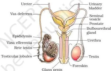
Testes and the seminiferous tubules
- The testes sit outside the abdominal cavity in the scrotum, which keeps them \(2\)–\(2.5^\circ\text{C}\) lower than body temperature — a condition necessary for spermatogenesis.
- Each testis is oval, about 4–5 cm long and 2–3 cm wide, and holds about 250 testicular lobules; each lobule contains one to three coiled seminiferous tubules where sperms are produced (NCERT, p. 27).
As the section below shows, each seminiferous tubule is lined inside by two cell types: male germ cells (spermatogonia), which undergo meiotic division and eventually form sperms, and Sertoli cells, which provide nutrition to the germ cells. Outside the tubules, in the interstitial spaces, lie the Leydig cells, which synthesise and secrete the testicular hormones called androgens.
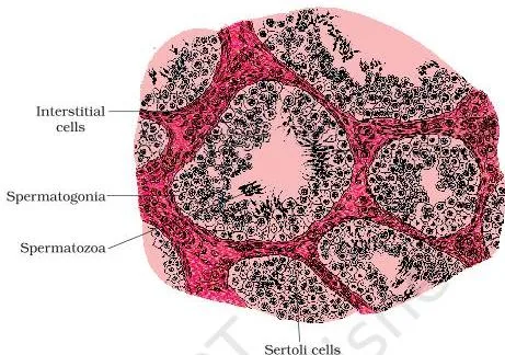
The sperm duct pathway
The seminiferous tubules open into the rete testis, which leads to the vasa efferentia, then to the epididymis along the posterior surface of the testis, and on to the vas deferens. The vas deferens ascends into the abdomen, loops over the urinary bladder, receives a duct from the seminal vesicle, and opens into the urethra as the ejaculatory duct.
The urethra runs from the bladder through the penis to the external opening, the urethral meatus (NCERT, p. 27).
Memory aid: Ravi Very Eagerly Visits Every University — Rete testis → Vasa efferentia → Epididymis → Vas deferens → Ejaculatory duct → Urethra.
Accessory glands and external genitalia
- Accessory glands: paired seminal vesicles, a prostate and paired bulbourethral glands. Their secretions form the seminal plasma, rich in fructose, calcium and certain enzymes. Bulbourethral secretions also lubricate the penis (NCERT, p. 28).
- Penis: the external genitalia, made of special tissue that erects to facilitate insemination. Its enlarged end, the glans penis, is covered by a loose fold of skin called the foreskin.
Female Reproductive System: Ovaries, Oviducts, Uterus and Mammary Glands
The female reproductive system consists of a pair of ovaries, a pair of oviducts, the uterus, cervix, vagina and external genitalia, plus a pair of mammary glands — all integrated to support ovulation, fertilisation, pregnancy, birth and child care (NCERT, p. 28).
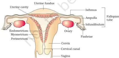
Ovaries and oviducts
- Ovaries are the primary female sex organs — they produce the ovum and several steroid (ovarian) hormones. Each ovary is about 2–4 cm long and is connected to the pelvic wall and uterus by ligaments.
- Oviduct (fallopian tube): about 10–12 cm long, running from the periphery of each ovary to the uterus. The funnel-shaped end near the ovary is the infundibulum, whose finger-like fimbriae collect the ovum after ovulation. The infundibulum leads to the wider ampulla, and the last part, the isthmus, has a narrow lumen and joins the uterus (NCERT, p. 29).
Uterus and its three layers
The uterus (womb) is single and shaped like an inverted pear. It opens into the vagina through a narrow cervix; the cervical canal together with the vagina forms the birth canal (NCERT, p. 30). Its wall has three layers you must know by heart:
- Perimetrium — the outer thin membranous layer.
- Myometrium — the middle thick layer of smooth muscle; it contracts strongly during delivery of the baby.
- Endometrium — the inner glandular layer lining the uterine cavity; it undergoes cyclical changes during the menstrual cycle.
External genitalia and the hymen myth
The external genitalia include five parts: mons pubis (a cushion of fatty tissue covered by skin and pubic hair), labia majora (fleshy folds surrounding the vaginal opening), labia minora (paired folds under the labia majora), hymen (a membrane that partially covers the vaginal opening) and clitoris (a tiny finger-like structure at the upper junction of the labia minora, above the urethral opening).
Handle the hymen carefully: NCERT states explicitly that it is often torn during first coitus, but it can also break from a sudden fall, insertion of a tampon, or sports such as cycling — and it can persist even after coitus. The presence or absence of a hymen is not a reliable indicator of virginity or sexual experience (NCERT, p. 30).
Mammary glands
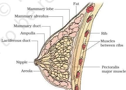
Milk follows a fixed route through the breast: clusters of milk-secreting cells called alveoli → mammary tubules → mammary ducts → a wider mammary ampulla → lactiferous duct, which opens at the nipple. Each breast has 15–20 mammary lobes (NCERT, pp. 30–31).
Gametogenesis: Spermatogenesis and Oogenesis
Gametogenesis is the production of gametes — sperms in the testis and ova in the ovaries (NCERT, p. 31). The two processes share the meiosis machinery but differ in almost every outcome. If the terms haploid and diploid need refreshing, the Sexual Reproduction in Flowering Plants notes cover the same cell biology in plants.
Spermatogenesis
Spermatogenesis begins at puberty. Go through it as a numbered chain:
- Spermatogonia (\(2n = 46\)) on the inside wall of the seminiferous tubules multiply by mitosis.
- Some become primary spermatocytes (\(2n = 46\)) and enter meiosis.
- Meiosis I: each primary spermatocyte forms two equal, haploid secondary spermatocytes (\(n = 23\)).
- Meiosis II: each secondary spermatocyte forms two haploid spermatids (\(n = 23\)) — four spermatids per primary spermatocyte.
- Spermiogenesis: spermatids transform into spermatozoa (sperms).
- Spermiation: sperms are released from the seminiferous tubules (NCERT, p. 31).
The hormonal drive runs: GnRH (from the hypothalamus) → anterior pituitary → LH and FSH. LH acts on Leydig cells to stimulate androgen synthesis and secretion; androgens then stimulate spermatogenesis. FSH acts on Sertoli cells to stimulate factors that help spermiogenesis (NCERT, pp. 31–32).
Examine the sperm’s structure in the figure below: it is a microscopic cell with a head, neck, middle piece and tail, all enveloped by a plasma membrane. The head carries an elongated haploid nucleus, capped by the acrosome, which is filled with enzymes that help fertilise the ovum.
The middle piece is packed with mitochondria that produce energy for tail movement, which drives sperm motility.
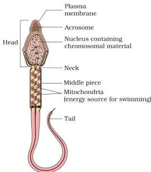
Two fertility numbers go with the sperm: a human male ejaculates about 200–300 million sperms per coitus, and for normal fertility at least 60% must have normal shape and size and at least 40% must show vigorous motility (NCERT, p. 32).
Oogenesis
Oogenesis produces the mature female gamete and is markedly different from spermatogenesis (NCERT, p. 32):
- Oogonia (\(2n\)) form in the fetal ovary; no more are added after birth. They enter prophase I and arrest there, becoming primary oocytes.
- Each primary oocyte gets surrounded by a layer of granulosa cells — a primary follicle. Most follicles degenerate between birth and puberty, leaving only 60,000–80,000 primary follicles per ovary at puberty.
- More granulosa layers and a theca turn a primary follicle into a secondary follicle.
- The secondary follicle becomes a tertiary follicle, marked by a fluid-filled cavity called the antrum; its theca organises into theca interna and theca externa.
- Inside the tertiary follicle the primary oocyte completes meiosis I — an unequal division producing one large haploid secondary oocyte (which keeps the nutrient-rich cytoplasm) and a tiny first polar body (NCERT, p. 33).
- The tertiary follicle matures into the Graafian follicle; the secondary oocyte surrounds itself with a new membrane, the zona pellucida.
- The Graafian follicle ruptures and releases the secondary oocyte: this is ovulation.
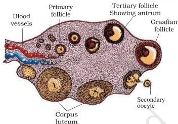
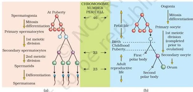
Figure 2.7 shows follicles at different stages in the ovary; Figure 2.8 places the two processes side by side. The table below is the revision core of both.
Spermatogenesis versus oogenesis
| Feature | Spermatogenesis | Oogenesis |
|---|---|---|
| Where it happens | Seminiferous tubules of the testis | Ovary |
| When it starts | At puberty | During embryonic development, in the fetal ovary |
| Continuation in old age | Continues even in old men | Ceases around age ~50 (menopause) |
| Type of meiotic division | Equal — four haploid spermatids | Unequal — one large secondary oocyte plus polar bodies |
| Polar bodies | None | First polar body after meiosis I; second after meiosis II |
| When meiosis completes | Both divisions complete inside the seminiferous tubule | Meiosis I completes in the tertiary follicle; meiosis II completes only after a sperm enters |
| Gametes per precursor | 4 sperms per primary spermatocyte | 1 functional ovum per primary oocyte |
This table is the answer skeleton for any “differentiate spermatogenesis and oogenesis” demand in the chapter’s exercises.
Key Numbers and Percentages: The Chapter’s Data Bank
This chapter has no formulas or equations. The quantitative facts — counts, sizes, durations and percentages — play that role, so these two tables are your formula list.
| Fact | Value | NCERT page |
|---|---|---|
| Scrotal temperature | \(2\)–\(2.5^\circ\text{C}\) below body temperature | p. 27 |
| Testis size | 4–5 cm × 2–3 cm | p. 27 |
| Testicular lobules per testis | About 250 | p. 27 |
| Oviduct length | 10–12 cm | p. 29 |
| Mammary lobes per breast | 15–20 | p. 31 |
| Sperms per ejaculate | 200–300 million | p. 32 |
| Sperms with normal shape and size (for fertility) | At least 60% | p. 32 |
| Sperms with vigorous motility (for fertility) | At least 40% | p. 32 |
| Primary follicles left at puberty | 60,000–80,000 per ovary | p. 32 |
| Menstrual cycle length | About 28/29 days | p. 33 |
| Menstrual flow duration | 3–5 days | p. 34 |
| LH/FSH peak | Mid-cycle, about day 14 | p. 35 |
| Menopause | Around 50 years of age | p. 35 |
| Chromosomes: zygote / gametes | 46 / 23 | pp. 31, 32, 36 |
| Gestation period | About 9 months | p. 38 |
The second data bank table covers the month-wise development of the foetus (NCERT, p. 38). The pregnancy section below refers back to it instead of repeating the list.
| Stage of pregnancy | Foetal milestone |
|---|---|
| End of 1 month | Heart is formed; heartbeat first detectable by stethoscope |
| End of 2nd month | Limbs and digits develop |
| End of 12 weeks (first trimester) | Most major organ systems formed; limbs and external genital organs well developed |
| 5th month | First movements of the foetus; hair appears on the head |
| End of about 24 weeks (second trimester) | Body covered with fine hair; eyelids separate; eyelashes formed |
| End of 9 months | Fully developed and ready for delivery |
The Menstrual Cycle: Phases, Hormones and Events
The menstrual cycle is the reproductive cycle of female primates — monkeys, apes and humans. In human females it repeats at an average interval of about 28/29 days, with one ovum released around the middle of each cycle (NCERT, pp. 33–34).
Figure 2.9 is the most information-dense diagram in the chapter: the upper rows plot the hormone curves (LH, FSH, estrogen, progesterone), the middle row the ovarian events, and the bottom row the endometrial changes. Practise reading any one row and predicting the other two.
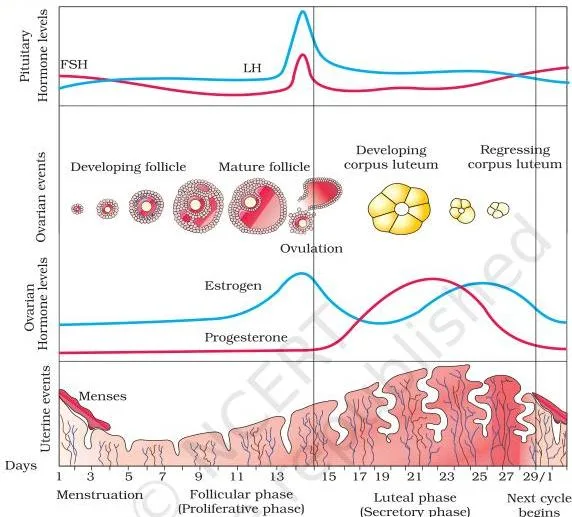
| Phase | Days | Main hormones | Ovarian event | Uterine event |
|---|---|---|---|---|
| Menstrual | 3–5 days | Low levels of all ovarian hormones | Previous corpus luteum has degenerated | Endometrial lining and blood vessels break down; menstrual flow out through the vagina |
| Follicular | After menses to ~day 13 | FSH and LH rise gradually; estrogens rise | Primary follicles grow into a fully mature Graafian follicle | Endometrium regenerates by proliferation |
| Ovulatory | About day 14 | LH surge — rapid peak of LH (with FSH) | LH surge ruptures the Graafian follicle; ovum released (ovulation) | — |
| Luteal | ~day 15 to ~28 | Progesterone in large amounts | Remaining parts of the Graafian follicle become the corpus luteum | Endometrium maintained — ready for implantation |
Two definition points anchor the cycle: menarche is the first menstruation at puberty; menopause is the cessation of cyclic menstruation around age 50. Cyclic menstruation between menarche and menopause is the indicator of a normal reproductive phase (NCERT, p. 35).
Note the textbook’s caution: menstruation occurs only when the released ovum is not fertilised, so its absence may indicate pregnancy — but it can also be caused by stress or poor health (NCERT, p. 34).
Real-life application: the mid-cycle LH surge is exactly the signal that commercial ovulation predictor kits detect — a rise in LH tells a woman ovulation is approaching. In the classic 28/29-day cycle, day 14 is the usual fertile marker, so cycle-day counting is how the surge is located in practice.
NCERT’s Menstrual Hygiene box (p. 35) adds practical rules: bathe and clean yourself regularly; use sanitary napkins or clean homemade pads; change the pad every 4–5 hours; wrap used pads in paper and dispose of them properly — never in toilet drainpipes or open areas; and wash hands with soap after handling.
Fertilisation and Implantation: From Sperm Entry to Embedding
During coitus, semen is released into the vagina — insemination. The motile sperms swim through the cervix, enter the uterus and finally reach the ampullary region of the fallopian tube (NCERT, p. 35).
- Site of fertilisation: the ampullary region only. Fertilisation occurs only if the ovum and sperms are transported there simultaneously — the textbook’s stated reason why not all copulations lead to fertilisation and pregnancy.
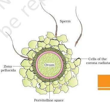
As Figure 2.10 shows, a sperm comes in contact with the zona pellucida of the ovum. This contact induces changes in the membrane that block the entry of additional sperms, ensuring that only one sperm fertilises an ovum. Secretions of the acrosome then help the sperm enter the ovum’s cytoplasm through the zona pellucida and the plasma membrane.
Sperm entry triggers the completion of the second meiotic division of the secondary oocyte. This division is also unequal, producing a second polar body and a haploid ovum (ootid). The haploid nucleus of the sperm and that of the ovum soon fuse to form a diploid zygote with 46 chromosomes (NCERT, p. 36).
Sex determination happens here
The sex of the baby is decided at fertilisation. The human female chromosome pattern is XX and the male is XY, so every ovum carries an X chromosome, while sperms are 50:50 — 50% carry X and 50% carry Y. An XX zygote develops into a female baby; an XY zygote develops into a male baby.
Scientifically, it is correct to say the father determines the sex of the baby, not the mother (NCERT, p. 36).
Cleavage to implantation
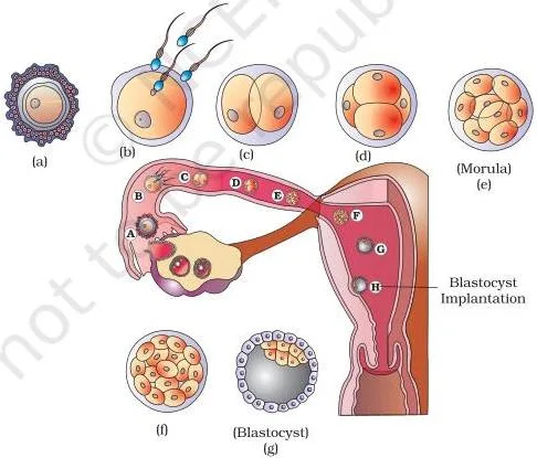
Figure 2.11 traces the whole passage. As the zygote moves through the isthmus of the oviduct towards the uterus, mitotic division begins — cleavage — forming 2, 4, 8 and 16 daughter cells called blastomeres. The embryo with 8–16 blastomeres is the morula.
As it moves further into the uterus, the morula transforms into the blastocyst, whose cells are arranged into an outer layer, the trophoblast, and an inner group of cells attached to it, the inner cell mass (NCERT, pp. 36–37).
The trophoblast attaches to the endometrium; uterine cells divide rapidly and cover the blastocyst, which becomes embedded in the endometrium. This embedding is implantation, and it leads to pregnancy.
Pregnancy and Embryonic Development: Placenta to Foetus
After implantation, finger-like projections called chorionic villi appear on the trophoblast, surrounded by uterine tissue and maternal blood. The villi and the uterine tissue interdigitate to form a structural and functional unit between the developing embryo (foetus) and the maternal body — the placenta (NCERT, p. 37).
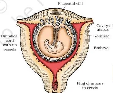
- Umbilical cord: connects the placenta to the embryo and transports substances to and from it.
- Exchange function: the placenta supplies oxygen and nutrients to the embryo and removes carbon dioxide and excretory/waste materials.
- Endocrine function: the placenta secretes hCG (human chorionic gonadotropin), hPL (human placental lactogen), estrogens and progestogens. In the later phase of pregnancy, relaxin is secreted by the ovary. hCG, hPL and relaxin are produced in women only during pregnancy.
- Supporting hormones: levels of estrogens, progestogens, cortisol, prolactin and thyroxine rise several-fold in maternal blood to support foetal growth, maternal metabolic changes and maintenance of pregnancy.
Immediately after implantation, the inner cell mass differentiates into an outer ectoderm and an inner endoderm, with a mesoderm appearing between them. All adult tissues and organs arise from these three layers. The inner cell mass also contains stem cells, which have the potency to give rise to all tissues and organs (NCERT, p. 38).
Foetal development is month-wise, and the complete milestone list is in the data bank table above — one glance at that table revises the entire month sequence.
Parturition and Lactation: Birth and Breastfeeding
The average duration of human pregnancy is about 9 months — the gestation period. The delivery of the foetus (childbirth) is parturition, induced by a complex neuroendocrine mechanism (NCERT, p. 38). Follow it as a positive-feedback loop:
- Signals originate from the fully developed foetus and the placenta, inducing mild uterine contractions — the foetal ejection reflex.
- This reflex triggers the release of oxytocin from the maternal pituitary.
- Oxytocin acts on uterine muscle, causing stronger uterine contractions.
- Stronger contractions stimulate further oxytocin secretion.
- The loop continues with stronger and stronger contractions until the baby is expelled through the birth canal.
- Soon after the infant is delivered, the placenta is also expelled from the uterus.
Because this loop runs the whole event, doctors exploit it directly: injecting oxytocin induces or strengthens labour — the natural mechanism, supplied from outside.
Lactation is the production of milk by the mammary glands, which differentiate during pregnancy and begin producing milk towards its end. The milk produced during the first few days of lactation is colostrum, which contains several antibodies absolutely essential for developing the newborn’s resistance. This is why doctors recommend breastfeeding during the initial period of infant growth (NCERT, p. 38).
Once this chapter is clear, the Reproductive Health notes — the next chapter — build directly on this anatomy and endocrinology.
Definitions Table: Terms You Must Write Exactly
Begin every answer from a correct definition. Two pairs need special care: spermiogenesis vs spermiation and seminal plasma vs semen — mixing either pair is exactly where marks are commonly lost.
| Term | Meaning | Context / example |
|---|---|---|
| Gametogenesis | Formation of gametes — sperms in males, ova in females | Occurs in the testes and ovaries |
| Spermatogenesis | Production of sperms from spermatogonia, beginning at puberty | Inside the seminiferous tubules |
| Spermiogenesis | Transformation of spermatids into spermatozoa (sperms) | A shape change, with no cell division |
| Spermiation | Release of sperms from the seminiferous tubules | Follows spermiogenesis |
| Oogenesis | Formation of a mature female gamete | Starts in the fetal ovary |
| Ovulation | Rupture of the Graafian follicle, releasing the secondary oocyte | Induced by the LH surge |
| Menarche | First menstruation, at puberty | Marks the start of the reproductive phase |
| Menopause | Cessation of cyclic menstruation, around age 50 | Marks the end of the reproductive phase |
| Menstrual cycle | The ~28/29-day reproductive cycle of female primates | Runs from one menstruation to the next |
| Corpus luteum | Structure formed from the remaining Graafian follicle after ovulation | Secretes progesterone to maintain the endometrium |
| Seminal plasma | The glandular fluid — secretions of the seminal vesicles, prostate and bulbourethral glands | Rich in fructose, calcium and certain enzymes |
| Semen | Seminal plasma plus sperms | The fluid ejaculated at coitus |
| Insemination | Transfer of sperms into the female genital tract | Entry of semen into the vagina |
| Fertilisation | Fusion of a sperm with an ovum | Occurs in the ampullary region of the oviduct |
| Cleavage | Repeated mitotic divisions of the zygote | Forms 2, 4, 8, 16 blastomeres |
| Blastomeres | Daughter cells produced by cleavage | 8–16 blastomeres form the morula |
| Morula | Embryo with 8–16 blastomeres | A solid ball of cells |
| Blastocyst | Later embryo with an outer trophoblast and an inner cell mass | Implants in the endometrium |
| Trophoblast | Outer cell layer of the blastocyst | Attaches to the endometrium; gives rise to chorionic villi |
| Inner cell mass | Inner group of cells attached to the trophoblast | Differentiates into the embryo; contains stem cells |
| Implantation | Embedding of the blastocyst in the endometrium | Leads to pregnancy |
| Placenta | Unit formed by interdigitation of chorionic villi and uterine tissue | Exchanges oxygen, nutrients and wastes; secretes hormones |
| Gestation | Duration of embryonic development in the uterus | About 9 months in humans |
| Parturition | Delivery of the foetus (childbirth) | Driven by oxytocin; the placenta is expelled after the baby |
| Lactation | Production of milk by the mammary glands towards the end of pregnancy | Feeds the newborn |
| Colostrum | Milk produced in the first few days of lactation | Contains antibodies essential for newborn immunity |
Worked Reasoning Examples: Chromosomes and Egg Counts
This chapter has no calculations, so these are stepwise reasoning models — the two patterns this chapter’s exercises generate. The numbers here are original and are not the values of any NCERT exercise.
Example 1: Chromosome accounting from spermatocyte to zygote
Method: meiosis I is the reduction division — it halves the chromosome number. Meiosis II keeps the chromosome number unchanged.
- A spermatogonium and a primary spermatocyte are diploid: \(2n = 46\) chromosomes (NCERT, p. 31).
- After meiosis I, each secondary spermatocyte has half the count: \(n = 23\) chromosomes.
- After meiosis II, each spermatid still has \(n = 23\) — meiosis II does not reduce the number.
- Each sperm, therefore, carries 23 chromosomes.
- Fertilisation adds the ovum’s 23 chromosomes to the sperm’s 23: the zygote restores \(2n = 46\) (NCERT, p. 36).
- Yield contrast: one primary spermatocyte produces 4 spermatids, while one primary oocyte produces only 1 functional ovum (plus polar bodies).
Final answer: sperms and ova carry 23 chromosomes each; the zygote has 46. Meiosis I is the only division that reduces the count.
Example 2: Counting eggs from the type of offspring
Method: identical multiples come from one zygote that split into two or more embryos; fraternal multiples come from separate ova fertilised separately.
Scenario: a woman gives birth to identical triplets. How many eggs were released?
- Identical offspring share one origin — a single zygote.
- That zygote formed from one egg and one sperm.
- So exactly 1 egg was released; the zygote split into three embryos.
Now change the condition: if the triplets were fraternal, each came from its own egg, so 3 eggs were released.
Extension: a female dog gives birth to 5 puppies. Since each puppy in a litter develops from its own fertilised ovum, at least 5 ova were released. The true number could be higher only if some embryos were lost before birth.
Final answer: identical triplets → 1 egg; fraternal triplets → 3 eggs; a 5-puppy litter → at least 5 ova.
Master the method, and any version of the question — with different offspring numbers — follows the same logic.
Common Mistakes and How to Avoid Them
Each row below is a real confusion this chapter reliably produces. Learn the corrected half and you remove the most common source of lost marks.
| Students write … | Correct is … | How to check your answer |
|---|---|---|
| “Androgens are produced by Sertoli cells.” | Leydig cells (interstitial cells) secrete androgens; Sertoli cells nourish germ cells and, under FSH, release factors that help spermiogenesis. | Ask: which cells sit outside the tubules in the interstitial spaces? Leydig cells — that is where androgens come from (NCERT, p. 27). |
| “Spermiogenesis is the release of sperms from the tubules.” | Spermiogenesis is the transformation of spermatids into spermatozoa; the release is spermiation. | “-genesis” means making or transforming; “spermiation” is sperms leaving the tubules (NCERT, p. 31). |
| “The ovum completes meiosis before ovulation.” | The secondary oocyte is arrested after meiosis I; meiosis II completes only after a sperm enters, forming the second polar body and the haploid ovum. | The second polar body appears only at fertilisation, never before it (NCERT, pp. 32, 36). |
| “The mother determines the baby’s sex.” | The father does — the ovum always carries X, while the sperm carries X or Y in a 50:50 ratio. | Zygote XX → female; XY → male (NCERT, p. 36). |
| “Menstrual flow is breakdown of the follicle.” | It is breakdown of the endometrial lining and its blood vessels. | The flow leaves through the vagina from the uterus — not from the ovary (NCERT, p. 34). |
| “Morula and blastocyst are the same stage.” | Morula is a solid ball of 8–16 blastomeres; the blastocyst is later — it has a fluid cavity, an outer trophoblast and an inner cell mass. | The blastocyst forms only after the morula reaches the uterus (NCERT, p. 36). |
| “A broken hymen proves loss of virginity.” | The hymen is not a reliable indicator of virginity or sexual experience — it can break from falls, tampons or sport, and it can persist after coitus. | NCERT states this directly on p. 30. |
| “The corpus luteum mainly secretes estrogen.” | It secretes large amounts of progesterone, which maintains the endometrium needed for implantation. | Progesterone is the pregnancy-maintenance hormone of the luteal phase (NCERT, p. 35). |
How to Write Answers for This Chapter
These response strategies apply to the NCERT exercise set on pages 39–40 of the chapter. They tell you how to structure answers — not what the answers are.
- Diagram questions: the exercises ask for labelled diagrams of the male system, female system, sperm, a section through the ovary and a Graafian follicle. Draw in pencil, use straight label lines, and include the anchor structures. For the male system, include scrotum, testis, epididymis, vas deferens, seminal vesicle, prostate and urethra. For the female, include ovary, oviduct with fimbriae, uterus with its layers, cervix and vagina.
- Two-functions questions (testis and ovary): write gamete production first, then hormone secretion. Both are primary sex organs, so a complete answer names both functions.
- Paired definitions: define each term separately, then add one line of contrast — spermiogenesis vs spermiation; semen vs seminal plasma.
- Sequence questions: present pathways as numbered chains in the exact order — the sperm duct path and the ovum path each start at their site of production.
- Hormone-chain questions: answer as source → hormone → target → effect. Example frame: GnRH → anterior pituitary → LH/FSH → Leydig/Sertoli cells → androgens / spermiogenesis factors.
- True/false corrections: rewrite the entire corrected statement, never just “false.” A false statement is fixed by writing the full true statement — for instance, replacing a wrong source of androgens with the correct Leydig cells.
One-Page Revision Summary
Block 1 — The event flow: gametogenesis → insemination → fertilisation → zygote → cleavage → morula → blastocyst → implantation → gestation → parturition → lactation.
Block 2 — Three pathways:
- Sperm duct path: rete testis → vasa efferentia → epididymis → vas deferens → ejaculatory duct → urethra.
- Sperm travel after insemination: vagina → cervix → uterus → ampulla.
- Ovum path: ovary → infundibulum (via fimbriae) → ampulla.
Block 3 — Hormone map: revise this table and you have the control of the whole chapter.
| Hormone | Source | Target | Effect |
|---|---|---|---|
| GnRH | Hypothalamus | Anterior pituitary | Stimulates secretion of LH and FSH |
| LH | Anterior pituitary | Leydig cells; Graafian follicle | Androgen synthesis and secretion; LH surge induces ovulation |
| FSH | Anterior pituitary | Sertoli cells; growing follicles | Factors that help spermiogenesis; follicular development and estrogen secretion |
| Androgens | Leydig cells of the testis | Reproductive tissues | Stimulate spermatogenesis; maintain accessory duct and gland function |
| Estrogens | Growing ovarian follicles | Uterus | Endometrial regeneration |
| Progesterone | Corpus luteum | Endometrium | Maintains the endometrium for implantation |
| hCG | Placenta | — | Produced in women only during pregnancy |
| hPL | Placenta | — | Produced in women only during pregnancy |
| Relaxin | Ovary (later phase of pregnancy) | — | Produced in women only during pregnancy |
| Oxytocin | Maternal pituitary | Uterine muscle | Stronger uterine contractions → parturition |
Block 4 — Chromosome journey: 46 (spermatogonium / primary spermatocyte) → 23 (sperm) → 46 (zygote, after fusing with the ovum’s 23). Ova also carry 23. XX → female baby; XY → male baby. For all the chapters around this one, browse the Class 12 notes hub or the full CBSE notes library.
Frequently Asked Questions
Why is the scrotum located outside the abdominal cavity?
The scrotum keeps the testes at a temperature \(2\)–\(2.5^\circ\text{C}\) lower than normal body temperature, which is necessary for spermatogenesis (NCERT, p. 27). Inside the abdominal cavity, sperm formation could not occur properly.
Where exactly does fertilisation take place in the female reproductive tract?
Fertilisation takes place in the ampullary region of the fallopian tube. It happens only if the ovum and sperms reach the ampulla simultaneously — which is why not every coitus leads to fertilisation and pregnancy (NCERT, p. 35).
Why does only one sperm fertilise the ovum?
When a sperm comes in contact with the zona pellucida of the ovum, it induces changes in the membrane that block the entry of additional sperms. This mechanism ensures that only one sperm can fertilise an ovum (NCERT, p. 35).
Why is the sex of the baby determined by the father and not the mother?
The ovum always carries an X chromosome, while sperms carry X or Y in a 50:50 ratio. If a sperm carrying X fertilises the ovum, the zygote is XX (female); if the sperm carries Y, the zygote is XY (male). The father’s sperm therefore decides the sex (NCERT, p. 36).
Why is colostrum essential for a newborn?
Colostrum is the milk produced during the first few days of lactation and it contains several antibodies that are absolutely essential for developing the newborn’s resistance. This is why doctors recommend breastfeeding during the initial period of infant growth (NCERT, p. 38).
What hormone is injected to induce delivery and why?
Oxytocin is the hormone injected. It acts on the uterine muscle to produce stronger uterine contractions, working through the same positive-feedback mechanism that drives natural parturition (NCERT, p. 38).
Reference: NCERT Class 12 Biology textbook, chapter Human Reproduction.
Explore Class 12 Biology Notes
- Class 12 Biology Notes
- Class 12 CBSE Notes
- CBSE Notes for Classes 1 to 12
- Previous: Sexual Reproduction in Flowering Plants
- Next: Reproductive Health
Class 12 Biology on LearnCBSE:
Related chapters:
- Principles of Inheritance and Variation notes
- Evolution notes
- Human Health and Disease notes