
Genetic disorders are conditions caused by changes in the genetic material — a mutated gene, a broken chromosome, or the wrong number of chromosomes. NCERT Class 11 Biotechnology Chapter 8 groups them into three blocks: chromosomal abnormalities and syndromes, monogenic disorders with pedigree mapping, and polygenic disorders with mitochondrial inheritance.
These genetic disorder class 11 notes are a compact revision version of the chapter for the 2026-27 session, with the definitions, karyotypes, inheritance patterns and worked examples you need to revise quickly.
The notes follow the rationalised NCERT Class 11 Biotechnology textbook; the official PDF is available at ncert.nic.in. Page references in brackets point into that book. This page is part of the Class 11 Biotechnology notes on this site — read the syndrome table first, then the inheritance patterns, and finish with the revision summary if you are short on time.
How This Chapter Is Organised
The chapter builds from whole chromosomes to single genes to multiple genes. Learn the blocks in the order shown below — the karyotype logic of Block 8.1 reappears inside the pedigree logic of Block 8.2.
| Block | Pages | What it covers | Revision order |
|---|---|---|---|
| 8.1 Chromosomal Abnormalities and Syndromes | NCERT pp. 217–222 | Structural vs numerical abnormalities; aneuploidy and polyploidy; Down, Klinefelter and Turner syndromes | Start here — every later block assumes you can read a karyotype |
| 8.2 Monogenic Disorders and Pedigree Mapping | NCERT pp. 222–227 | Single-gene diseases, pedigree symbols, autosomal and X-linked inheritance patterns | Needs Block 8.1 |
| 8.3 Polygenic Disorders and Mitochondrial Inheritance | NCERT pp. 227–231 | Diabetes, hypertension, coronary heart disease, maternal mitochondrial inheritance | Needs Block 8.2 |
Master Block 8.1 before Block 8.2: the Down, Klinefelter and Turner karyotypes use the same notation you apply when interpreting pedigrees later. For the wider revision set, see the Class 11 notes index, and browse every subject’s quick notes in the CBSE notes library.
Chromosomal Abnormalities: Structural and Numerical
Normal human body cells are diploid — 23 pairs of chromosomes — while haploid gametes carry one copy of each chromosome. Radiation, diet or internal genetic conditions can damage chromosomes or change their number (NCERT, p. 217). This gives two broad categories:
- Structural chromosomal abnormality — the structure of a chromosome changes.
- Numerical chromosomal abnormality — the number of chromosomes changes.
Numerical abnormalities: aneuploidy and polyploidy
When one chromosome of a pair is missing, the condition is monosomy (\(2n-1\)); when a chromosome is present in three copies, it is trisomy (\(2n+1\)). Both are forms of aneuploidy. When the entire chromosome set is multiplied, the condition is polyploidy (NCERT, p. 217).
| Type | What happens | Examples from the chapter |
|---|---|---|
| Monosomy | One chromosome of a pair is absent — \(2n-1\) | Monosomy of chromosome 1 |
| Trisomy | A chromosome is present in three copies — \(2n+1\) | Trisomy of chromosome X |
| Aneuploidy | Umbrella term covering monosomy and trisomy | Both conditions above |
| Polyploidy | The whole chromosome set is multiplied | 69 (\(23 \times 3\)) or 92 (\(23 \times 4\)) chromosomes |
Polyploidy appears in our food because artificial breeding of plants created polyploid varieties: bread wheat is hexaploid, cabbage and mustard are tetraploid, banana and apple are triploid, and strawberry and sugarcane are octoploid (NCERT, pp. 217–218).
Structural abnormalities and the length memory device
The four structural abnormalities all start with a chromosome that breaks and rejoins abnormally. Fig. 8.1 shows the same chromosome in each of the four arrangements — watch what happens to the chromosome’s length in every case, because that is the fastest way to keep the types apart.
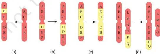
| Abnormality | What happens | Effect on the chromosome | Chapter example |
|---|---|---|---|
| Deletion | A segment breaks away and is lost; two deleted ends can rejoin to form a ring chromosome | Chromosome shortens | Retinoblastoma (chromosome 13) |
| Duplication | A segment of the chromosome is repeated | Chromosome lengthens | Charcot-Marie-Tooth disease (chromosome 17) |
| Inversion | A segment breaks away, reverses by 180 degrees and reattaches in place | Length unchanged; gene order reversed | RCAD syndrome (chromosome 17) |
| Translocation | A segment breaks away and attaches to another chromosome | Segment moves; Robertsonian type may reduce chromosome number | Reciprocal: Burkitt’s lymphoma (chromosomes 8 and 14) |
In reciprocal translocation, two chromosomes exchange segments with each other — this is what happens between chromosomes 8 and 14 in Burkitt’s lymphoma. In Robertsonian translocation, a segment attaches to another chromosome without mutual exchange, which can lower the cell’s chromosome count (NCERT, pp. 218–219).
Memory device: deletion shortens, duplication lengthens, inversion flips (length stays the same), translocation moves a segment to another chromosome.
Down, Klinefelter and Turner: Three Syndromes in One Table
These three syndromes are the chapter’s numerical chromosomal disorders — each is caused by an extra or a missing chromosome, and each is identified by its karyotype (the organised display of an individual’s chromosomes) before its symptoms (NCERT, p. 219).
- Down syndrome — trisomy 21, caused by non-disjunction (chromosomes failing to separate at cell division); risk rises with maternal age.
- Klinefelter syndrome — an extra X chromosome in males.
- Turner syndrome — a missing X chromosome in females.
The table below puts all the facts side by side — the NCERT text spreads these details across separate passages.
| Feature | Down syndrome | Klinefelter syndrome | Turner syndrome |
|---|---|---|---|
| Incidence | About 1 in 800 live births | About 1 in 1000 newborn males | 1 in 2500 newborn girls; frequent in miscarriages and stillbirths |
| Karyotype | 47,XX,+21 (female) or 47,XY,+21 (male) — trisomy 21 | 47,XXY — extra X in males | 45,X — monosomy X in females |
| Origin of the error | Non-disjunction (chromosomes fail to separate); more than 85% of Down babies are born to mothers over 35 | Extra X from failure of the X pair to separate during gamete formation; XX ovum fertilised by a Y sperm | Meiotic error during ovum formation gives an ovum with no X; it fuses with an X-bearing sperm |
| Key symptoms | Flat face, protruding tongue, short neck, short limbs, single deep palmar crease, low IQ, muscular hypotonia, underdeveloped gonads | Unusually tall, reduced facial and body hair, small testes, enlarged breasts, coarse voice | Short stature, webbed neck, small breasts, low-set ears, swollen hands and feet, underdeveloped ovaries, absent menstruation |
| Diagnosis / treatment | Karyotype shows the extra chromosome 21; speech therapy, physiotherapy and nutritional supplements | Barr body test of buccal smear shows one Barr body; testosterone therapy and psychological counselling | Prenatal: amniocentesis or chorionic villus sampling; at puberty, absent Barr body; controlled androgen and estrogen therapy |
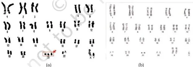
The karyogram in Fig. 8.2(a) shows a Down syndrome individual’s chromosomes arranged for counting — the third copy of chromosome 21 is the signature of trisomy 21. Fig. 8.2(b) shows a Klinefelter karyogram, where the sex-chromosome line reads XXY instead of XY.
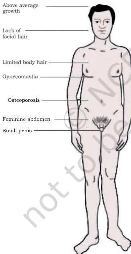
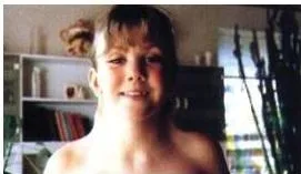
Figs. 8.3 and 8.4 show how Klinefelter and Turner syndromes present physically. These features become noticeable at different times — Klinefelter differences strengthen at puberty, while Turner is often suspected earlier from short stature and webbed neck.
The textbook’s box on famous people with syndromes (NCERT, p. 222) profiles achievers in fashion, medicine and the arts who have Down, Klinefelter or Turner syndrome. Its point: a genetic diagnosis does not define a person’s potential.
Definitions Table: Syndrome, Disease and Inheritance Vocabulary
Every term below appears in the chapter or in its exercise list. Learn each meaning together with its example — the example is what makes the definition usable in a cross.
| Term | Meaning | Example from the chapter |
|---|---|---|
| Syndrome | A group of symptoms that consistently occur together | Down syndrome: flat face, short neck and low IQ together |
| Disease | Abnormal physiological response to internal or external factors | Fever caused by microbes |
| Aneuploidy | Deviation where one or a few chromosomes are absent or present in extra copies | Monosomy and trisomy |
| Monosomy | One chromosome of a pair is missing — \(2n-1\) | Monosomy X in Turner syndrome |
| Trisomy | A chromosome is present in three copies — \(2n+1\) | Trisomy 21 in Down syndrome |
| Polyploidy | The entire set of chromosomes is multiplied | Bread wheat (hexaploid) |
| Structural abnormality | A change in the structure of a chromosome | Deletion, duplication, inversion, translocation |
| Numerical abnormality | A change in chromosome number | Aneuploidy and polyploidy |
| Carrier | A person with one defective recessive allele who does not show the disease | An S/s individual in sickle cell anaemia |
| Monogenic disorder | A disease caused by an error in a single gene | Sickle cell anaemia, cystic fibrosis |
| Polygenic disorder | A disease caused by the combined action of more than one gene | Diabetes, hypertension |
| Dominant allele | An allele whose effect shows even when only one copy is present | Allele D in achondroplasia |
| Recessive allele | An allele whose effect shows only when both copies carry it | Allele s in sickle cell anaemia |
| Homozygous | Both alleles of a gene are the same | s/s — affected in sickle cell anaemia |
| Heterozygous | The two alleles of a gene are different | S/s — carrier in sickle cell anaemia |
| Phenotype | The observable trait or set of physical features | Sickle-shaped red blood cells |
| Genotype | The allele pair an individual carries | s/s, S/s or S/S |
Monogenic Disorders and Pedigree Mapping
A monogenic disease is caused by an error in a single gene; over 10,000 human diseases are estimated to be monogenic (NCERT, p. 222). These diseases are inherited according to Mendel’s laws, so the allele notation from the inheritance chapter applies directly. Sometimes the mutation is spontaneous — then there is no previous family history (NCERT, p. 223).
One defective gene can behave in two ways: a single mutation in one gene causes sickle cell anaemia, while more than 200 different mutations in the same gene can all produce cystic fibrosis.
If the allele notation feels rusty, refresh it in the Basic Principles of Inheritance notes — the pedigree patterns below are Mendel’s laws drawn as a family tree.
Carriers first
In a recessive disorder, a person with one defective allele and one normal allele is a carrier — healthy, but able to pass the defective allele to children. Statistically, every human carries about five or more defective recessive genes (NCERT, p. 223).
Pedigree symbols
Pedigree analysis interprets the information in a family tree; it is useful when more than one person in a family has the disease. The textbook uses fixed symbols (NCERT, p. 223):
| Symbol | Meaning |
|---|---|
| Square | Male |
| Circle | Female |
| Shaded square or circle | Affected individual |
| Two symbols joined by a line | Mating |
| Offspring in birth order; I and II are generations; offspring numbered II-1, II-2 | Family structure across generations |
| Joined twin symbols | Identical twins vs non-identical twins |
The four inheritance patterns
| Pattern | Genotype required | Who is affected | Chapter examples |
|---|---|---|---|
| Autosomal recessive | Two copies of the defective allele (homozygous, e.g. s/s) | Both sexes; carriers (S/s) do not show the disease | Sickle cell anaemia, cystic fibrosis, Tay-Sachs, phenylketonuria |
| Autosomal dominant | One copy of the abnormal dominant allele (e.g. D/d) | Both sexes; heterozygotes show the disease | Achondroplasia, Huntington’s disease |
| X-linked recessive | Defective allele on the single X in males; females are usually carriers | Mainly males | Haemophilia, Duchenne muscular dystrophy |
| X-linked dominant | One dominant allele on the X | Both sexes; affected father passes it to all daughters but no sons | Hypophosphatemia, Alport syndrome |
Sickle cell anaemia is the chapter’s worked example of an autosomal recessive disease. It is caused by a mutation in the haemoglobin-β gene on chromosome 11; after giving up oxygen, the defective haemoglobin molecules cluster into rod-like structures that make red blood cells stiff and sickle-shaped (NCERT, p. 224).
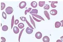
In the blood smear of Fig. 8.6 the red blood cells have collapsed into a sickle shape — the stiffened cells are what the disease phenotype looks like under the microscope.
If the sickle cell allele is designated s and the normal allele S, then affected people have the genotype s/s, while unaffected people are S/S or S/s (NCERT, p. 224).
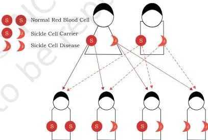
Fig. 8.7 shows the projected cross when both parents are carriers (S/s × S/s). Each parent passes either S or s, so the children can be S/S (normal), S/s (carrier, normal-looking) or s/s (affected). The disease appears only in the homozygous s/s child — that is the meaning of a recessive disorder.
Where sickle cell is found: the disease is common among people whose ancestors came from sub-Saharan Africa, South America, Cuba, Central America, Saudi Arabia, India and Mediterranean countries. In India it is common on the Deccan plateau of central India, with a smaller focus in northern Kerala and Tamil Nadu (NCERT, pp. 224–225).
X-linked recessive inheritance changes the pattern. In a carrier mother (XX), the affected gene lies on one X chromosome, so usually only males (XY) are affected.
Because sons get the Y from the father and the X from the mother, an affected father passes his X to all daughters — who become carriers — and his Y to all sons, who stay unaffected (NCERT, pp. 225–226).
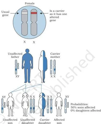
Fig. 8.9 shows an X-linked recessive pedigree: a carrier mother passes her affected X to about half her sons, who become affected, so the disorder appears in males through carrier females.
Haemophilia (mutation in the coagulation factor VIII gene, type A, or factor IX gene, type B) and Duchenne muscular dystrophy (mutation in the dystrophin gene) are both X-linked recessive (NCERT, p. 226).
X-linked dominant disorders behave differently because the allele is dominant, not hidden. An affected male passes the mutant dominant gene to all his daughters but to none of his sons. An affected female married to an unaffected male passes the condition to half of her sons and half of her daughters (NCERT, p. 227).
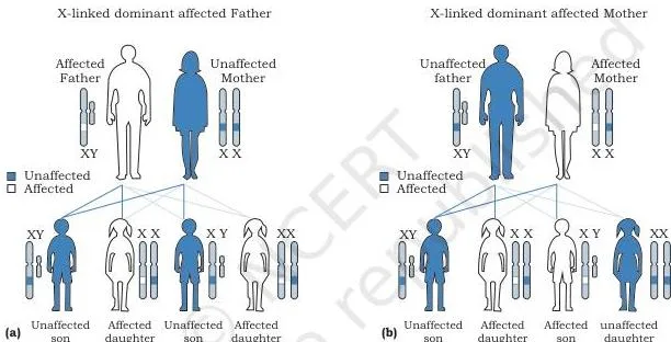
The two pedigrees in Fig. 8.10 contrast these outcomes. In (a), through the affected father, every daughter is affected and no son is affected. In (b), through the affected mother, roughly half of each sex inherits the condition. Hypophosphatemia (a vitamin-D-resistant rickets) and Alport syndrome (progressive hearing loss with kidney disease) are the chapter’s examples.
Karyotype Notation and Inheritance Ratios
This chapter has no algebraic formulas. Its formula-like tools are karyotype lines and Mendelian ratios — learn to read both, and every problem in the chapter reduces to one of them.
Reading a karyotype line is like reading an address: the first number is the total chromosome count, the letters name the sex-chromosome pair, and an entry such as +21 flags an extra parcel on chromosome 21.
| Part of the line | What it tells you |
|---|---|
| Number before the comma | Total number of chromosomes in the cell (46 is normal) |
| Sex chromosomes after the comma | XX = female, XY = male; unusual lines like XXY or a lone X appear here |
| +n after the sex chromosomes | An extra autosome — chromosome n is present in three copies |
| 45,X | 45 chromosomes with only one sex chromosome — a missing X |
The four karyotype lines you must recognise instantly:
- 47,XX,+21 — female with Down syndrome
- 47,XY,+21 — male with Down syndrome
- 47,XXY — male with Klinefelter syndrome
- 45,X — female with Turner syndrome
Standard ratios that behave like formulas:
- Autosomal recessive cross of two carriers (Aa × Aa): \(\frac{1}{4}\) affected (aa), \(\frac{1}{2}\) carriers (Aa), \(\frac{1}{4}\) unaffected (AA).
- X-linked recessive, affected father: all daughters become carriers, all sons are unaffected — sons get the Y, daughters get the X.
- X-linked dominant, affected father: all daughters affected, no sons affected.
- Mitochondrial inheritance: all children get their mitochondria from the mother, and disease severity depends on the ratio of normal to abnormal mitochondria in the cells.
The worked examples below apply these tools to actual problems.
Worked Examples: Karyotype Reading and Two Genetic Crosses
Three problems, each using the chapter’s own notation and ratios. The method is named first, then every step is shown.
Example 1: Reading the karyotype line 47,XY,+21
- Step 1: Read the number before the comma — 47 means the cell has 47 chromosomes instead of the normal 46.
- Step 2: Read the sex chromosomes — XY identifies a male.
- Step 3: Read the +21 — an extra copy of chromosome 21, so chromosome 21 is trisomic.
- Step 4: Combine the parts: 47 chromosomes, male sex chromosomes, trisomy 21.
Final answer: 47,XY,+21 is the karyotype of a male with Down syndrome; the female equivalent is 47,XX,+21.
Example 2: Autosomal recessive cross — two carriers of phenylketonuria
Method: monohybrid cross of two heterozygous carriers, using the disease allele p and the normal allele P. Phenylketonuria is caused by a mutation in the phenylalanine hydroxylase gene that raises phenylalanine levels in the blood (NCERT, p. 225).
Step 1: Assign alleles: P = normal allele, p = phenylketonuria allele.
Both parents are carriers, genotype P/p.
- Step 1: Each parent produces two types of gametes in equal proportion: P and p.
- Step 2: Combine the gametes in a Punnett square:
| Gametes | P | p |
|---|---|---|
| P | PP | Pp |
| p | Pp | pp |
- Step 1: Read the four zygote genotypes: PP once, Pp twice, pp once.
- Step 2: Convert to probabilities: \(\frac{1}{4}\) (25%) affected p/p, \(\frac{1}{2}\) (50%) carriers P/p, \(\frac{1}{4}\) (25%) unaffected P/P.
- Step 3: Check the phenotype logic: only p/p shows the disease because the disease allele is recessive; carriers look normal.
Final answer: Two PKU carriers have a 1 in 4 chance of an affected child, a 1 in 2 chance of a carrier child, and a 1 in 4 chance of a child with two normal alleles.
Example 3: Autosomal dominant cross — achondroplasia with a lethal homozygote
Method: dominant monohybrid cross, then exclude the lethal genotype from the surviving births. In achondroplasia, normal people are d/d, mildly affected people are D/d, and severely affected D/D is often lethal — so most surviving cases are heterozygotes (NCERT, p. 225).
Step 1: Assign alleles: D = dominant mutant allele causing achondroplasia, d = normal allele.
Both parents are affected heterozygotes, D/d.
Step 2: Cross D/d × D/d.
The possible zygotes are DD, Dd, dD and dd in equal proportions.
- Step 1: Apply the lethality rule: the DD zygote is often lethal, so it is removed from the count of live births.
- Step 2: Among the surviving births, the remaining classes are D/d (appears twice) and d/d (appears once).
- Step 3: State the ratio: about 2 affected (D/d) to 1 normal (d/d) among live children.
Final answer: When two achondroplasia heterozygotes marry, about 2 in 3 live-born children have achondroplasia and about 1 in 3 is normal — a 2:1 ratio among survivors instead of the usual 3:1, because the DD class is usually lethal.
Polygenic Disorders and Mitochondrial Inheritance
A polygenic disorder is caused by the defect or combined action of more than one gene, so it cannot be explained as a monogenic disease. The classical examples are hypertension, coronary heart disease and diabetes (NCERT, p. 227).
Diabetes: Type 1 vs Type 2
Diabetes mellitus is a group of disorders marked by persistent high blood sugar (hyperglycaemia). The two common forms differ in cause and treatment:
| Feature | Type 1 diabetes (IDDM) | Type 2 diabetes (NIDDM) |
|---|---|---|
| Cause | Destruction of the beta cells of the pancreas by an immunological abnormality | Impaired insulin secretion from beta cells plus peripheral insulin resistance |
| Share of cases | About 10% | About 90% — the commonest form |
| Key management point stated | Life-long treatment with insulin injections is needed | Insulin normally transports glucose from blood into muscle and other cells for energy |
Hypertension categories
Persistent high blood pressure is a major risk factor for kidney disease, heart disease and stroke. The textbook’s categories (NCERT, p. 228):
| Category | Blood pressure (mm Hg) |
|---|---|
| Normal | Below 120 systolic / below 80 diastolic |
| Elevated | 120–129 / below 80 |
| Stage 1 hypertension | 130–139 or 80–89 |
| Stage 2 hypertension | 140 or above systolic, or 90 or above diastolic |
Coronary heart disease as a chain
- Fatty material builds up gradually on the wall of the coronary artery — this build-up is atherosclerosis.
- The narrowed coronary artery cannot supply enough oxygen-rich blood to the continuously working heart muscle.
- That reduced blood supply is ischemia — which is why coronary heart disease was earlier called ischaemic heart disease (NCERT, p. 229).
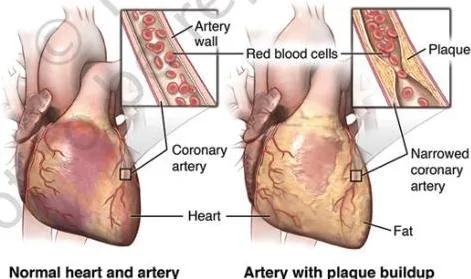
Fig. 8.11 contrasts the normal artery in (a) with the plaque-narrowed artery in (b). The fatty layer visible on the artery wall is the atherosclerosis that narrows the vessel and produces ischemia.
Mitochondrial inheritance
Mitochondria are cytoplasmic organelles that produce energy as ATP. They carry their own genes, which code for critical enzymes of energy production; a mutation in these genes can disrupt ATP production and cellular function (NCERT, p. 229).
- Cells of the brain, heart, kidneys, muscle and liver are metabolically active and contain large numbers of mitochondria.
- In a mitochondrial gene defect, symptoms and severity depend on the ratio of normal to abnormal mitochondria in the cell — if all mitochondria are affected, survival is not possible.
- Mitochondrial DNA is circular, and in animals mitochondria are the only organelle besides the nucleus that carries DNA and genes.
- The sperm contains very few mitochondria, so mitochondrial genes in the offspring come from the mother. A father with a mitochondrial gene defect cannot transmit the disease to his offspring (NCERT, pp. 229–231).
A simple way to remember it: the egg is the new cell’s power source; the sperm is mostly a delivery vehicle for the nucleus. The mitochondria — and their genes — always arrive with the egg.
Common Mistakes Students Make in Genetic Disorders
These six confusions cause the most wrong answers in this chapter. Each row gives the error, the correct rule, and a quick check.
| Students write… | Correct is… | How to check |
|---|---|---|
| X-linked recessive disorders affect boys and girls equally | They mostly affect males, because males have only one X chromosome — one defective allele is enough. A female needs the defective allele on both X chromosomes | Count X chromosomes: an XY male has no second X to mask the defect |
| Turner syndrome karyotype is 45,XO | It is 45,X — there is no O chromosome in humans; the line says 45 chromosomes with a single X | Read the line literally: 45,X names 45 chromosomes and one sex chromosome |
| A carrier has the disease | A carrier has one defective recessive allele and one normal allele, so the disease does not appear | An S/s genotype is healthy-looking but can pass s to children |
| Klinefelter syndrome is inherited from the father | It arises from non-disjunction during gamete formation — an XX ovum fertilised by a Y sperm — and is not transmitted genetically | Remember the textbook rule: a Klinefelter newborn cannot have a Klinefelter father |
| A father can pass a mitochondrial disease to his children | Mitochondria are maternal — the sperm contributes very few, so an affected father cannot transmit the disease | Trace the mitochondria: they arrive with the egg, not the sperm |
| Down syndrome risk has nothing to do with the mother’s age | Risk increases with maternal age — more than 85% of Down syndrome babies are born to mothers over 35 | In any family history, note the mother’s age at pregnancy |
Exam Notes: Structuring Answers for the Chapter Exercises
The chapter ends with five exercises. These skeletons organise a complete answer for each; the content comes from the tables above.
Exercise 1 (define six terms). Give each term a one-sentence meaning plus a chapter example — exactly the pattern of the definitions table: dominant allele (D in achondroplasia), recessive allele (s in sickle cell anaemia), homozygous (s/s — affected), heterozygous (S/s — carrier), phenotype (sickle-shaped red blood cells), genotype (the allele pair an individual carries).
Exercises 2–4 (the three syndromes). Use the same three-part structure for each syndrome:
- Origin: the karyotype line (47,XX,+21 or 47,XY,+21; 47,XXY; 45,X) and the cell-division error behind it — trisomy 21 from non-disjunction, an extra X from failure of the X pair to separate, or a missing X. State whether the condition is inherited.
- Clinical symptoms: list the characteristic physical features from the syndrome table.
- Diagnosis and treatment: name the test (karyotype, Barr body test, amniocentesis or chorionic villus sampling) and the treatment approach.
For Klinefelter and Turner, a complete answer also states the Barr body finding: one Barr body appears in Klinefelter, and no Barr body appears in Turner.
Exercise 5 (structural chromosomal abnormalities). Write one paragraph per abnormality. For each of deletion, duplication, inversion and translocation, state what happens to the chromosome and give one disease example.
Under translocation, add the two subtypes: reciprocal translocation involves mutual exchange of segments between two chromosomes (Burkitt’s lymphoma, chromosomes 8 and 14), while Robertsonian translocation attaches a segment without mutual exchange and may reduce the chromosome number.
Revision Summary: Quick Tables for the Night Before
Table A — the three syndromes at a glance.
| Syndrome | Karyotype | One-line origin | One-line symptoms |
|---|---|---|---|
| Down | 47,XX,+21 / 47,XY,+21 | Trisomy 21 from non-disjunction; maternal age over 35 raises risk | Flat face, protruding tongue, short neck, palmar crease, low IQ |
| Klinefelter | 47,XXY | Extra X from failure of the X pair to separate in gamete formation | Tall, small testes, enlarged breasts, coarse voice |
| Turner | 45,X | Monosomy X from an ovum that lost its X chromosome | Short stature, webbed neck, small breasts, absent menstruation |
Table B — inheritance patterns.
| Pattern | Rule to remember | Examples |
|---|---|---|
| Autosomal recessive | Needs two mutant alleles; carriers look normal | Sickle cell anaemia, cystic fibrosis, Tay-Sachs, PKU |
| Autosomal dominant | One mutant allele is enough; heterozygotes are affected | Achondroplasia, Huntington’s disease |
| X-linked recessive | Males affected through carrier mothers; affected fathers never pass to sons | Haemophilia, Duchenne muscular dystrophy |
| X-linked dominant | Affected father passes to all daughters, no sons | Hypophosphatemia, Alport syndrome |
Table C — disorder classes.
| Disorder class | Level of error | Examples |
|---|---|---|
| Chromosomal | Structure or number of whole chromosomes | Down, Klinefelter, Turner; retinoblastoma, Burkitt’s lymphoma |
| Monogenic | Error in a single gene | Sickle cell anaemia, cystic fibrosis, achondroplasia, haemophilia |
| Polygenic | Combined action of more than one gene | Diabetes, hypertension, coronary heart disease |
| Mitochondrial | Genes in mitochondrial DNA | Diseases of ATP production; severity set by the normal-to-abnormal mitochondrial ratio |
60-second summary. Chromosomes suffer two kinds of damage — structural (deletion, duplication, inversion, translocation) and numerical (aneuploidy, polyploidy). Numerical errors produce the three named syndromes: trisomy 21 (Down), 47,XXY (Klinefelter) and monosomy X (Turner). Monogenic disorders are single-gene diseases that follow Mendel’s laws — autosomal recessive, autosomal dominant, X-linked recessive and X-linked dominant — and pedigrees track them through families.
Polygenic disorders combine several genes (diabetes, hypertension, coronary heart disease), and mitochondrial disorders pass only through the mother, with severity set by the ratio of normal to abnormal mitochondria (NCERT, p. 231).
FAQs: Genetic Disorder Class 11 Doubts
What is the difference between a disease and a syndrome?
A disease is an abnormal physiological response to internal or external factors — the textbook’s example is fever caused by microbes. A syndrome is a specific collection of signs or symptoms that consistently occur together, such as the flat face, short neck and low IQ that appear together in Down syndrome.
In short: the disease is the faulty response; the syndrome is the set of features that travel together.
Why are males more often affected by X-linked recessive disorders like haemophilia?
Because males have only one X chromosome, a single defective allele on that X is enough to cause the disorder. Females have two X chromosomes, so a defective allele on one X is usually masked by a normal allele on the other — they become carriers instead of being affected.
Why is Klinefelter syndrome not inherited from the father?
Klinefelter syndrome arises from non-disjunction during gamete formation — an XX ovum fertilised by a Y sperm gives an XXY zygote. The extra X is not transmitted genetically, so a Klinefelter newborn cannot have a Klinefelter father.
What does 47,XX,+21 mean in a karyotype?
It means the cell has 47 chromosomes instead of 46: the sex chromosomes are XX, so the person is female, and +21 means there is an extra copy of chromosome 21 (trisomy 21) — the karyotype of Down syndrome in a female. The male equivalent is 47,XY,+21.
Why is mitochondrial DNA inherited only from the mother?
Because the sperm contains very few mitochondria, the mitochondria of the zygote — and therefore all mitochondrial genes — come from the egg. A father with a mitochondrial gene defect cannot transmit the disease to his offspring.
Reference: NCERT Class 11 Biotechnology textbook, chapter Genetic Disorder.
Explore Class 11 Biotechnology Notes
- Class 11 Biotechnology Notes
- Class 11 CBSE Notes
- CBSE Notes for Classes 1 to 12
- Previous: Basic Principles of Inheritance
Class 11 Biotechnology on LearnCBSE:
Related chapters:
- Cellular Processes notes