
If you want cellular processes class 11 notes that are complete but quick to finish, you are in the right place. This page condenses NCERT Class 11 Biotechnology Chapter 5 into revision notes covering cell signaling, metabolic pathways, the cell cycle, apoptosis, cell differentiation and cell migration — with the chapter’s own diagrams, comparison tables, and worked ATP and carbon counts.
These notes follow the 2026-27 session. Reference: NCERT Class 11 Biotechnology textbook, chapter Cellular Processes.
The page follows the chapter’s own sequence: signaling first, then metabolism, then cell cycle, apoptosis, differentiation and migration. Think of it as trigger → fuel → outcomes. Signaling tells the cell what to do, metabolism pays for it with ATP, and division, death, specialisation and movement are what the cell does with that information.
Every page number below refers to the rationalised NCERT Class 11 Biotechnology textbook; you can verify each equation in the official NCERT chapter PDF. They are part of the wider Class 11 notes archive, the Class 11 Biotechnology notes hub, and all CBSE notes by class.
The Six Topics of Cellular Processes at a Glance
Before the details, fix the map of the chapter in your head. Each of the six sections answers exactly one question.
| Chapter topic | The question it answers | Textbook pages |
|---|---|---|
| Cell Signaling | How does a cell sense a signal and decide how to reply? | 103–104 |
| Metabolic Pathways | Where does a cell get energy and building blocks? | 104–126 |
| Cell Cycle | How does one cell grow and divide into two? | 126–134 |
| Apoptosis | Why do cells die on purpose? | 135–136 |
| Cell Differentiation | How do cells with the same DNA become specialised? | 136–139 |
| Cell Migration | How do cells move around the body? | 139–141 |
Signaling comes first because it is the trigger. Metabolism is the fuel that makes every response affordable. The cell cycle, apoptosis, differentiation and migration are the outcomes — what a cell actually does after it senses a signal and spends energy on it.
Cell Signaling: How Cells Talk to Each Other
Cells constantly receive and interpret environmental signals — light, heat, sound, touch — and answer them in real time. Signalling pathways specify cell fates during development, and cells talk to neighbours by sending and receiving chemical messages released into the extracellular space (NCERT, p. 103).
Two definitions carry this section:
- Receptor — a protein located on the cell surface, or inside the cytoplasm or nucleus, that a signal molecule docks onto.
- Ligand — the chemical messenger to which a receptor responds.
The binding between a receptor and its ligand is highly specific. A cell responds to a chemical messenger only if it carries the corresponding receptor — not otherwise (NCERT, p. 103-104). When the ligand binds, the receptor changes shape; this conformational change starts a message relay inside the cell and alters its activities.
| Signaling type | Distance | How it works | Example |
|---|---|---|---|
| Paracrine | Short | A chemical released into the extracellular space is sensed instantly by nearby cells | Communication between neurons |
| Autocrine | Self | A cell secretes a ligand and also carries the receptor for that same ligand | Cancer cells making their own growth factors |
| Endocrine | Long | The ligand is released into the bloodstream and travels to a distant target cell | Hormones |
The cancer cell is the memorable autocrine case. Cancer cells grow without control and therefore need more growth factors than normal cells. Instead of depending on outside supplies, they synthesise their own growth factors and possess the receptors for them (NCERT, p. 104) — a cell signalling itself.
Metabolism: Anabolic and Catabolic Pathways
Metabolism is the process through which living organisms take up and use the free energy needed for life. Organisms differ in how they get that energy: phototrophs (plants, some bacteria) convert sunlight into chemical energy, chemotrophs oxidise organic or inorganic chemical compounds, and heterotrophs (animals) obtain energy indirectly from plants through food (NCERT, p. 104-105).
Central to all these energy transactions is ATP, the cell’s energy currency. The trick that makes life possible is coupling: the exergonic reactions of nutrient oxidation are linked to the endergonic processes needed to maintain the living state (NCERT, p. 105). Energy released by breaking food down is what pays for building, moving and synthesising.
| Feature | Anabolic pathways | Catabolic pathways |
|---|---|---|
| Direction | Small molecules → large, complex molecules | Large molecules → small molecules |
| Energy | Endergonic (consumes energy) | Exergonic (releases energy) |
| Products | Glucose, fats, proteins, DNA | CO₂, H₂O, reducing equivalents, ATP |
| Example | Synthesis of glucose, proteins or DNA | Glycolysis (breakdown of glucose) |
The useful energy and reducing equivalents produced in catabolism are exactly what anabolism spends to build complex structures from simple ones (NCERT, p. 106). That single statement is the chapter’s energy rule.
Because every pathway has a home, keep this location map ready — it is the fastest way to revise where each process runs.
| Pathway or step | Cellular location | Textbook page |
|---|---|---|
| Glycolysis (all enzymes) | Cytosol | p. 110 |
| Pyruvate → acetyl CoA (PDH complex) | Mitochondria | p. 114 |
| Citric acid cycle | Mitochondrial matrix | p. 115 |
| Succinate dehydrogenase step of TCA | Inner mitochondrial membrane | p. 116 |
| Electron transport chain and ATP synthase | Inner mitochondrial membrane | p. 116-118 |
| Light reactions of photosynthesis | Thylakoid membranes | p. 119 |
| Calvin cycle | Stroma of the chloroplast | p. 122 |
| C4: PEP carboxylase step | Mesophyll cells | p. 125-126 |
| C4: RuBisCO and Calvin step | Bundle sheath cells | p. 126 |
Carbohydrate, Lipid and Amino Acid Metabolism: The Three Fuel Tracks
Glucose is the metabolic fuel for most animal tissues. The carbohydrate track starts when glucose enters glycolysis to form pyruvate. With oxygen, pyruvate enters the mitochondrial matrix, is converted to acetyl CoA, and is completely oxidised in the citric acid cycle to CO₂ and H₂O. Without oxygen, pyruvate becomes lactic acid (NCERT, p. 106). Fig 5.1 shows that whole flow.
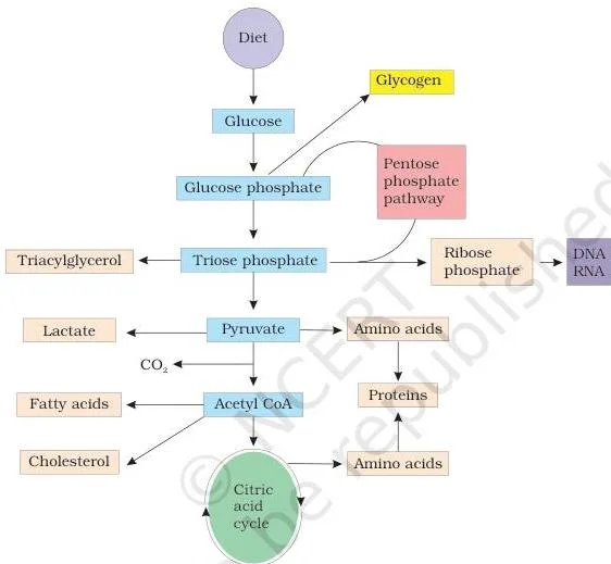
The intermediates of glycolysis are not merely on the way to pyruvate — they branch into other needs (NCERT, p. 106-107):
- Glucose 6-phosphate → glycogen, stored in animals.
- Pentose phosphate pathway → NADPH for fatty acid synthesis, and ribose for nucleotides and nucleic acids.
- Triose phosphate → the glycerol part of triacylglycerol.
- Acetyl CoA → fatty acids and cholesterol; cholesterol then forms all other steroids.
- Pyruvate and TCA intermediates → carbon skeletons for amino acid synthesis.
- In starvation, when glycogen reserves run out, lactic acid, amino acids and glycerol rebuild glucose by gluconeogenesis.
Lipids matter most when glucose is scarce. Brain, heart and red blood cells depend almost exclusively on glucose; in the fasting state, less glucose-dependent tissues such as muscle and liver switch to long-chain fatty acids (NCERT, p. 107).
Fig 5.2 shows the lipid track: fatty acids are oxidised to acetyl CoA through beta-oxidation, or esterified with glycerol to form triacylglycerol, the main fat reserve in adipose tissue.
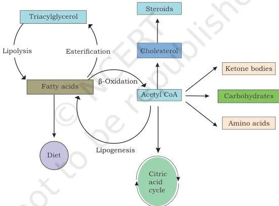
Acetyl CoA made by beta-oxidation has three fates (NCERT, p. 108):
- Oxidised to CO₂ and H₂O through the citric acid cycle.
- Used as a precursor for cholesterol, from which all other steroids (hormones, bile pigments) are made.
- Converted into ketone bodies — acetone, acetoacetate and 3-hydroxybutyrate — which are an alternative fuel in prolonged fasting.
Protein provides the third track. Protein synthesis needs 20 standard amino acids. Non-essential amino acids are made in the body by transamination — amino nitrogen is transferred from one amino acid to a carbon skeleton to form another. Essential amino acids cannot be made this way and must come from the diet.
In deamination, the amino nitrogen is excreted as urea (NCERT, p. 109). Fig 5.3 compresses the whole track.
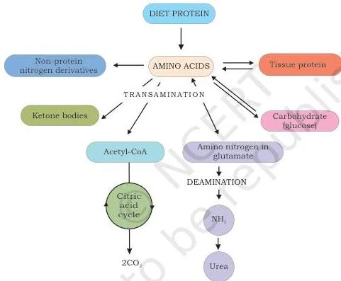
The carbon skeletons left after transamination are useful: they are oxidised to CO₂ via the citric acid cycle, converted to glucose through gluconeogenesis, or turned into ketone bodies that can be oxidised or used for fatty acid synthesis (NCERT, p. 109). Some amino acids additionally feed synthesis of plant and animal hormones, purines, pyrimidines and neurotransmitters.
Glycolysis: The Universal 10-Step Pathway
Glycolysis — also called the Embden-Meyerhof-Parnas (EMP) pathway — is a universal pathway present in all living cells, and every enzyme of it works in the cytosol (NCERT, p. 110). Use Fig 5.4 as the spine of the pathway.
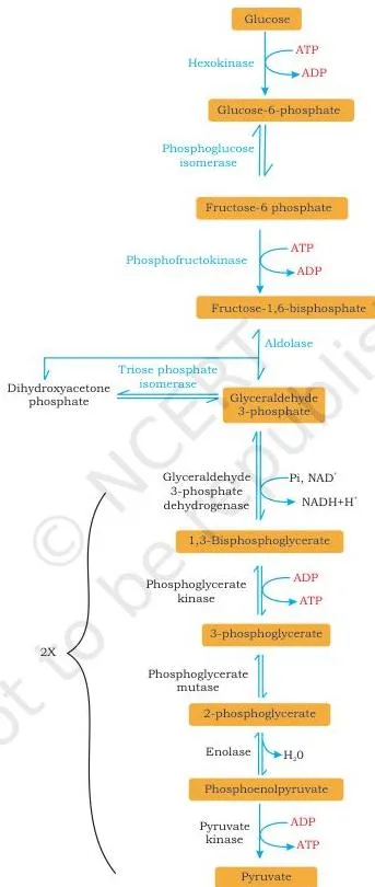
Read Fig 5.4 in three phases. Investment phase: glucose (6C) is phosphorylated to glucose 6-phosphate by hexokinase, with ATP as the phosphate donor. The step is irreversible, and hexokinase is inhibited allosterically by its own product. Glucose 6-phosphate is isomerised to fructose 6-phosphate, then phosphorylated again by phosphofructokinase (PFK) to fructose 1,6-bisphosphate — also irreversible and allosterically regulated.
Aldolase then splits the 6-carbon molecule into two triose phosphates, glyceraldehyde 3-phosphate and dihydroxyacetone phosphate, which are interconverted by triose phosphate isomerase (NCERT, p. 110-111).
Payoff phase: glyceraldehyde 3-phosphate is oxidised to 1,3-bisphosphoglycerate by a NAD-dependent dehydrogenase. Phosphoglycerate kinase then transfers phosphate to ADP, forming ATP — the first substrate-level phosphorylation. Because each glucose makes two trioses, this step credits 2 ATP per glucose. The chain continues: 3-phosphoglycerate → 2-phosphoglycerate (mutase) → phosphoenolpyruvate or PEP (enolase) → pyruvate (NCERT, p. 111-112).
The final step, catalysed by pyruvate kinase, is the second substrate-level phosphorylation, also worth 2 ATP per glucose. Of the 10 reactions, three are exergonic and irreversible — the regulatory enzymes hexokinase, PFK and pyruvate kinase — and those three are the major control sites of glycolysis (NCERT, p. 112).
Net result per glucose: the pathway generates 4 ATP but invests 2, so the net gain is 2 ATP plus 2 NADH.
\[ \text{Glucose} + 2NAD^+ + 2ADP + 2P_i \rightarrow 2\text{Pyruvate} + 2NADH + 2H^+ + 2ATP + 2H_2O \]
NCERT states the same accounting in words: four ATP are generated per glucose and two are utilised, so net ATP production is two (NCERT, p. 112).
All organisms share the same glycolytic sequence from glucose to pyruvate; what differs is the fate of pyruvate (NCERT, p. 112-113). Fig 5.5 shows the three branches.
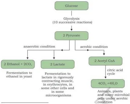
- Anaerobic muscle: pyruvate is reduced by NADH to lactate (homolactic fermentation), catalysed by lactate dehydrogenase.
- Anaerobic yeast: pyruvate is decarboxylated to acetaldehyde and CO₂ (pyruvate decarboxylase, needing coenzyme TPP), then acetaldehyde is reduced to ethanol by alcohol dehydrogenase, which reoxidises NADH to NAD⁺.
- Aerobic: pyruvate is converted to acetyl CoA and fully oxidised through the citric acid cycle to CO₂ and H₂O.
Why ferment at all? Glycolysis needs NAD⁺, which cells hold only in limited amounts. Fermentation recycles NADH back to NAD⁺ so glycolysis can keep running (NCERT, p. 113). Fermentation yields just 2 ATP per glucose.
Citric Acid Cycle and Electron Transport Chain: The ATP Engine
The citric acid cycle (Krebs cycle, or TCA cycle) is the final common pathway for carbohydrates, lipids and proteins, because glucose, fatty acids and most amino acids all feed into acetyl CoA or TCA intermediates (NCERT, p. 114). In eukaryotes it runs in the mitochondrial matrix — unlike glycolysis, which stays in the cytosol.
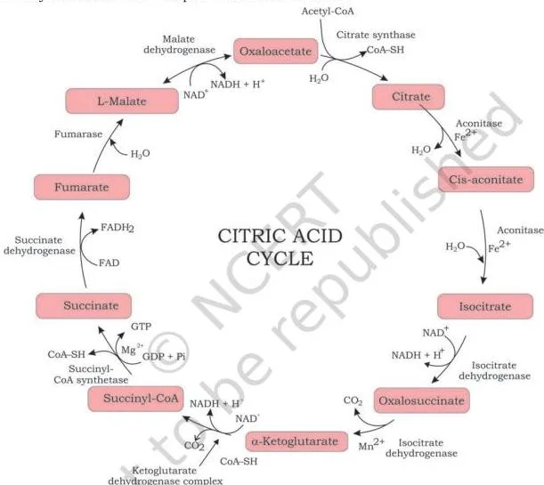
Follow Fig 5.6 around one turn. Acetyl CoA (2C) condenses with oxaloacetate (4C) to form citrate (6C). Citrate isomerises to isocitrate, which is oxidised and decarboxylated to alpha-ketoglutarate (5C) with the first CO₂ released. Alpha-ketoglutarate is oxidatively decarboxylated to succinyl CoA (4C) with a second CO₂ — an irreversible step.
Succinyl CoA is converted to succinate in the only substrate-level phosphorylation of the cycle (GDP → GTP → ATP). Succinate is oxidised to fumarate by succinate dehydrogenase (which uses FAD); fumarate becomes malate, and malate is oxidised back to oxaloacetate, closing the cycle (NCERT, p. 115-116).
Per acetyl CoA, the cycle harvests 3 NADH, 1 FADH₂, 1 GTP and 2 CO₂ (NCERT, p. 116). One structural fact is worth locking in: succinate dehydrogenase is the only TCA enzyme bound to the inner mitochondrial membrane; all the others float free in the matrix.
The overall reaction of one turn is:
\[ \text{Acetyl CoA} + 3NAD^+ + FAD + GDP + P_i + 2H_2O \rightarrow 2CO_2 + 3NADH + 3H^+ + FADH_2 + GTP + CoA \]
Electron Transport Chain and Oxidative Phosphorylation
The NADH and FADH₂ produced in the TCA and other catabolic pathways are reoxidised by the electron transport chain in the inner mitochondrial membrane (NCERT, p. 116-117). Fig 5.7 names the players.
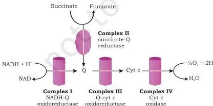
The four complexes are NADH-Q-oxidoreductase (complex I), succinate-Q-reductase (complex II), Q-cytochrome C oxidoreductase (complex III) and cytochrome C oxidase (complex IV). Complex II does not pump protons.
The lipid-soluble carrier ubiquinone (coenzyme Q) shuttles electrons from complex I to complex III; FADH₂ enters at complex II and passes to complex III; and complex IV finally reduces oxygen to water (NCERT, p. 117).
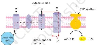
Fig 5.8 shows the payoff. Electron flow pumps protons out of the matrix, building a proton motive force — a pH gradient plus a membrane potential. When protons flow back through ATP synthase, ATP is made from ADP and inorganic phosphate. That is oxidative phosphorylation (NCERT, p. 118).
This textbook’s ATP values: one NADH generates enough gradient for 2.5 ATP (1 from complex I, 0.5 from complex III, 1 from complex IV), while FADH₂ delivers electrons only at complex III and therefore earns 1.5 ATP (NCERT, p. 118). Use these numbers — never the older 3 and 2.
Photosynthesis: From Light to Sugar
Photosynthesis traps solar energy and converts it to chemical energy. The basic reaction:
\[ 6CO_2 + 12H_2O \xrightarrow[\text{Chlorophyll}]{\text{light}} C_6H_{12}O_6 + 6O_2 + 6H_2O \]
The process splits into two acts. The light reactions (the photochemical phase) capture solar energy, split water and release oxygen, and build ATP and NADPH + H⁺. The dark reactions (Calvin cycle) use that ATP and NADPH to reduce CO₂ into carbohydrate (NCERT, p. 118-119). Light reactions run in the thylakoid membranes; the Calvin cycle runs in the stroma.
Light reactions
Fig 5.9 traces the electron path. Two photosystems respond at slightly different wavelengths: PSII at 680 nm and PSI at 700 nm. Under normal conditions PSII is activated first, despite its name.
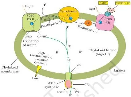
The route is: PSII → pheophytin → plastoquinone (PQ) → cytochrome b₆f → plastocyanin (PC) → PSI → phylloquinone → ferredoxin (Fd) → ferredoxin-NADP reductase (FNR), which finally reduces NADP⁺ to NADPH + H⁺ in the stroma (NCERT, p. 120).
When PSII loses an electron, the positively charged P680⁺ pulls replacement electrons from water. The oxygen evolving complex, on the luminal surface of the thylakoid, splits two water molecules:
\[ 2H_2O \xrightarrow{\text{oxygen evolving complex}} 4H^+ + 4e^- + O_2 \]
So the oxygen released in photosynthesis comes from water — one O₂ for every four electrons sent down the chain (NCERT, p. 120-121). This full route is non-cyclic photophosphorylation: it makes ATP and NADPH and releases O₂.
Plastoquinone picks up protons from the stroma and releases them into the lumen through cytochrome b₆f, building a proton gradient; ATP synthase (CF₀ channel with the CF₁ catalytic head) makes ATP as protons flow back (NCERT, p. 121).
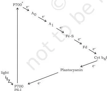
Sometimes only PSI works. In cyclic photophosphorylation, the photoexcited electron leaves PSI, passes through phylloquinone and cytochrome b₆f (which pumps protons), and returns to PSI through plastocyanin (Fig 5.10). The only product is ATP — no NADPH, no O₂ (NCERT, p. 121-122).
Dark reactions: the Calvin cycle
The dark reactions run in the stroma of the chloroplast (cytosol in cyanobacteria). They are also called CO₂ assimilation or CO₂ fixation, since CO₂ is the sole carbon source that is turned into trioses and hexoses leading to sugar (NCERT, p. 122). They need the ATP and NADPH made by light; they do not need darkness.
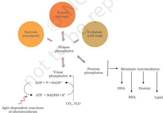
The cycle, worked out by Melvin Calvin and his co-workers in 1950, is best learned in its three stages (NCERT, p. 123-124), shown in Fig 5.12.
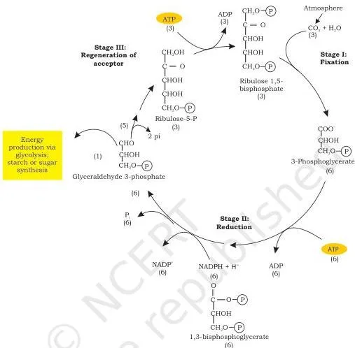
Stage I — fixation: CO₂ condenses with the five-carbon acceptor ribulose 1,5-bisphosphate (RuBP) to form two molecules of 3-phosphoglycerate (3-PGA). The enzyme is RuBisCO (ribulose 1,5-bisphosphate carboxylase/oxygenase), which possesses both carboxylase and oxygenase activities. Because three CO₂ are fixed to three RuBP, six molecules of 3-PGA are produced.
The first product has three carbons, which is why this is the C3 cycle.
Stage II — reduction: the six 3-PGA molecules are converted to six molecules of glyceraldehyde 3-phosphate. This uses 6 ATP (to make 1,3-bisphosphoglycerate) and 6 NADPH + H⁺ for the reduction.
Stage III — regeneration: one of the six G3P molecules is diverted for glycolysis, sugar or starch synthesis; the remaining five G3P (15 carbons) are rearranged into three ribulose 5-phosphate (15 carbons), releasing 2 Pi, and 3 ATP convert these into three ribulose 1,5-bisphosphate, ready to accept CO₂ again.
C4 pathway
Maize, sorghum and sugarcane follow a C4 pathway and have roughly 50% higher photosynthetic efficiency than C3 plants (NCERT, p. 125). Their leaves show Kranz anatomy: mesophyll cells clustered in a ring around bundle sheath cells, with more chloroplasts in the bundle sheath than in the mesophyll.
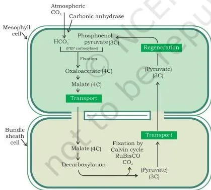
Fig 5.13 is a two-cell handoff. In mesophyll cells, phosphoenol pyruvate (PEP, a 3-carbon acceptor) is carboxylated by PEP carboxylase to oxaloacetate (4 carbons — hence C4). Oxaloacetate forms malate, which is transported to bundle sheath cells. There malate is decarboxylated to release CO₂ and pyruvate.
The CO₂ is refixed by RuBisCO into the Calvin cycle; pyruvate returns to the mesophyll and regenerates PEP (NCERT, p. 125-126).
| Feature | C3 plants | C4 plants |
|---|---|---|
| First CO₂ acceptor | Ribulose 1,5-bisphosphate (RuBP, 5C) | Phosphoenol pyruvate (PEP, 3C) |
| Carboxylating enzyme | RuBisCO | PEP carboxylase first; RuBisCO second |
| RuBisCO location | Mesophyll cells | Bundle sheath cells |
| Kranz anatomy | Absent | Present: mesophyll ring around bundle sheath |
| Examples | Majority of plants | Maize, sorghum, sugarcane |
The key sentence to remember: in C3 plants RuBisCO lives in the mesophyll and is the first CO₂ acceptor; in C4 plants PEP carboxylase fixes CO₂ first in the mesophyll, and RuBisCO in the bundle sheath receives the released CO₂ (NCERT, p. 126).
Cell Cycle and Mitosis: How One Cell Becomes Two
Cells divide because they must — if they did not, they would die. A typical eukaryotic cell takes about 24 hours to divide, though this duration varies between organisms (NCERT, p. 126-127). The cell cycle has two broad phases: interphase and the mitotic (M) phase.
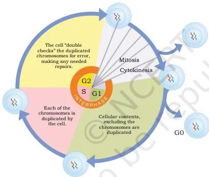
Fig 5.14 shows the whole cycle. Interphase contains three sub-phases:
- G1 (Gap 1): the cell is metabolically active and grows continuously, building RNA, organelles and molecular building blocks; DNA is not replicated.
- S (Synthesis): DNA is synthesised in the nucleus. The amount of DNA doubles from 2C to 4C, but the chromosome number stays unchanged — a diploid 2n cell remains 2n. The centriole also duplicates in the cytoplasm, ready for chromosome segregation.
- G2 (Gap 2): more proteins and organelles are synthesised; the cell is prepared to enter M phase (NCERT, p. 127-128).
Not every cell can divide. Some cells exit the cycle from G1 into the quiescent stage (G0), an inactive stage for cells that will not divide again — cardiac cells and neurons are the classic examples. These G0 cells are often called differentiated cells.
The trap: they are not “resting” in any idle sense; they have simply withdrawn from the cycle (NCERT, p. 128).
Mitosis: the four stages
Mitosis is equational division: the number of chromosomes in parent and daughter cells is the same (NCERT, p. 128-129). Karyokinesis (nuclear division) has four stages, followed by cytokinesis.
- Prophase: diffused chromatin condenses into chromosomes; centrioles (duplicated in S phase) move to opposite poles, initiating the mitotic spindle; Golgi apparatus, ER, nucleolus and nuclear membrane begin to disappear.
- Metaphase: the nuclear envelope is fully gone and chromosomes are at their most condensed — the best stage to study chromosome morphology. Sister chromatids are held together at the centromere, which carries disc-shaped kinetochores, the attachment sites for spindle fibres. All chromosomes align on the metaphase plate, one chromatid of each connected to each pole.
- Anaphase: chromatids are pulled apart toward opposite poles.
- Telophase: the nuclear envelope reforms, the nucleolus reappears, and chromosomes at the poles begin to decondense (NCERT, p. 129).
Cytokinesis then divides the cytoplasm. Animal cells form a furrow in the plasma membrane that deepens into a contractile ring of actin filaments. Plant cells, boxed in by an inextensible cell wall, instead synthesise new cell wall material, starting with a precursor called the cell plate (NCERT, p. 130).
Meiosis: Halving the Chromosome Number
Meiosis produces haploid gametes from diploid cells; the gametes fuse at fertilisation to restore diploidy. There is a single interphase followed by two nuclear divisions: meiosis I is reductional (chromosome number is halved) and meiosis II is equational (the number stays the same). One meiotic cycle ends in four haploid cells (NCERT, p. 130-131).
Meiosis differs from mitosis in two crucial respects: it halves the chromosome number, and recombination shuffles genes between the chromosomes of each pair (NCERT, p. 131).
Prophase I — the longest phase
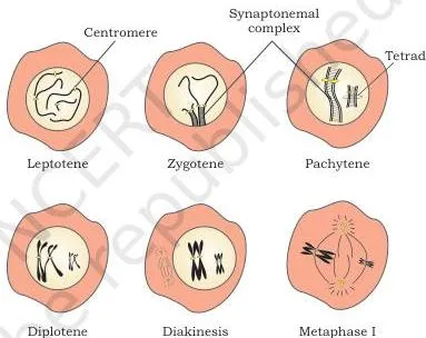
Prophase I dominates the meiotic cycle. Fig 5.16 shows its five substages in order (NCERT, p. 131-132):
- Leptotene: chromosomes appear as thin threads; each consists of two sister chromatids.
- Zygotene: homologous chromosomes pair up (synapsis) with the help of the synaptonemal complex; the pair is called a bivalent or tetrad.
- Pachytene: recombinational nodules appear — the sites where crossing over occurs between non-sister chromatids of homologous chromosomes. This exchange of genetic material is a source of variation.
- Diplotene: the synaptonemal complex degenerates and homologues separate, except at the crossover sites called chiasmata. In the oocytes of some vertebrates this stage can last months or years.
- Diakinesis: chiasmata are terminalised, chromosomes become fully condensed, the meiotic spindle assembles, and the nuclear envelope breaks down with the nucleolus disappearing.
Memory device for the order: Leptotene, Zygotene, Pachytene, Diplotene, Diakinesis — “Lazy Zebras Play Drums Daily”.
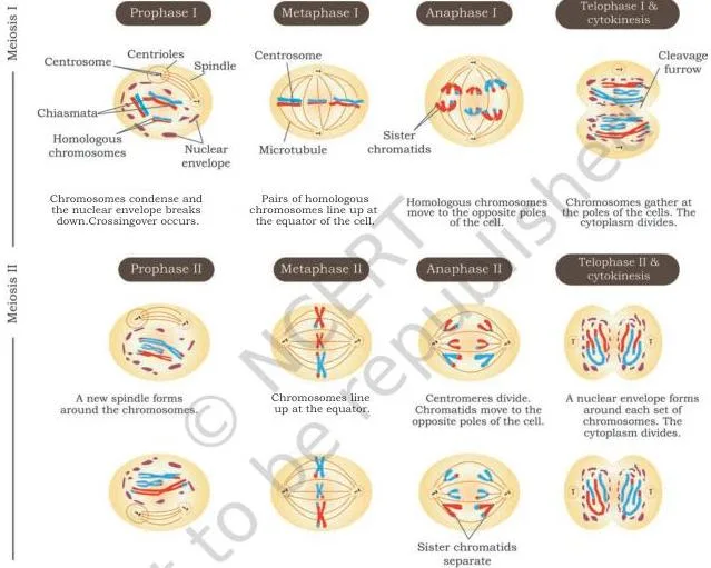
Fig 5.17 shows how the two divisions follow each other. In meiosis I, metaphase I aligns bivalents on the equatorial plate; anaphase I separates homologous chromosomes while sister chromatids stay attached at their centromeres; telophase I and cytokinesis complete the first division, followed by a short interkinesis that involves no further DNA replication.
Meiosis II then repeats prophase, metaphase, anaphase and telophase — this time anaphase II separates sister chromatids, producing four haploid cells (NCERT, p. 132-134).
Meiosis matters for four reasons (NCERT, p. 134):
- It keeps the same chromosome number (n) in all sexually reproducing organisms.
- It restricts chromosome number and maintains stability of the species.
- Crossing over between homologous chromosomes is a significant source of genetic variation.
- The four sister chromatids segregate into four different daughter cells, making them genetically different.
The genetic variation created by crossing over is the raw material for the inheritance patterns you will revise next in the Basic Principles of Inheritance notes.
The textbook’s Table 5.1 compares mitosis and meiosis in nine rows; the rows below are the ones comparative answers need (NCERT, p. 134-135).
| Feature | Mitosis | Meiosis |
|---|---|---|
| Where it occurs | Somatic cells of the body | Germ cells |
| Type of organism | Sexually and asexually reproducing organisms | Only sexually reproducing organisms |
| Nuclear divisions | One | Two (meiosis I and meiosis II) |
| Chromosome number in daughter cells | Same as the mother cell | Half of the mother cell |
| Crossing over | Does not occur | Occurs between non-sister chromatids of homologous chromosomes |
| Daughter cells produced | Two | Four |
Apoptosis: Programmed Cell Death vs Necrosis
Start with your own hands. During embryonic development your fingers are joined web-like; the cells between them die by apoptosis, creating the gaps. Without that death, we would have webbed hands (NCERT, p. 135).
Apoptosis (programmed cell death) is a highly controlled, energy-dependent process that does not occur randomly. Once a cell is committed to it, the fate cannot be reversed. It protects us from viral infections and cancer — and when apoptosis fails to work efficiently, unregulated growth and division can result in cancer (NCERT, p. 135).
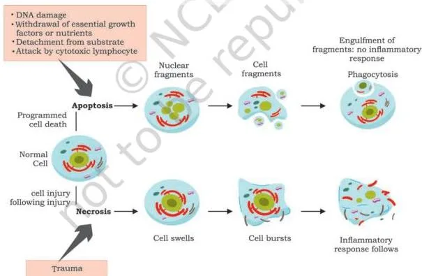
Fig 5.18 contrasts the two deaths. Apoptosis shows DNA fragmentation, blebbing of the plasma membrane, breakdown of the nuclear envelope, and increased susceptibility of DNA to DNAase. The cell finally splits into small membrane-enclosed fragments that nearby macrophages phagocytose — so no contents spill out and no inflammation follows (NCERT, p. 136).
Necrosis is a different event: a traumatic, unplanned death caused by physical injury or exposure to toxic substances. It is unregulated, shows no DNA fragmentation or blebbing, and the cell’s contents are released into the surroundings, triggering inflammation (NCERT, p. 136).
| Feature | Apoptosis | Necrosis |
|---|---|---|
| Regulation | Highly controlled and programmed | Unregulated, accidental |
| Energy | Energy-dependent | Not a regulated energy-dependent process |
| DNA fragmentation | Present | Absent |
| Plasma membrane blebbing | Present | Absent |
| Inflammation | No — fragments are phagocytosed by macrophages | Yes — cell contents are released |
| Typical cause | Development, defence against viruses and cancer | Physical injury, toxic substances |
Cell Differentiation and Stem Cell Potency
Here is the puzzle: the trillions of cells in your body carry exactly the same DNA, yet a neuron is specialised to conduct impulses and a red blood cell to transport oxygen. The answer lies in the way genes are expressed — the unique combination of genes switched on at a given time dictates each cell’s structure and function (NCERT, p. 136).
Cell differentiation is the process by which an unspecialised cell becomes specialised. Epithelial cells, RBCs, WBCs, cardiac cells, neurons and muscle cells are all differentiated cells (NCERT, p. 136-137).
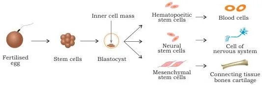
Fig 5.19 shows the route: a fertilised egg gives rise to many kinds of specialised cells (NCERT, p. 138). Not every body cell is differentiated — populations of unspecialised stem cells persist even in adults. A stem cell divides to produce two daughters: one remains a stem cell and the other becomes differentiated (NCERT, p. 137).
Stem cells come from two major sources: embryos (usually the inner cell mass of a week-old blastocyst) and adult tissues such as bone marrow, blood vessels, brain, skeletal muscle and liver. Adult stem cells stay in a quiescent, non-dividing state until tissue injury activates them (NCERT, p. 137).
The ability of a stem cell to differentiate into other cell types is its cell potency.
| Potency | What it can form | Differentiation potential | Example |
|---|---|---|---|
| Totipotent | All cell types — embryonic and extraembryonic tissues | Highest | Zygote, asexual spores |
| Pluripotent | Most body tissues via the three germ layers (endoderm, mesoderm, ectoderm); not extraembryonic tissues like placenta or yolk sac | Less than totipotent | Inner cell mass cells |
| Multipotent | A limited range of tissues | Even lower | Haematopoietic stem cells → RBCs, WBCs, platelets |
One modern detail is worth carrying forward: in 2006, Shinya Yamanaka of Japan developed induced pluripotent stem cell (iPSC) technology, which uses adult tissue cells and induces pluripotency by expressing specific transcription factors. This bypasses the need for embryos and gives patient-matched pluripotent cells, avoiding the risk of transplant rejection (NCERT, p. 138).
Cell Migration: How Cells Move
Cell migration is the movement of cells from one place to another — from amoeba to mammals, and across environments from natural soil to Petri dishes. It occurs during embryogenesis (creating new layers), organogenesis, regeneration, feeding and immune responses (NCERT, p. 139).
Cells may move as single units or in groups, depending on cytoskeleton organisation, the extracellular matrix, adhesion strength and migratory signals.
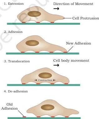
Fig 5.20 breaks migration into five coordinated stages (NCERT, p. 139-140):
- Polarisation: the cell develops a definite front and rear, guided by directional signals that can be chemical, electric-field-based, mechanical, or based on substrate concentration.
- Protrusion of the leading edge: membrane extensions such as pseudopodia form and extend, driven by branching and polymerisation of actin filaments.
- Adhesion: protrusions attach to the substratum; adhesions are sites of molecular communication between cell and substrate, assembling at the front and disassembling at the rear.
- Translocation of the cell body: the cell body moves forward as the lagging edge separates from the substratum.
- Retraction of the rear: rear adhesions are dismantled (by down-regulation through phosphatases or by proteolysis of adhesive contacts by proteases), and the trailing edge pulls in — otherwise the cell would be ripped apart under tension.
Migration is not just a developmental story. It maintains homeostasis: tissue regeneration and repair, and the inflammation of injury or infection, both depend on it — immune cells migrate from lymph nodes to the circulation, ready to respond when an inflammatory reaction is triggered.
Failure of cells to migrate can therefore cause severe defects such as autoimmune diseases and defective wound repair. Migration also drives metastasis — cancer cells, having lost cell-cell interactions and gained motility, spread from the primary tumour (NCERT, p. 140-141).
Key Terms from Cellular Processes: A Quick-Reference Table
Every exam-facing definition in one lookup table. Page numbers refer to the NCERT textbook.
| Term | Meaning | Page |
|---|---|---|
| Receptor | Protein on the cell surface or inside the cytoplasm or nucleus that a signal molecule docks onto | 103 |
| Ligand | The chemical messenger that binds to its specific receptor | 103 |
| Paracrine signaling | Short-distance signaling: the ligand diffuses through extracellular space to nearby cells | 104 |
| Autocrine signaling | A cell secretes a ligand and also carries the receptor for it — a cell signalling itself | 104 |
| Endocrine signaling | Long-distance signaling: the ligand travels through the bloodstream to reach target cells | 104 |
| Anabolism | Pathways that build large complex molecules from small ones; endergonic, they consume energy | 105 |
| Catabolism | Pathways that break down large molecules; exergonic, they release energy and make ATP and reducing equivalents | 106 |
| Glycolysis | The cytosolic pathway that converts glucose to pyruvate (EMP pathway) | 110 |
| Gluconeogenesis | Making glucose from non-carbohydrate precursors such as lactic acid, amino acids and glycerol | 106-107 |
| Transamination | Transfer of amino nitrogen from one amino acid to a carbon skeleton to form another amino acid | 109 |
| Deamination | Removal of amino nitrogen, which is excreted from the body as urea | 109 |
| Substrate-level phosphorylation | Direct transfer of phosphate to ADP to form ATP, seen at phosphoglycerate kinase and pyruvate kinase | 111-112 |
| Oxidative phosphorylation | ATP synthesis driven by the proton gradient built by the electron transport chain | 117-118 |
| Quiescent stage (G0) | A stage outside the cell cycle that non-dividing cells enter from G1; neurons and cardiac cells are examples | 128 |
| Reductional division | Meiosis I, in which the chromosome number is halved | 130 |
| Equational division | Mitosis and meiosis II, in which the chromosome number stays the same | 129, 130-131 |
| Apoptosis | Programmed cell death: controlled, energy-dependent and part of normal development | 135 |
| Necrosis | Traumatic, unregulated cell death from injury or toxins; it triggers inflammation | 136 |
| Cell differentiation | The process by which an unspecialised cell becomes specialised | 136 |
| Cell potency | The ability of a stem cell to differentiate into other cell types | 137 |
| Totipotent | Able to form all embryonic and extraembryonic tissues; the zygote is the example | 137 |
| Pluripotent | Able to form the three germ layers and most body tissues, but not extraembryonic tissues | 138 |
| Multipotent | Able to form only a limited range of tissues, like haematopoietic stem cells forming blood cells | 138 |
| Metastasis | The spread of cancer cells from the primary tumour to other places | 141 |
| Kranz anatomy | C4 leaf structure with mesophyll cells clustered in a ring around bundle sheath cells | 125 |
| Crossing over | Exchange of genetic material between non-sister chromatids of homologous chromosomes in pachytene | 131 |
Equations and Energy Statements You Must Know
| Equation | Where it happens | What it tells you |
|---|---|---|
| \( \text{Glucose} + 2NAD^+ + 2ADP + 2P_i \rightarrow 2\text{Pyruvate} + 2NADH + 2H^+ + 2ATP + 2H_2O \) | Cytosol of all living cells | Net glycolysis: 4 ATP are made but 2 are invested, so net = 2 ATP plus 2 NADH per glucose (p. 112) |
| \( \text{Glucose} + 2ADP + 2P_i \rightarrow 2\text{Lactate} + 2ATP + 2H_2O + 2H^+ \) | Anaerobic muscle (cytosol) | Homolactic fermentation regenerates NAD⁺ so glycolysis continues; yield is 2 ATP per glucose (p. 113) |
| \( \text{Pyruvate} + CoA + NAD^+ \rightarrow \text{Acetyl CoA} + CO_2 + NADH + H^+ \) | Mitochondria (pyruvate dehydrogenase complex) | The irreversible link between glycolysis and the citric acid cycle (p. 114) |
| \( \text{Acetyl CoA} + 3NAD^+ + FAD + GDP + P_i + 2H_2O \rightarrow 2CO_2 + 3NADH + 3H^+ + FADH_2 + GTP + CoA \) | Mitochondrial matrix | One TCA turn: 3 NADH, 1 FADH₂, 1 GTP and 2 CO₂ per acetyl CoA (p. 116) |
| \( 6CO_2 + 12H_2O \xrightarrow[\text{Chlorophyll}]{\text{light}} C_6H_{12}O_6 + 6O_2 + 6H_2O \) | Chloroplast | Overall photosynthesis: CO₂ is reduced to carbohydrate; the O₂ released comes from water (p. 118-119) |
| \( 2H_2O \xrightarrow{\text{oxygen evolving complex}} 4H^+ + 4e^- + O_2 \) | Oxygen evolving complex on the luminal thylakoid surface | Water splitting: one O₂ is produced for every four electrons sent down the electron transport chain (p. 120-121) |
Worked Examples: Counting ATP and Carbon
Worked Example 1: The Glycolysis ATP Account (per glucose)
Step 1 — Method: use energy accounting — separate the ATP invested from the ATP paid back.
Step 2 — Investment: hexokinase spends 1 ATP to phosphorylate glucose, and phosphofructokinase spends 1 ATP to make fructose 1,6-bisphosphate.
Invested = 2 ATP per glucose.
Step 3 — Payoff by substrate-level phosphorylation: each glucose yields two triose phosphates, so both payoff steps run twice.
Phosphoglycerate kinase pays 2 ATP (one per triose) and pyruvate kinase pays 2 ATP.
Generated = 4 ATP per glucose.
\[ \text{Net ATP} = 4\ \text{generated} – 2\ \text{invested} = 2\ \text{ATP per glucose} \]
Step 4 — Reducing power: glyceraldehyde-3-phosphate dehydrogenase makes 1 NADH per triose, so the tally also includes 2 NADH per glucose.
Final answer: glycolysis yields a net of 2 ATP and 2 NADH per glucose, from a gross production of 4 ATP (NCERT, p. 112).
Worked Example 2: The Aerobic ATP Tally (per glucose)
Step 1 — Method: multiply each reduced coenzyme by this book’s value — 2.5 ATP per NADH and 1.5 ATP per FADH₂ — then add direct ATP and GTP.
Step 2 — Glycolysis: net ATP = 2 ATP.
Step 3 — NADH: complete aerobic oxidation produces 10 NADH per glucose (2 from glycolysis, 2 from the pyruvate dehydrogenase step for two pyruvates, 6 from two turns of the TCA cycle).
\[ 10 \times 2.5 = 25\ \text{ATP} \]
Step 4 — FADH₂: two turns of the TCA cycle give 2 FADH₂ per glucose.
\[ 2 \times 1.5 = 3\ \text{ATP} \]
Step 5 — GTP: the TCA makes 1 GTP per turn; with two turns this is 2 ATP-equivalents.
\[ \text{Total} = 2 + 25 + 3 + 2 = 32\ \text{ATP per glucose} \]
Final answer: 32 ATP per glucose. These are the rationalised NCERT values (NCERT, p. 118) — not the older 3 ATP per NADH and 2 ATP per FADH₂ figures, which do not match this syllabus.
Worked Example 3: The Calvin Cycle Carbon and Energy Budget (per 3 CO₂)
Step 1 — Method: track both carbon atoms and energy through the three stages: fixation, reduction, regeneration.
Step 2 — Fixation: 3 CO₂ condense with 3 ribulose 1,5-bisphosphate to form 6 molecules of 3-phosphoglycerate (3-PGA); RuBisCO catalyses this carboxylation.
\[ 3CO_2 + 3\text{RuBP} \rightarrow 6\ \text{3-PGA} \]
Step 3 — Reduction: 6 ATP convert the 6 PGA to 1,3-bisphosphoglycerate, and 6 NADPH reduce these to 6 molecules of glyceraldehyde 3-phosphate (G3P).
Step 4 — Export: one G3P leaves the cycle for sugar synthesis, starch or glycolysis.
Step 5 — Regeneration: the remaining 5 G3P (15 carbons) reform 3 ribulose-5-phosphate (15 carbons), releasing 2 Pi, and 3 ATP convert them to 3 ribulose 1,5-bisphosphate.
\[ \text{Energy spent per 3 CO}_2\ \text{fixed} = 6\ \text{ATP (reduction)} + 3\ \text{ATP (regeneration)} = 9\ \text{ATP} \]
\[ \text{Plus } 6\ \text{NADPH}; \quad \text{net export} = 1\ \text{G3P (a triose phosphate)} \]
Final answer: for every 3 CO₂ fixed, the Calvin cycle spends 9 ATP and 6 NADPH and exports one triose phosphate, G3P (NCERT, p. 123-124).
Common Mistakes in Cellular Processes and Their Corrections
| Students write | Correct is | Why |
|---|---|---|
| S phase doubles the chromosome number | S phase doubles the DNA content (2C → 4C); the chromosome number stays the same (2n stays 2n) | Each chromosome now has two sister chromatids, but the number of chromosomes is unchanged (NCERT, p. 128) |
| Interphase is a resting phase | Interphase is a phase of growth, metabolic activity and preparation | The cell accumulates RNA, organelles and proteins, and replicates DNA in S; “resting” only means it is not visibly dividing (NCERT, p. 127-128) |
| PSI is the first photosystem | Under normal conditions PSII is activated first | PSII absorbs at 680 nm and feeds electrons into the chain that ends at PSI (700 nm) (NCERT, p. 120) |
| Dark reactions happen only at night | Dark reactions are light-independent, not night-only | They consume ATP and NADPH made by light, so they normally run in daylight (NCERT, p. 122) |
| NADH gives 3 ATP and FADH₂ gives 2 ATP | This book uses 2.5 ATP per NADH and 1.5 ATP per FADH₂ | FADH₂ enters the chain at complex III, skipping the proton-pumping contribution of complex I (NCERT, p. 118) |
| Glycolysis nets 4 ATP | Gross is 4 ATP, net is 2 ATP | Two ATP are invested at hexokinase and phosphofructokinase before the payoff (NCERT, p. 112) |
| Apoptosis and necrosis are the same | Apoptosis is regulated, energy-dependent and non-inflammatory; necrosis is traumatic and inflammatory | Necrosis shows no DNA fragmentation or blebbing and releases cell contents that trigger inflammation (NCERT, p. 135-136) |
| Meiosis II halves the chromosome number | Meiosis I is reductional; meiosis II is equational | Homologous chromosomes separate in meiosis I; only sister chromatids separate in meiosis II (NCERT, p. 130-131) |
How to Write Answers to This Chapter’s Exercise Types
These are notes on answer structure, not solved exercises. The chapter’s exercise set (NCERT, p. 143-144) repeats a few formats; here is what a complete answer to each contains.
- Comparative questions (apoptosis vs necrosis, autocrine vs paracrine, anabolic vs catabolic, totipotent vs pluripotent): use a paired point-by-point table with one feature per row. Open with the defining difference — regulation for apoptosis vs necrosis, who receives the signal for autocrine vs paracrine, energy direction for anabolic vs catabolic, and range of tissues for totipotent vs pluripotent.
- Potency questions (how stem cells differ from blood cells): define cell potency first (the ability to differentiate into other cell types), then state which type is more potent and why — stem cells are unspecialised and keep potency, while blood cells are fully differentiated.
- Match-the-following: recall the exact function attached to each term — centromere holds the two sister chromatids together; kinetochore is the site of attachment of chromosomes to spindle fibres; zygotene is the pairing of homologous chromosomes; pachytene is the stage of crossing over; meiosis I is reductional division; meiosis II is equational division.
- Cyclic vs non-cyclic photophosphorylation: structure as a table — photosystems used (PSI alone vs PSII + PSI), electron source and fate, and products (ATP only vs ATP + NADPH + O₂).
- Calvin cycle descriptions: organise by the three stages — fixation (RuBisCO; 3 CO₂ + 3 RuBP → 6 PGA), reduction (6 ATP + 6 NADPH → 6 G3P), regeneration (3 ATP; 5 G3P → 3 RuBP). Name the first stable product, 3-PGA, and the enzyme RuBisCO.
- Dark reaction questions: lead with light independence — the reactions do not need light energy directly and can run in daylight because they use ATP and NADPH; they are called dark reactions only because light is not required.
Rapid Revision: One-Line Takeaways
- A cell answers a signal only if it carries the matching receptor — specificity decides the response.
- Catabolism pays for anabolism: energy released by breaking molecules down drives the building of new ones.
- Glycolysis is cytosolic and nets 2 ATP + 2 NADH per glucose (gross 4 ATP, 2 invested).
- The TCA cycle plus electron transport chain make the bulk of aerobic ATP — 2.5 ATP per NADH and 1.5 ATP per FADH₂ in this book.
- Photosynthesis splits water, makes ATP and NADPH in the light reactions, then fixes CO₂ to sugar in the Calvin cycle; the O₂ released comes from water.
- Interphase = G1 + S + G2; DNA doubles only in S (2C → 4C), while the chromosome number stays 2n.
- Mitosis is equational; meiosis I is reductional and meiosis II is equational.
- Apoptosis is orderly, energy-dependent cell suicide; necrosis is traumatic, messy death that inflames.
- Stem cell potency narrows from totipotent to pluripotent to multipotent.
- Cell migration powers gastrulation and organogenesis, tissue repair and inflammation, and — when hijacked — cancer metastasis.
Two memory devices close the chapter. The prophase I substage order is Leptotene, Zygotene, Pachytene, Diplotene, Diakinesis. And the three irreversible glycolytic enzymes — hexokinase, phosphofructokinase and pyruvate kinase — fit the phrase “He Pushes Pyruvate”: H for hexokinase, P for phosphofructokinase, P for pyruvate kinase — the three one-way doors of glycolysis that end at pyruvate.
FAQs on Cellular Processes: ATP Yields, Dark Reactions and Stem Cells
Why is interphase called the resting phase if the cell is working so hard?
It is a misnomer carried over from early microscopy: a cell in interphase is not visibly dividing, so the phase was labelled “resting”. In reality interphase is a period of intense activity, not idleness — G1 builds RNA and organelles, S replicates DNA (2C → 4C), and G2 synthesises more proteins and organelles.
What actually rests is a cell that has left the cycle into the quiescent G0 stage. When the textbook uses “resting phase” for interphase, it only means “between divisions” (NCERT, p. 127-128).
What is the real difference between cyclic and non-cyclic photophosphorylation?
Non-cyclic flow uses both photosystems: PSII starts the chain, water is split, and the electrons end at NADP⁺. The products are ATP, NADPH and O₂. Cyclic flow uses PSI alone — the excited electron leaves PSI, passes through cytochrome b₆f (which pumps protons) and returns to PSI through plastocyanin.
The only product is ATP; there is no NADPH and no oxygen (NCERT, p. 120-122).
Does the dark reaction of photosynthesis happen only at night?
No. “Dark reaction” means light-independent, not night-only. The Calvin cycle needs ATP and NADPH, which the light reactions supply, so it normally runs in daylight alongside them. What it does not need is light energy directly. Saying it cannot occur in daylight is wrong (NCERT, p. 122).
Why do C4 plants need Kranz anatomy while C3 plants do not?
C4 plants separate CO₂ fixation from the Calvin cycle in space. Mesophyll cells, which contain PEP carboxylase, fix CO₂ into a four-carbon compound that travels to bundle sheath cells; there CO₂ is released and refixed by RuBisCO. Kranz anatomy — mesophyll cells clustered in a ring around bundle sheath cells — makes this two-cell route possible.
C3 plants fix CO₂ directly in the mesophyll with RuBisCO and need no such structure (NCERT, p. 125-126).
What is the difference between totipotent, pluripotent and multipotent stem cells?
Totipotent cells can form all cell types, embryonic and extraembryonic — the zygote is the example. Pluripotent cells form the three germ layers and most body tissues but not extraembryonic tissues such as placenta or yolk sac — the inner cell mass is the example. Multipotent cells form only a limited range of tissues — haematopoietic stem cells give rise to blood cells.
Potency decreases in that order (NCERT, p. 137-138).
Why does this chapter say NADH gives 2.5 ATP and FADH₂ gives 1.5 ATP?
Because the values track where electrons enter the chain. Electron flow through complex I, complex III and complex IV builds enough gradient for 1, 0.5 and 1 ATP respectively — together 2.5 ATP per NADH. FADH₂ delivers its electrons at complex III (via complex II), skipping complex I, so it earns only 1.5 ATP (NCERT, p. 118).
Older texts rounded these to 3 and 2; this rationalised NCERT chapter uses 2.5 and 1.5, so use those in your answers.
Reference: NCERT Class 11 Biotechnology textbook, chapter Cellular Processes.
Explore Class 11 Biotechnology Notes
- Class 11 Biotechnology Notes
- Class 11 CBSE Notes
- CBSE Notes for Classes 1 to 12
- Next: Basic Principles of Inheritance
Class 11 Biotechnology on LearnCBSE:
Related chapters:
- Genetic Disorder notes
Official source: download the NCERT textbook free from ncert.nic.in.