
These notes summarise NCERT Class 11 Biology Chapter 14, Breathing and Exchange of Gases, as a revision-ready study page for the current syllabus; they follow the 2026-27 session.
The chapter’s core problem is simple: every cell needs oxygen to break down glucose, amino acids and fatty acids for energy, and the carbon dioxide released is harmful, so O2 must keep entering and CO2 must keep leaving the body (NCERT, p. 183).
Below you get the respiratory organs of different animal groups, the human air passage, the mechanism of breathing, the lung volumes and capacities, gas exchange and transport, regulation of respiration, and the disorders — with tables, memory devices, worked calculations and a map of the textbook’s own exercises.
This page is part of the Class 11 Biology notes collection; for the other chapters in the class, see the complete Class 11 notes list.
Breathing and Respiration: What This Chapter Covers
Respiration is not a single event. The textbook lists five steps (NCERT, p. 185):
- Pulmonary ventilation or breathing — atmospheric air is drawn in and CO2-rich alveolar air is released out.
- Diffusion of gases (\( O_2 \) and \( CO_2 \)) across the alveolar membrane.
- Transport of gases by the blood.
- Diffusion of O2 and CO2 between blood and tissues.
- Utilisation of O2 by the cells for catabolic reactions, with CO2 released — cellular respiration, covered in Chapter 12 of the same book.
This chapter deals with steps (i) to (iv); step (v), the chemistry inside cells, belongs to the chapter on cellular respiration. Removing CO2 is one half of the body’s waste disposal — the other metabolic wastes are handled by systems described in the notes on Excretory Products and Their Elimination.
Respiratory Organs Across Animals
Which respiratory surface an animal uses depends mainly on its habitat and level of organisation (NCERT, p. 183). The body-plan levels behind these surfaces are covered in the Animal Kingdom notes.
| Group of animals | Respiratory surface / mechanism |
|---|---|
| Sponges, coelenterates, flatworms (lower invertebrates) | Simple diffusion over the entire body surface |
| Earthworms | Moist cuticle |
| Insects | Network of tracheal tubes |
| Most aquatic arthropods and molluscs | Gills (branchial respiration) |
| Terrestrial forms | Lungs (pulmonary respiration) |
| Fishes | Gills |
| Amphibians, reptiles, birds and mammals | Lungs |
| Frogs (amphibians) | Lungs, plus moist skin (cutaneous respiration) |
One compact takeaway: simple aquatic animals exchange over the whole body surface, insects pipe air directly to the tissues through tracheal tubes, and vertebrates use gills in water or lungs on land — with frogs adding their moist skin.
Human Respiratory System: Pathway and Parts
Air follows one fixed passage from the outside to the exchange surfaces (NCERT, p. 184):
- External nostrils → nasal chamber through the nasal passage; the chamber opens into the pharynx, a portion of which is the common passage for food and air.
- Pharynx → larynx, a cartilaginous sound box; during swallowing the glottis is covered by the thin elastic epiglottis so food cannot enter the larynx.
- Larynx → trachea, a straight tube that divides at the level of the 5th thoracic vertebra into right and left primary bronchi.
- Primary bronchi → secondary and tertiary bronchi → bronchioles → terminal bronchioles.
- Each terminal bronchiole gives rise to thin, irregular-walled, vascularised bags called alveoli — the exchange site.
Keep the order with a mnemonic: ‘Never Play Lazy Tennis, Breathe Big Air’ = Nostrils, Pharynx, Larynx, Trachea, Bronchi, Bronchioles, Alveoli.
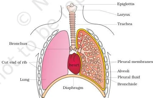
As Figure 14.1 shows, the branching network of bronchi, bronchioles and alveoli makes up the lungs, with the sectional view of the left lung exposing the branching airways.
The whole system is supported: the trachea, bronchi and initial bronchioles carry incomplete cartilaginous rings, and each lung is covered by a double-layered pleura whose pleural fluid reduces friction on the lung surface (NCERT, p. 184).
Functionally the system splits into two parts (NCERT, p. 185):
| Part | Extent | Function |
|---|---|---|
| Conducting part | External nostrils up to the terminal bronchioles | Transports atmospheric air to the alveoli, clears it of foreign particles, humidifies it and brings it to body temperature |
| Respiratory (exchange) part | Alveoli and their ducts | Site of the actual diffusion of \( O_2 \) and \( CO_2 \) between blood and atmospheric air |
The lungs sit in an air-tight thoracic chamber — vertebral column dorsally, sternum ventrally, ribs laterally, and a dome-shaped diaphragm on the lower side (NCERT, p. 185). Because the chamber is airtight, any change in its volume is reflected in the lung cavity; we cannot alter pulmonary volume directly, so this arrangement is essential for breathing.
Mechanism of Breathing: Inspiration and Expiration
Air moves into and out of the lungs only when a pressure gradient exists between the lungs and the atmosphere (NCERT, p. 185). Inspiration occurs when the intra-pulmonary pressure is less than atmospheric pressure — a negative pressure in the lungs. Expiration occurs when the intra-pulmonary pressure is higher than atmospheric pressure.
The diaphragm and the external and internal intercostal muscles create this gradient. Contraction of the diaphragm enlarges the thoracic chamber in the antero-posterior axis; contraction of the external intercostals lifts the ribs and sternum and enlarges it in the dorso-ventral axis. The bigger thoracic volume pulls the pulmonary volume up, pressure falls, and air rushes in — inspiration.
Relaxation of the diaphragm and intercostals reverses everything, pressure rises slightly above atmospheric, and air is expelled — expiration (NCERT, p. 185–186).
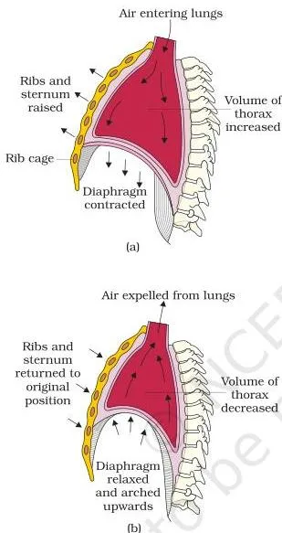
Figure 14.2 shows the two stages side by side: panel (a) is inspiration, when the thoracic and pulmonary volumes enlarge and intra-pulmonary pressure falls below atmospheric; panel (b) is expiration, after the diaphragm and intercostals relax and the pressure rises slightly above atmospheric.
| Feature | Inspiration | Expiration |
|---|---|---|
| Muscle action | Diaphragm contracts; external intercostals contract and lift the ribs and sternum | Diaphragm and intercostals relax and return to normal position |
| Thoracic volume | Increases in the antero-posterior and dorso-ventral axes | Decreases |
| Pulmonary volume | Increases | Decreases |
| Intra-pulmonary pressure | Falls below atmospheric; air is drawn in | Rises slightly above atmospheric; air is expelled |
We can strengthen both acts using additional muscles in the abdomen, which is how forced inspiration and expiration work. On average a healthy human breathes 12–16 times per minute, and the volume of air involved can be estimated with a spirometer, which is used in clinical assessment of pulmonary function (NCERT, p. 186).
Respiratory Volumes and Capacities: The Numbers and Formulas
Four volumes describe single breaths; five capacities are derived by adding volumes and are used in clinical diagnosis (NCERT, p. 186–187).
The Four Respiratory Volumes
| Volume | What it measures | Approximate value |
|---|---|---|
| Tidal Volume (TV) | Air inspired or expired during a normal respiration | ~500 mL |
| Inspiratory Reserve Volume (IRV) | Extra air a person can draw in by a forcible inspiration beyond the tidal volume | 2500–3000 mL |
| Expiratory Reserve Volume (ERV) | Extra air a person can force out by a forcible expiration beyond the tidal volume | 1000–1100 mL |
| Residual Volume (RV) | Air that remains in the lungs even after a forcible expiration | 1100–1200 mL |
The residual volume is the air you cannot breathe out no matter how hard you try — it stays behind even after a maximum forced expiration (NCERT, p. 187).
The Five Pulmonary Capacities and Their Formulas
| Capacity | Definition | Formula |
|---|---|---|
| Inspiratory Capacity (IC) | Total air a person can inspire after a normal expiration | \( TV + IRV \) |
| Expiratory Capacity (EC) | Total air a person can expire after a normal inspiration | \( TV + ERV \) |
| Functional Residual Capacity (FRC) | Air that remains in the lungs after a normal expiration | \( ERV + RV \) |
| Vital Capacity (VC) | Maximum air a person can breathe in after a forced expiration, or breathe out after a forced inspiration | \( ERV + TV + IRV \) |
| Total Lung Capacity (TLC) | Total air accommodated in the lungs at the end of a forced inspiration | \( VC + RV \) = \( RV + ERV + TV + IRV \) |
Remember the order of the capacities with the mnemonic ‘I F V T — I Feel Very Tired’: Inspiratory Capacity, Functional Residual Capacity, Vital Capacity, Total Lung Capacity (NCERT, p. 187).
Exchange of Gases: Partial Pressure Gradients
Exchange happens at two sites — the alveoli (primary site) and the body tissues. Both gases cross by simple diffusion, driven mainly by the pressure or concentration gradient; the gas’s solubility and the thickness of the diffusion membrane also affect the rate (NCERT, p. 187).
Partial pressure is the pressure contributed by an individual gas in a mixture, written \( pO_2 \) for oxygen and \( pCO_2 \) for carbon dioxide. NCERT Table 14.1 gives the partial pressures in mm Hg at five locations:
| Gas | Atmospheric air | Alveoli | Deoxygenated blood | Oxygenated blood | Tissues |
|---|---|---|---|---|---|
| \( O_2 \) | 159 | 104 | 40 | 95 | 40 |
| \( CO_2 \) | 0.3 | 40 | 45 | 40 | 45 |
Reading the numbers (NCERT, p. 187–188): \( O_2 \) runs 159 → 104 → 40 → 95 → 40, so it is always higher on the entry side — the alveolus holds 104 against the deoxygenated blood’s 40, and oxygenated blood holds 95 against the tissues’ 40. \( O_2 \) therefore diffuses from alveoli to blood and from blood to tissues. \( CO_2 \)
walks the gradient backwards: deoxygenated blood carries 45 against the alveolus’s 40, and the tissues sit at 45 against the arriving oxygenated blood’s 40, so \( CO_2 \) diffuses from tissues to blood and from blood to alveoli.
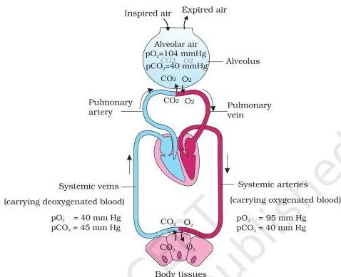
As Figure 14.3 shows, the same pattern repeats at both ends of the journey: at the alveolus, \( O_2 \) enters the blood while \( CO_2 \) leaves it; at the tissues the directions reverse.
Two factors modify the rate. First, solubility: \( CO_2 \) is 20–25 times more soluble than \( O_2 \), so far more \( CO_2 \) diffuses per unit difference in partial pressure. Second, the thickness of the diffusion membrane. The diffusion membrane has just three layers (NCERT, p. 188):
- the thin squamous epithelium of the alveolus,
- the endothelium of the alveolar capillaries, and
- the basement substance between them — a thin basement membrane supporting the epithelium plus the basement membrane surrounding the capillary endothelial cells.
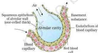
Figure 14.4 zooms into one alveolus with its pulmonary capillary — the membrane the gases must cross. Its total thickness is much less than a millimetre. So every factor in the body — gradient direction, solubility, thinness — favours \( O_2 \) diffusion from alveoli to tissues and \( CO_2 \) diffusion from tissues to alveoli (NCERT, p. 188).
Transport of Gases: How Blood Carries Oxygen and Carbon Dioxide
Blood is the medium of transport for both gases. The headline splits (NCERT, p. 189):
- \( O_2 \): about 97% carried by red blood cells; the remaining 3% dissolved in plasma.
- \( CO_2 \): about 70% carried as bicarbonate, 20–25% as carbamino-haemoglobin in RBCs, and about 7% dissolved in plasma.
Transport of Oxygen
Haemoglobin is a red, iron-containing pigment in the RBCs. Oxygen binds to it in a reversible manner to form oxyhaemoglobin, and each haemoglobin molecule can carry a maximum of four \( O_2 \) molecules (NCERT, p. 189). Binding is governed mainly by \( pO_2 \), but \( pCO_2 \), hydrogen-ion concentration and temperature interfere with it. This gives the loading and unloading sites their logic:
- Alveoli — high \( pO_2 \), low \( pCO_2 \), lesser \( H^+ \), lower temperature: all factors favour formation of oxyhaemoglobin (loading).
- Tissues — low \( pO_2 \), high \( pCO_2 \), high \( H^+ \), higher temperature: all factors favour dissociation of oxygen from oxyhaemoglobin (unloading).
So \( O_2 \) gets bound at the lung surface and dissociates at the tissues; every 100 mL of oxygenated blood delivers about 5 mL of \( O_2 \) to the tissues under normal physiological conditions (NCERT, p. 189).
Transport of Carbon Dioxide
About 70% of \( CO_2 \) travels as bicarbonate (\( HCO_3^- \)). RBCs contain a very high concentration of the enzyme carbonic anhydrase, with minute amounts in plasma too; this enzyme facilitates the reaction in both directions (NCERT, p. 189–190):
\[ CO_2 + H_2O \underset{\text{carbonic anhydrase}}{\rightleftharpoons} H_2CO_3 \underset{\text{carbonic anhydrase}}{\rightleftharpoons} HCO_3^- + H^+ \]
At the tissue site, where catabolism keeps \( pCO_2 \) high, \( CO_2 \) diffuses into the blood and the reaction runs forward, trapping \( CO_2 \) as \( HCO_3^- \) and \( H^+ \). At the alveoli, where \( pCO_2 \) is low, the reaction reverses, releasing \( CO_2 \) and \( H_2O \) — and the \( CO_2 \) is breathed out.
The remaining 20–25% binds to haemoglobin as carbamino-haemoglobin: binding is favoured by high \( pCO_2 \) and low \( pO_2 \) (tissues) and released where \( pCO_2 \) is low and \( pO_2 \) is high (alveoli). Every 100 mL of deoxygenated blood delivers about 4 mL of \( CO_2 \) to the alveoli (NCERT, p. 190).
The two gases use different carriers; this side-by-side is the one to revise from:
| Feature | Oxygen | Carbon dioxide |
|---|---|---|
| Main transported form | Oxyhaemoglobin in RBCs | Bicarbonate in plasma and RBCs |
| Share of total transport | ~97% (3% dissolved in plasma) | ~70% bicarbonate; 20–25% carbamino-haemoglobin; ~7% dissolved |
| Conditions favouring binding | Alveoli: high \( pO_2 \), low \( pCO_2 \), low \( H^+ \), lower temperature | Tissues: high \( pCO_2 \), low \( pO_2 \) (for carbamino-haemoglobin) |
| Where release happens | Tissues: low \( pO_2 \), high \( pCO_2 \), high \( H^+ \), higher temperature | Alveoli: low \( pCO_2 \), high \( pO_2 \) |
| Delivery per 100 mL blood | ~5 mL \( O_2 \) from oxygenated blood | ~4 mL \( CO_2 \) from deoxygenated blood |
Oxygen Dissociation Curve: Reading the S-Shape
The oxygen dissociation curve plots the percentage saturation of haemoglobin with \( O_2 \) against \( pO_2 \), and its shape is sigmoid, or S-shaped (NCERT, p. 189).
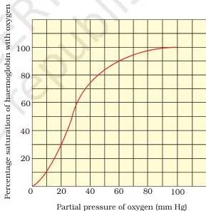
Reading Figure 14.5 from left to right: at very low \( pO_2 \) saturation rises slowly; through the middle range it climbs steeply, so a small rise in \( pO_2 \) loads a lot of oxygen; near the top it flattens again as haemoglobin approaches full saturation.
The curve is highly useful for studying how factors like \( pCO_2 \) and \( H^+ \) concentration affect the binding of \( O_2 \) with haemoglobin (NCERT, p. 189).
Its practical message matches the mechanism section: at the alveoli the conditions are right for oxyhaemoglobin to form, and at the tissues they are right for it to dissociate — oxygen loads at the lung surface and unloads at the tissues. A memory hook: the tissues are hot, acidic and \( CO_2 \)-rich — exactly where haemoglobin lets go of \( O_2 \).
If a question asks you to suggest a reason for the sigmoid pattern, as NCERT Exercise Q11 does, a sound line of reasoning is this: each haemoglobin carries up to four \( O_2 \) molecules, and the binding sites work cooperatively — after the first \( O_2 \) binds, the remaining sites accept further \( O_2 \) more readily.
That cooperation produces the steep middle section and the flattened ends of the S.
Regulation of Respiration: Control Centres in the Brain
Human beings can maintain and moderate the respiratory rhythm to suit the demands of the body tissues; this is done by the neural system (NCERT, p. 190). The chain of control:
| Structure | Location | Function |
|---|---|---|
| Respiratory rhythm centre | Medulla region of the brain | Primarily responsible for maintaining the rhythm |
| Pneumotaxic centre | Pons region of the brain | Moderates the rhythm centre; reduces the duration of inspiration and alters the respiratory rate |
| Chemosensitive area | Adjacent to the rhythm centre | Highly sensitive to \( CO_2 \) and hydrogen ions; signals the rhythm centre to adjust breathing so these substances are eliminated |
| Peripheral receptors | Aortic arch and carotid artery | Recognise changes in \( CO_2 \) and \( H^+ \) and send signals to the rhythm centre |
The counterintuitive fact to memorise: the role of oxygen in the regulation of respiratory rhythm is quite insignificant (NCERT, p. 190). What the brain senses is \( CO_2 \) and \( H^+ \), not low \( O_2 \).
Respiratory Disorders: What Goes Wrong
Three disorders are covered; each links a cause to an effect on the respiratory passage or surface (NCERT, p. 190–191):
| Disorder | Cause | Effect |
|---|---|---|
| Asthma | Inflammation of the bronchi and bronchioles | Difficulty in breathing, with wheezing |
| Emphysema | Alveolar walls are damaged; a major cause is cigarette smoking | Respiratory surface is decreased |
| Occupational respiratory disorders | Long exposure to heavy dust in grinding or stone-breaking industries | Inflammation leads to fibrosis (proliferation of fibrous tissue) and serious lung damage; workers should wear protective masks |
Notice how emphysema connects back to the exchange section: a smaller respiratory surface means less area for diffusion, so the same gradient moves less gas.
Worked Examples: Breathing Volume Calculations
The values below are illustrative — the NCERT exercises use their own numbers — but the method is exactly what you apply to them.
Example 1 — Minute ventilation (air moved per minute and per hour).
Method: Air moved per minute (minute ventilation) = tidal volume × breathing rate.
A student breathes 14 times per minute with a tidal volume of 450 mL.
Step 1: Air per minute = \( 450 \times 14 = 6300\ \text{mL} = 6.3\ \text{L} \).
\[ \text{Air per minute} = 450\ \text{mL} \times 14 = 6300\ \text{mL} = 6.3\ \text{L} \]
Step 2: Air per hour = air per minute × 60 = \( 6300 \times 60 = 378{,}000\ \text{mL} = 378\ \text{L} \).
\[ \text{Air per hour} = 6300\ \text{mL} \times 60 = 378{,}000\ \text{mL} = 378\ \text{L} \]
Final answer: The student moves 6.3 L of air per minute and 378 L per hour.
This is the same skill NCERT Exercise Q14 asks for (tidal volume over an hour), just with different data.
Example 2 — Computing capacities from given volumes.
Method: Capacities are sums of volumes: \( IC = TV + IRV \); \( FRC = ERV + RV \); \( VC = TV + IRV + ERV \); \( TLC = VC + RV \).
Given \( TV = 480\ \text{mL} \), \( IRV = 2600\ \text{mL} \), \( ERV = 1050\ \text{mL} \), \( RV = 1150\ \text{mL} \).
Step 1 — Inspiratory Capacity: \( IC = TV + IRV = 480 + 2600 = 3080\ \text{mL} \).
\[ IC = 480 + 2600 = 3080\ \text{mL} \]
Step 2 — Functional Residual Capacity: \( FRC = ERV + RV = 1050 + 1150 = 2200\ \text{mL} \).
\[ FRC = 1050 + 1150 = 2200\ \text{mL} \]
Step 3 — Vital Capacity: \( VC = TV + IRV + ERV = 480 + 2600 + 1050 = 4130\ \text{mL} \).
\[ VC = 480 + 2600 + 1050 = 4130\ \text{mL} \]
Step 4 — Total Lung Capacity: \( TLC = VC + RV = 4130 + 1150 = 5280\ \text{mL} \).
\[ TLC = 4130 + 1150 = 5280\ \text{mL} \]
Final answers: \( IC = 3080\ \text{mL} \), \( FRC = 2200\ \text{mL} \), \( VC = 4130\ \text{mL} \), \( TLC = 5280\ \text{mL} \).
Key Terms and Definitions at a Glance
The chapter’s key vocabulary in one table; each meaning is written so you can use it directly in an answer:
| Term | Meaning | Example or context |
|---|---|---|
| Breathing | Process of drawing atmospheric air in and releasing \( CO_2 \)-rich alveolar air out | Step (i) of respiration — pulmonary ventilation |
| Inspiration | Drawing air in when intra-pulmonary pressure falls below atmospheric pressure | Diaphragm contracts |
| Expiration | Releasing alveolar air when intra-pulmonary pressure rises above atmospheric pressure | Diaphragm and intercostals relax |
| Tidal volume | Air inspired or expired in one normal breath | ~500 mL |
| Vital capacity | Maximum air breathed in after a forced expiration, or breathed out after a forced inspiration | \( ERV + TV + IRV \) |
| Total lung capacity | Total air in the lungs at the end of a forced inspiration | \( VC + RV \) |
| Partial pressure | Pressure contributed by one gas in a mixture; written \( pO_2 \) or \( pCO_2 \) | Alveolar \( pO_2 \) = 104 mm Hg |
| Oxyhaemoglobin | Haemoglobin with reversibly bound \( O_2 \) | Forms at the alveoli, dissociates at the tissues |
| Carbamino-haemoglobin | Haemoglobin carrying \( CO_2 \) | Carries 20–25% of \( CO_2 \) |
| Carbonic anhydrase | Enzyme catalysing \( CO_2 + H_2O \rightleftharpoons H_2CO_3 \rightleftharpoons HCO_3^- + H^+ \) | Very high concentration in RBCs |
| Conducting part | Passage from external nostrils to terminal bronchioles | Transports, humidifies and warms the air |
| Respiratory (exchange) part | Alveoli and their ducts | Site of actual \( O_2 \)/\( CO_2 \) diffusion |
| Chemosensitive area | Region beside the rhythm centre sensitive to \( CO_2 \) and \( H^+ \) | Activates the rhythm centre when levels rise |
| Epiglottis | Cartilaginous flap that covers the glottis during swallowing | Prevents food from entering the larynx |
Common Mistakes Students Make
Six classic traps from this chapter; each row gives the correction, the reason, and a quick self-check:
| Mistake | Correction | Reason / quick check |
|---|---|---|
| “Breathing and respiration are the same.” | Breathing is only step (i), pulmonary ventilation; respiration covers all five steps, ending with cellular use of \( O_2 \). | Count the five steps: steps (ii)–(v) are respiration but not breathing. |
| “Expiration is always a passive process.” | Normal expiration is passive muscle relaxation, but forced expiration uses additional abdominal muscles. | The mechanism section lists abdominal muscles for strengthening both inspiration and expiration. |
| “Residual volume is the air left after a normal expiration.” | RV is the air left after a forcible expiration; FRC (\( ERV + RV \)) is what remains after a normal expiration. | Ask: which volume stays after breathing out as hard as possible? That is RV. |
| “Most \( CO_2 \) travels as carbamino-haemoglobin.” | About 70% travels as bicarbonate; only 20–25% as carbamino-haemoglobin. | Memorise the split: 70 bicarbonate / 20–25 carbamino / ~7 dissolved. |
| “Low oxygen is what makes us breathe faster.” | The rhythm centre responds mainly to \( CO_2 \) and \( H^+ \); the role of \( O_2 \) in regulating respiratory rhythm is quite insignificant. | Read the chemosensitive area’s sensitivities — \( CO_2 \) and \( H^+ \), not \( O_2 \). |
| “The alveoli are part of the conducting system.” | The conducting part ends at the terminal bronchioles; alveoli and their ducts form the respiratory part. | Check the boundary: the conducting part runs from the external nostrils to the terminal bronchioles. |
Misconception autopsy: low oxygen is not the breathing trigger
This is the chapter’s most counterintuitive statement, so it deserves a full autopsy. The confusion is natural: sprinting leaves you breathless and gasping, so it feels as if low \( O_2 \) forces you to breathe harder.
But look at the regulation pathway (NCERT, p. 190): the chemosensitive area adjacent to the rhythm centre is sensitive to \( CO_2 \) and hydrogen ions, and a rise in these — not a fall in \( O_2 \) — activates the adjustment. The receptors at the aortic arch and carotid artery behave the same way.
The textbook states the conclusion plainly: the role of oxygen in the regulation of respiratory rhythm is quite insignificant. In any answer on regulation, name \( CO_2 \) and \( H^+ \) as the sensed signals and keep \( O_2 \) out of the mechanism.
Exam Notes: How to Write the NCERT Exercise Answers
The 14 NCERT exercises (NCERT, p. 192) fall into a few answer types. For each type, here is what a complete answer must contain — apply the recipe to the question number given:
| Question | Answer type | What a complete answer needs |
|---|---|---|
| Q1, Q2, Q8, Q11 | Definition + short answer | Open with the definition, then add one line of mechanism or significance. For Q1, link vital capacity to clinical assessment of pulmonary function. For Q2, name the capacity (functional residual capacity) and write \( ERV + RV \). For Q8, say that \( pCO_2 \) interferes with \( O_2 \)-haemoglobin binding. For Q11, define the curve and end with a suggested reason (the four binding sites and cooperative binding). |
| Q3, Q10 | Single-fact | One precise line. Q10: the network of tracheal tubes. Q3: diffusion happens only in the alveolar region because the diffusion membrane there is thin and the concentration/pressure gradient exists only between alveoli and blood. |
| Q4 | Explanation | List all three \( CO_2 \) mechanisms with their shares — bicarbonate (~70%), carbamino-haemoglobin (20–25%), dissolved in plasma (~7%) — plus the carbonic anhydrase reaction. |
| Q5 | Data reading | Compare the Atmospheric air and Alveoli columns of Table 14.1: which \( pO_2 \) is higher and which \( pCO_2 \) is lower? Pick the option that matches both comparisons. |
| Q6 | Process | Write the sequence: diaphragm and external intercostals contract → thoracic volume increases in both axes → pulmonary volume increases → intra-pulmonary pressure falls below atmospheric → air moves in (inspiration). |
| Q7 | Process / system | Name the rhythm centre (medulla), the pneumotaxic centre (pons) and the chemosensitive area, then add the peripheral receptors and the quite insignificant role of \( O_2 \). |
| Q9 | Application | Apply the gradient idea: at altitude, atmospheric \( pO_2 \) is lower, so the gradient driving \( O_2 \) into the blood is reduced and breathing speeds up and deepens. |
| Q12 | Discussion | The chapter does not define hypoxia; build from the \( pO_2 \) idea in Table 14.1 and the hill-climbing scenario of Q9 — lower atmospheric \( pO_2 \) reduces the oxygen gradient into the blood. |
| Q13 (a)–(c) | Comparison | Answer in a two-column table: define each term in one line, then state the difference. |
| Q14 | Calculation | Show working with units: tidal volume × breathing rate per minute, then × 60 for the hour. |
Two general tips. For any definition question, the complete answer names the term first and then the quantity or function that defines it — never just an example. For any process question, keep the pressure rule visible: air moves only when a pressure gradient exists between the lungs and the atmosphere.
You can verify the exercise list and wording in the official NCERT Class 11 Biology textbook PDF (Chapter 14) at ncert.nic.in. Once you have finished revising, you can browse all CBSE notes on this site for the other chapters.
Revision Summary: The Chapter in One Page
Last-night revision needs just two things from this chapter: the numbers and the process.
Numbers to memorise
| Quantity | Value to recall |
|---|---|
| Breathing rate of a healthy human | 12–16 breaths per minute |
| Tidal volume | ~500 mL per normal breath |
| Inspiratory reserve volume | 2500–3000 mL |
| Expiratory reserve volume | 1000–1100 mL |
| Residual volume | 1100–1200 mL |
| Oxygen transport | ~97% as oxyhaemoglobin; 3% dissolved in plasma |
| Carbon dioxide transport | ~70% as bicarbonate; 20–25% carbamino-haemoglobin; ~7% dissolved |
| Oxygen delivery | ~5 mL per 100 mL of oxygenated blood |
| Carbon dioxide delivery | ~4 mL per 100 mL of deoxygenated blood |
| Solubility of \( CO_2 \) vs \( O_2 \) | \( CO_2 \) is 20–25 times more soluble than \( O_2 \) |
The five steps as a process map
- Ventilation — lungs: pressure gradient moves air in and out.
- Diffusion across the alveolar membrane — alveoli: driven by partial-pressure gradients.
- Transport by blood — haemoglobin and bicarbonate carry the gases.
- Diffusion between blood and tissues — tissues: \( O_2 \) unloads, \( CO_2 \) loads.
- Cellular utilisation — cells: catabolic reactions use \( O_2 \) and release \( CO_2 \) (covered in Chapter 12).
One-sentence takeaway: \( O_2 \) flows down its partial-pressure gradient from the alveoli to the tissues, while \( CO_2 \) flows the opposite way, from the tissues to the alveoli.
FAQs
What is the difference between breathing and respiration in Class 11 Biology?
Breathing is step (i) of respiration — pulmonary ventilation, the mechanical drawing in of air and release of alveolar air. Respiration is the whole chain (steps i–v): ventilation, diffusion across the alveolar membrane, transport by blood, diffusion into tissues, and the cellular utilisation of \( O_2 \) that releases \( CO_2 \).
Why is the oxygen dissociation curve sigmoid in shape?
A suggested reason is cooperative binding: each haemoglobin molecule carries up to four \( O_2 \) molecules, and once the first \( O_2 \) binds, the remaining sites bind further \( O_2 \) more readily. That produces the steep middle of the S-curve, with the curve flattening as saturation approaches its maximum.
Why is the role of oxygen insignificant in regulating respiration?
The respiratory rhythm centre in the medulla and the adjacent chemosensitive area respond mainly to \( CO_2 \) and hydrogen ions. When these rise, the centre adjusts breathing to eliminate them; the chapter concludes that the role of oxygen in regulating respiratory rhythm is quite insignificant.
What is the volume of air remaining in the lungs after a normal breathing?
After a normal expiration, the air remaining is the functional residual capacity (FRC = \( ERV + RV \)). Do not confuse it with the residual volume — RV (1100–1200 mL) is what remains after a forcible expiration.
How is most of the carbon dioxide transported in blood?
About 70% is carried as bicarbonate (\( HCO_3^- \)), formed through the carbonic anhydrase reaction; 20–25% binds to haemoglobin as carbamino-haemoglobin, and about 7% is dissolved in plasma.
Why does a person going up a hill breathe faster?
At altitude, atmospheric \( pO_2 \) is lower, so the partial-pressure gradient driving \( O_2 \) into the blood is reduced. The respiratory process responds with faster, deeper breathing. Note the chapter’s nuance: although the threat is to the oxygen supply, the regulatory centres sense \( CO_2 \) and \( H^+ \), not \( O_2 \).
Reference: NCERT Class 11 Biology textbook, chapter 14 Breathing and Exchange of Gases.
Explore Class 11 Biology Notes
- Class 11 Biology Notes
- Class 11 CBSE Notes
- CBSE Notes for Classes 1 to 12
- Previous: Animal Kingdom
- Next: Excretory Products and Their Elimination
Class 11 Biology on LearnCBSE:
Related chapters:
- Biological Classification notes
Official source: download the NCERT textbook free from ncert.nic.in.