
These notes revise Biological Classification (Class 11 Biology, Chapter 2) end to end — from Aristotle’s first attempt at a scientific classification and the two kingdom system, through Whittaker’s five kingdoms, to the acellular viruses, viroids and prions, and to lichens, which the five kingdoms do not mention. They follow the 2026-27 session and are built from the rationalised NCERT Class 11 Biology textbook.
Use the page in two passes: read the sections in order once (each idea builds on the last), then the night before an exam, drop straight to the revision summary, the five-kingdom table and the comparison tables — everything needed to answer the chapter’s questions sits in those tables.
This page is part of the Class 11 Biology notes collection. If you want to follow the original text alongside these notes, the official NCERT PDF of this chapter is on the NCERT website at ncert.nic.in.
How Classification Systems Changed: Aristotle to Whittaker
Classification is older than biology. Humans grouped organisms instinctively, out of the need to use them for food, shelter and clothing — not for any scientific reason. Aristotle made the earliest attempt at a scientific basis, using simple morphological characters: he sorted plants into trees, shrubs and herbs, and animals into those with red blood and those without (NCERT, p. 10).
Linnaeus’s era produced the two kingdom system — everything went into Plantae or Animalia. It was simple and easy to understand, but it could not survive scrutiny (NCERT, p. 10):
| What the two kingdom system failed to separate | Consequence |
|---|---|
| Eukaryotes and prokaryotes | Bacteria and blue-green algae (prokaryotes) sat beside plants (eukaryotes) as if equal. |
| Unicellular and multicellular organisms | Chlamydomonas and Spirogyra were both filed under algae despite completely different body organisation. |
| Photosynthetic and non-photosynthetic organisms | Photosynthetic green algae and non-photosynthetic fungi shared the plant kingdom despite opposite nutrition. |
| Leftover organisms | A large number of organisms fitted neither kingdom, so the whole scheme was judged inadequate. |
Clearly, more characters were needed: cell structure, nature of the wall, mode of nutrition, habitat, methods of reproduction and evolutionary relationships (NCERT, p. 10). R.H. Whittaker’s answer in 1969 became the standard — five kingdoms named Monera, Protista, Fungi, Plantae and Animalia, judged by five criteria: cell structure, body organisation, mode of nutrition, reproduction and phylogenetic relationships (NCERT, p. 11).
A later three-domain system splits Kingdom Monera into two domains and leaves the eukaryotic kingdoms in a third domain, giving a six-kingdom classification (NCERT, p. 11).
Whittaker’s regrouping also fixed the absurdities of the old system (NCERT, pp. 11-12):
- Fungi left Plantae — fungal walls contain chitin while green plant walls are cellulosic, a wall-composition difference the old system ignored.
- All prokaryotes were gathered into Kingdom Monera.
- All single-celled eukaryotes were gathered into Kingdom Protista, so Chlamydomonas and Chlorella (earlier ‘plants’) now sit beside Paramoecium and Amoeba (earlier ‘animals’).
The lesson to carry forward: classification keeps changing as understanding of characteristics and evolutionary relationships improves. Modern systems aim to be phylogenetic — based on evolutionary relationships rather than only appearance (NCERT, p. 12).
Whittaker’s Five Kingdoms at a Glance
The chapter’s master table (Table 2.1) compares the five kingdoms on five characters. Here it is in condensed form (NCERT, p. 11):
| Character | Monera | Protista | Fungi | Plantae | Animalia |
|---|---|---|---|---|---|
| Cell type | Prokaryotic | Eukaryotic | Eukaryotic | Eukaryotic | Eukaryotic |
| Cell wall | Noncellulosic (polysaccharide + amino acid) | Present in some | Present, with chitin | Present (cellulose) | Absent |
| Nuclear membrane | Absent | Present | Present | Present | Present |
| Body organisation | Cellular | Cellular | Multicellular / loose tissue | Tissue / organ | Tissue / organ / organ system |
| Mode of nutrition | Autotrophic (chemosynthetic and photosynthetic) and heterotrophic (saprophytic / parasitic) | Autotrophic (photosynthetic) and heterotrophic | Heterotrophic (saprophytic / parasitic) | Autotrophic (photosynthetic) | Heterotrophic (holozoic / saprophytic etc.) |
Read the table diagonally. Monera is the odd row — prokaryotic, no nuclear membrane, noncellulosic wall. Fungi is the only kingdom with chitin walls. Animalia is the kingdom whose cells characteristically lack a cell wall; Mycoplasma is an exception. Body organisation is cellular in Monera and Protista; Fungi is multicellular/loose tissue except for unicellular yeasts.
This one table is the answer skeleton for any ‘compare the five kingdoms’ question.
Kingdom Monera: The Prokaryotic Kingdom
Bacteria are the sole members of Kingdom Monera and the most abundant micro-organisms on Earth — hundreds are present in a single handful of soil. They survive in extreme habitats such as hot springs, deserts, snow and deep oceans, and many live in or on other organisms as parasites (NCERT, p. 12).
Though structurally simple, they behave in very complex ways and show the most extensive metabolic diversity of any group (NCERT, p. 13).
Bacterial shapes
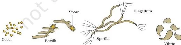
Bacteria are grouped into four categories by shape (NCERT, p. 12): the spherical Coccus (plural cocci), the rod-shaped Bacillus (plural bacilli), the comma-shaped Vibrio (plural vibrio) and the spiral Spirillum (plural spirilla), shown in Figure 2.1.
Archaebacteria
Archaebacteria specialise in the harshest habitats: extreme salty areas (halophiles), hot springs (thermoacidophiles) and marshy areas (methanogens). They differ from other bacteria in having a different cell wall structure, and this feature is what lets them survive such extremes. Methanogens live in the gut of ruminants such as cows and buffaloes and produce methane (biogas) from their dung (NCERT, p. 13).
Eubacteria
The thousands of eubacteria (‘true bacteria’) are characterised by a rigid cell wall and, if motile, a flagellum (NCERT, p. 13). They split into three nutritional camps:
- Cyanobacteria (blue-green algae) have chlorophyll a like green plants and are photosynthetic autotrophs. They may be unicellular, colonial or filamentous, and live in fresh water, marine water or on land. The colonies are wrapped in a gelatinous sheath and often form blooms in polluted water bodies. Some fix atmospheric nitrogen in specialised cells called heterocysts — Nostoc and Anabaena are the examples (NCERT, p. 13).
- Chemosynthetic autotrophs oxidise inorganic substances such as nitrates, nitrites and ammonia, using the released energy for ATP production. They play a major role in recycling nutrients such as nitrogen, phosphorus, iron and sulphur (NCERT, p. 13).
- Heterotrophic bacteria are the most abundant in nature; most are important decomposers. They help make curd from milk, produce antibiotics and fix nitrogen in legume roots; others are pathogens causing cholera, typhoid, tetanus and citrus canker (NCERT, pp. 13-14).
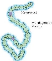
Figure 2.2 shows the filamentous form of a typical cyanobacterium — Nostoc — the shape in which these ‘blue-green algae’ build colonies and, through heterocysts, fix atmospheric nitrogen (NCERT, p. 13).
Reproduction and Mycoplasma
Bacteria reproduce mainly by fission (Figure 2.3). Under unfavourable conditions they produce spores, and they also show a sort of sexual reproduction through a primitive DNA transfer from one bacterium to another (NCERT, p. 14).
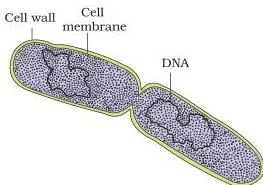
Figure 2.3 catches a bacterium mid-division — the visual for the statement that bacteria reproduce mainly by fission (NCERT, p. 14).
Mycoplasma are the outliers of the kingdom. They completely lack a cell wall, are the smallest living cells known, can survive without oxygen, and many are pathogenic in animals and plants. These are the survival facts the textbook gives — no mechanism beyond them (NCERT, p. 14).
Kingdom Protista: Five Groups in One Kingdom
Kingdom Protista takes in all single-celled eukaryotes, and the chapter is frank that the kingdom’s boundaries are not well defined — what is ‘a photosynthetic protistan’ to one biologist may be ‘a plant’ to another. Members are primarily aquatic and, being eukaryotes, their cells contain a well-defined nucleus and membrane-bound organelles; some carry flagella or cilia.
They reproduce asexually and sexually by a process involving cell fusion and zygote formation (NCERT, p. 14). Five groups are included in this book.
Chrysophytes
Chrysophytes comprise the diatoms and the golden algae (desmids). They are microscopic, float passively in water currents as plankton, and are mostly photosynthetic. The diatom’s wall is the famous part: two thin overlapping shells that fit together like a soap box, embedded with silica, making them practically indestructible.
Deposits of these walls accumulated over billions of years form diatomaceous earth — gritty, and therefore used in polishing and in the filtration of oils and syrups. Diatoms are the chief ‘producers’ in the oceans (NCERT, pp. 14-15).
Dinoflagellates
Dinoflagellates are mostly marine and photosynthetic, appearing yellow, green, brown, blue or red depending on their pigments. Their cell wall carries stiff cellulose plates, and most have two flagella — one longitudinal and one transverse, lying in a furrow between the plates.
When red dinoflagellates such as Gonyaulax multiply rapidly, the sea appears red — the red tide — and the toxins released by such numbers may kill marine animals such as fish (NCERT, p. 15).
Euglenoids
Euglenoids are mostly freshwater organisms of stagnant water. Instead of a cell wall they have a protein-rich pellicle, which makes the body flexible. They have two flagella — a short and a long one — and live a double life: photosynthetic in sunlight, but when deprived of light they behave like heterotrophs, predating on smaller organisms.
Their pigments are identical to those of higher plants. Example: Euglena (NCERT, p. 15).
Slime moulds
Slime moulds are saprophytic protists whose body moves along decaying twigs and leaves, engulfing organic material. Under suitable conditions they form an aggregation called a plasmodium, which can grow and spread over several feet. Under unfavourable conditions the plasmodium differentiates and forms fruiting bodies bearing spores at their tips.
These spores possess true walls, are extremely resistant, can survive for years even in adverse conditions, and are dispersed by air currents (NCERT, p. 15).
Protozoans
All protozoans are heterotrophs, living as predators or parasites, and they are believed to be primitive relatives of animals. The four major groups (NCERT, pp. 15-16):
- Amoeboid protozoans — move and capture prey by putting out pseudopodia (‘false feet’), as Amoeba does; marine forms carry silica shells, and Entamoeba is parasitic.
- Flagellated protozoans — free-living or parasitic, moving by flagella; Trypanosoma causes sleeping sickness.
- Ciliated protozoans — aquatic and actively moving, steered by thousands of cilia whose coordinated beating drives water laden with food into a gullet; example, Paramoecium.
- Sporozoans — have an infectious spore-like stage in their life cycle; Plasmodium, the malarial parasite, causes malaria.
Kingdom Fungi: Hyphae, Spores and the Sexual Cycle
Fungi form a unique kingdom of heterotrophic organisms. You meet them on moist bread and rotten fruit, in the mushroom you eat, and as the white spots on mustard leaves (a parasitic fungus). Unicellular yeasts make bread and beer; Penicillium gives us antibiotics; Puccinia causes wheat rust.
Fungi prefer warm, humid places — which is exactly why we keep food in the refrigerator (NCERT, p. 16).
Body structure and nutrition
Except the unicellular yeasts, fungi are filamentous. Their bodies consist of long, slender threads called hyphae; the network of hyphae is the mycelium. Some hyphae are continuous tubes filled with multinucleated cytoplasm — coenocytic hyphae — while others have septae (cross-walls). Fungal cell walls are composed of chitin and polysaccharides (NCERT, p. 16).
Most fungi are heterotrophic and absorb soluble organic matter from dead substrates — they are saprophytes. Those that depend on living plants and animals are parasites. Others live as symbionts: with algae as lichens and with plant roots as mycorrhiza (NCERT, p. 16).
Reproduction and the sexual cycle
Fungal reproduction runs along three routes (NCERT, pp. 16-17):
- Vegetative — by fragmentation, fission and budding.
- Asexual — by spores called conidia, sporangiospores or zoospores.
- Sexual — by oospores, ascospores and basidiospores, produced in distinct fruiting bodies.
The sexual cycle always passes through three steps (NCERT, p. 17):
- Plasmogamy — fusion of the protoplasms of two motile or non-motile gametes.
- Karyogamy — fusion of the two nuclei.
- Meiosis in the zygote, producing haploid spores.
The dikaryon is the subtle step in this cycle. In some fungi the fusion of two haploid cells immediately produces a diploid (2n) cell. But in ascomycetes and basidiomycetes an intervening stage occurs first — a cell with two nuclei per cell (n+n) — called a dikaryon, and the phase is the dikaryophase.
Only later do the parental nuclei fuse and the cells become diploid (NCERT, p. 17).
The four classes of fungi
The kingdom is divided into classes on the basis of mycelium morphology, mode of spore formation and fruiting bodies (NCERT, p. 17). The full comparison appears in the comparison-tables section; the essential lines are here:
- Phycomycetes — live in aquatic habitats, on decaying wood in moist, damp places, or as obligate parasites on plants. Mycelium is aseptate and coenocytic; asexual reproduction is by motile zoospores or non-motile aplanospores, produced endogenously in a sporangium; a zygospore forms by fusion of two gametes. Examples: Mucor, Rhizopus (the bread mould), Albugo (parasitic on mustard) (NCERT, p. 17).
- Ascomycetes (sac fungi) — mostly multicellular, rarely unicellular (yeast, Saccharomyces). Mycelium is branched and septate; asexual conidia form exogenously on conidiophores; sexual ascospores form endogenously in sac-like asci (singular ascus), arranged in fruiting bodies called ascocarps. Neurospora is used extensively in biochemical and genetic work, and morels and truffles are edible delicacies. Examples: Penicillium, Aspergillus, Claviceps (NCERT, pp. 17-18).
- Basidiomycetes — the mushrooms, bracket fungi and puffballs. Mycelium is branched and septate; asexual spores are generally absent (vegetative reproduction by fragmentation is common); plasmogamy happens by fusion of vegetative cells of different strains, and karyogamy and meiosis take place in the basidium, producing four basidiospores exogenously. The basidia are arranged in fruiting bodies called basidiocarps. Examples: Agaricus (mushroom), Ustilago (smut), Puccinia (rust) (NCERT, p. 18).
- Deuteromycetes (‘imperfect fungi’) — so called because only their asexual or vegetative phases are known; they reproduce only by conidia, and the mycelium is septate and branched. When their sexual stages were found, they were moved to ascomycetes or basidiomycetes. Many are decomposers of litter and help in mineral cycling. Examples: Alternaria, Colletotrichum, Trichoderma (NCERT, p. 18).
Kingdom Plantae and Kingdom Animalia: The Two Familiar Kingdoms
Kingdom Plantae includes all eukaryotic chlorophyll-containing organisms. A few members are partially heterotrophic: the insectivorous bladderwort and Venus fly trap, and the parasite Cuscuta (amarbel). Plant cells have chloroplasts and a cellulose cell wall, and the kingdom spans algae, bryophytes, pteridophytes, gymnosperms and angiosperms.
The plant life cycle shows alternation of generations — the diploid sporophytic and haploid gametophytic phases alternate, though their lengths and whether they are free-living differ between groups. The kingdom is developed fully in Chapter 3 (NCERT, p. 19).
Kingdom Animalia comprises heterotrophic eukaryotes that are multicellular and whose cells lack cell walls. They depend directly or indirectly on plants, digest food in an internal cavity, store food reserves as glycogen or fat, and feed by holozoic nutrition — by ingestion.
They follow a definite growth pattern into adults of definite shape and size; higher forms show elaborate sensory and neuromotor mechanisms, and most are capable of locomotion. Reproduction is sexual, by copulation followed by embryological development (NCERT, p. 19). The phylum-level treatment is Chapter 4, covered in the Animal Kingdom notes.
Viruses, Viroids, Prions and Lichens: Beyond the Five Kingdoms
Whittaker’s five kingdoms make no mention of the acellular organisms — viruses, viroids and prions — or of lichens. Viruses are excluded because they are not considered truly ‘living’ if being living means having a cell structure.
Outside a living cell a virus is an inert crystalline structure; once it infects a cell, it takes over the host’s machinery to replicate itself, killing the host. Viruses are therefore obligate parasites (NCERT, p. 19).
The discovery of viruses: a three-step timeline
| Year | Scientist | Contribution |
|---|---|---|
| 1892 | Dmitri Ivanowsky | Recognised microbes as the cause of tobacco mosaic disease; they passed through bacteria-proof filters, proving they were smaller than bacteria (NCERT, p. 20). |
| 1898 | M.W. Beijerinck | Showed that the extract of infected tobacco plants could infect healthy plants; named the new pathogen a virus and called the fluid Contagium vivum fluidum (‘infectious living fluid’) (NCERT, p. 20). |
| 1935 | W.M. Stanley | Showed that viruses could be crystallised and that the crystals consist largely of proteins (NCERT, p. 20). |
Structure and genetic material
A virus is a nucleoprotein — protein plus genetic material. That genetic material is either RNA or DNA, never both. The protein coat is the capsid, made of small subunits called capsomeres arranged in helical or polyhedral geometric forms. The type of genetic material follows the host (NCERT, p. 20):
- Plant viruses usually have single-stranded RNA.
- Animal viruses have single- or double-stranded RNA, or double-stranded DNA.
- Bacteriophages (viruses that infect bacteria) are usually double-stranded DNA viruses.
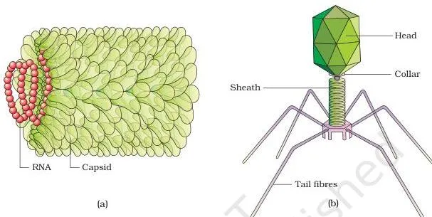
Figure 2.6 pairs the two viruses that matter most in this chapter: the Tobacco Mosaic Virus, the plant virus of Ivanowsky’s 1892 discovery, and a bacteriophage, the bacterial virus that is usually double-stranded DNA (NCERT, p. 20).
In humans, viruses cause mumps, smallpox, herpes, influenza and AIDS. In plants their symptoms are mosaic formation, leaf rolling and curling, yellowing and vein clearing, dwarfing and stunted growth (NCERT, p. 20).
Viroids, prions and lichens
- Viroids — discovered by T.O. Diener in 1971 as the cause of potato spindle tuber disease. A viroid is a free RNA of low molecular weight, lacking the protein coat that viruses have (NCERT, p. 21).
- Prions — infectious agents consisting of abnormally folded protein, similar in size to viruses. They cause bovine spongiform encephalopathy (BSE), commonly called mad cow disease in cattle, and its analogous variant Creutzfeldt-Jakob disease (CJD) in humans (NCERT, p. 21).
- Lichens — symbiotic (mutually useful) associations between an alga and a fungus. The algal partner is the phycobiont (autotrophic); the fungal partner is the mycobiont (heterotrophic). Algae prepare food for fungi; fungi provide shelter and absorb mineral nutrients and water for the algae. The association is so close that the two look like a single organism. Lichens are good pollution indicators — they do not grow in polluted areas (NCERT, p. 21).
Are viruses living? An evidence audit
Both sides of the argument rest on real observations. Here is the chapter’s own evidence, laid out as a balance sheet (NCERT, pp. 19-20):
| Evidence for ‘living’ | Evidence for ‘non-living’ |
|---|---|
| Replicates inside the host by taking over its machinery | Inert crystalline structure outside the living cell |
| Carries genetic material — RNA or DNA | Has no cell structure |
| Acts as an obligate parasite that kills the host | Can be crystallised; the crystals consist largely of proteins |
Verdict: the textbook settles the question by definition. If ‘living’ means having a cell structure, viruses are not truly living — they lack cell structure and sit inert outside a host — which is exactly why they find no place in the five kingdoms.
It is why the chapter itself closes the topic with the question: “Would you call viruses living or non-living?”
Key Terms Defined: From Heterocysts to Phycobiont
The chapter reuses one fixed vocabulary. This glossary compresses it into the two things you need per term — the meaning and a concrete example from the chapter.
| Term | Meaning | Example from the chapter |
|---|---|---|
| Autotroph | Makes its own food from inorganic substrates | Photosynthetic and chemosynthetic bacteria |
| Heterotroph | Depends on other organisms or dead organic matter for food | Most bacteria; all fungi |
| Saprophyte | Heterotroph that absorbs soluble organic matter from dead substrates | Mucor, Rhizopus |
| Parasite | Organism that lives on living plants or animals | Albugo (on mustard), Entamoeba |
| Symbiont | Partner in a mutually useful association | Alga in a lichen; mycorrhizal fungus on roots |
| Halophile | Archaebacterium of extreme salty areas | No genus named in the chapter |
| Thermoacidophile | Archaebacterium of hot springs | No genus named in the chapter |
| Methanogen | Archaebacterium that produces methane; lives in marshy areas and ruminant guts | Gut archaea of cows and buffaloes |
| Heterocyst | Specialised cyanobacterial cell that fixes atmospheric nitrogen | Nostoc, Anabaena |
| Plankton | Microscopic organisms floating passively in water currents | Diatoms |
| Diatomaceous earth | Billion-year deposits of silica diatom walls | Used in polishing; filtering oils and syrups |
| Red tide | Sea water reddened by rapid multiplication of red dinoflagellates | Gonyaulax; toxins can kill fish |
| Pellicle | Protein-rich flexible layer that replaces a cell wall | Euglena |
| Plasmodium | Slime-mould aggregation that creeps and spreads over surfaces | Slime moulds in suitable conditions |
| Pseudopodia | ‘False feet’ used for movement and capturing prey | Amoeba |
| Hypha / mycelium | Thread-like unit of the fungal body / the network of hyphae | Rhizopus bread mould |
| Coenocytic hypha | Continuous tube with many nuclei and no cross-walls | Phycomycetes |
| Dikaryon | Fungal cell with two nuclei per cell (n+n) before they fuse | Dikaryophase of ascomycetes, basidiomycetes |
| Plasmogamy / karyogamy | Fusion of protoplasms / fusion of nuclei | First two steps of the fungal sexual cycle |
| Conidia | Asexual spores formed exogenously on conidiophores | Ascomycetes, deuteromycetes |
| Ascus / basidium | Sac in which ascospores form / club on which basidiospores form | Ascomycetes / basidiomycetes |
| Capsid / capsomere | Protein coat of a virus / its structural subunits | TMV, bacteriophage |
| Viroid | Infectious free RNA of low molecular weight; no protein coat | Potato spindle tuber agent (Diener, 1971) |
| Prion | Infectious agent of abnormally folded protein, virus-sized | BSE in cattle; CJD in humans |
| Phycobiont / mycobiont | Algal / fungal partner of a lichen | Both partners in any lichen |
| Alternation of generations | Diploid sporophytic and haploid gametophytic phases alternating | Life cycle of plants |
Comparison Tables: Fungi, Protists and Acellular Agents
Biological Classification is a descriptive chapter, so it has no formulas — its ‘formula box’ is comparison tables. These three, plus the spore memory table and the mnemonics, are the fastest revision tools in the chapter.
Four classes of fungi compared
This table is the building block for any comparative answer on Kingdom Fungi (built from NCERT, pp. 17-18):
| Class | Mycelium | Asexual spores | Sexual spores and fruiting body | Examples |
|---|---|---|---|---|
| Phycomycetes | Aseptate, coenocytic | Zoospores (motile) or aplanospores (non-motile), endogenous in sporangia | Zygospore from fusion of two gametes | Mucor, Rhizopus, Albugo |
| Ascomycetes | Branched, septate | Conidia, exogenous on conidiophores | Ascospores endogenous in asci; asci in ascocarps | Penicillium, yeast (Saccharomyces), Aspergillus, Claviceps, Neurospora |
| Basidiomycetes | Branched, septate | Generally absent; vegetative reproduction by fragmentation | Four basidiospores exogenous on basidia; basidia in basidiocarps | Agaricus, Ustilago, Puccinia |
| Deuteromycetes | Branched, septate | Conidia only | Sexual stage unknown; moved to Ascomycetes or Basidiomycetes when found | Alternaria, Colletotrichum, Trichoderma |
Five groups of Protista compared
| Group | Key feature | Nutrition | Example(s) | Notable fact |
|---|---|---|---|---|
| Chrysophytes | Silica-embedded diatom shells like a soap box | Mostly photosynthetic | Diatoms, desmids | Source of diatomaceous earth; chief ocean producers |
| Dinoflagellates | Stiff cellulose plates; two flagella (longitudinal + transverse) | Photosynthetic, mostly marine | Gonyaulax | Red tides; toxins may kill fish |
| Euglenoids | Protein-rich pellicle makes the body flexible | Photosynthetic in light; heterotrophic in darkness | Euglena | Pigments identical to higher plants |
| Slime moulds | Plasmodium aggregation; fruiting bodies with spore-bearing tips | Saprophytic | — | Resistant true-walled spores spread by air |
| Protozoans | Four subgroups: amoeboid, flagellated, ciliated, sporozoans | Heterotrophic — predators or parasites | Amoeba, Entamoeba, Trypanosoma, Paramoecium, Plasmodium | Plasmodium causes malaria |
Viruses vs viroids vs prions
| Agent | Discovered by / year | Nature of the agent | Protein coat | Example disease |
|---|---|---|---|---|
| Virus | Ivanowsky (1892); crystallised by Stanley (1935) | Nucleoprotein; RNA or DNA, never both | Present — capsid of capsomeres | Mumps, smallpox, herpes, influenza, AIDS |
| Viroid | Diener (1971) | Free RNA of low molecular weight | Absent | Potato spindle tuber disease |
| Prion | Identified in modern medicine | Abnormally folded protein, virus-sized | The agent itself is protein | BSE (‘mad cow disease’) in cattle; CJD in humans |
Spore types memory table
Spores are the easiest fungal facts to mix up. Link each spore to its class and to whether it forms inside or outside the parent structure (NCERT, pp. 17-18):
| Spore | Fungal class | Formed inside or outside? |
|---|---|---|
| Zoospores (motile), aplanospores (non-motile) | Phycomycetes | Endogenous — inside the sporangium |
| Conidia | Ascomycetes, Deuteromycetes | Exogenous — on conidiophores |
| Ascospores | Ascomycetes | Endogenous — inside asci |
| Basidiospores (four per basidium) | Basidiomycetes | Exogenous — on basidia |
Mnemonics for this chapter
- Five kingdoms: ‘My Pretty Flowers Please Animals’ → Monera, Protista, Fungi, Plantae, Animalia.
- Four fungal classes: ‘Please Always Be Dexterous’ → Phycomycetes, Ascomycetes, Basidiomycetes, Deuteromycetes.
- Four protozoan groups: ‘All Fools Can Sing’ → Amoeboid, Flagellated, Ciliated, Sporozoans.
Worked Example: Classifying an Organism Step by Step
Method: Whittaker’s five criteria work as a decision key — ask about cell structure, body organisation, mode of nutrition, reproduction and phylogenetic relationships, in that order, and each answer narrows the kingdom.
The five-question decision key
- Is the cell prokaryotic or eukaryotic? Prokaryotic → Monera. Eukaryotic → one of Protista, Fungi, Plantae, Animalia.
- Is the body unicellular or multicellular? A single-celled eukaryote → Protista (or yeast, if fungal); multicellular → Fungi, Plantae or Animalia.
- What is the mode of nutrition? Autotrophic → Monera, Protista or Plantae; saprophytic or parasitic (heterotrophic) → Fungi; holozoic ingestion → Animalia.
- What is the cell wall? Chitin → Fungi; cellulose → Plantae; noncellulosic → Monera; silica-embedded, pellicle-like or none → Protista; no wall → Animalia (or Mycoplasma in Monera).
- What are the reproductive details? Spore types, fruiting bodies, heterocysts, flagella and the steps of the sexual cycle pin down the group — and often the genus.
Case (a): A unicellular, prokaryotic organism forming filamentous colonies in a gelatinous sheath, with heterocysts
- Step 1: The cell is prokaryotic (no nuclear membrane) → the organism belongs to Monera (criterion: cell type).
- Step 2: Filamentous colonies wrapped in a gelatinous sheath → the description matches cyanobacteria among eubacteria.
- Step 3: Heterocysts are the specialised nitrogen-fixing cells described only for cyanobacteria such as Nostoc and Anabaena (criterion: reproduction).
Verdict: Kingdom Monera — a cyanobacterium such as Nostoc or Anabaena.
Case (b): A unicellular eukaryote with a flexible protein-rich pellicle and two flagella, photosynthetic in light but predatory in darkness
- Step 1: Eukaryotic and single-celled → Protista (criteria: cell type + body organisation).
- Step 2: A pellicle in place of a cell wall is the signature of euglenoids — no other protist group has it (criterion: wall composition).
- Step 3: Two flagella (short + long) and dual nutrition — photosynthetic in sunlight, heterotrophic when deprived of it — complete the match with Euglena.
Verdict: Kingdom Protista — an euglenoid such as Euglena.
Case (c): A septate fungus with no asexual spores, plasmogamy by fusion of vegetative cells, and four exogenously formed basidiospores
- Step 1: Septate, branched mycelium → narrows the field to Ascomycetes, Basidiomycetes or Deuteromycetes (criterion: body organisation / mycelium type).
- Step 2: Asexual spores generally absent, with plasmogamy by fusion of vegetative cells of different strains → only Basidiomycetes fit (criterion: reproduction).
- Step 3: Karyogamy and meiosis in a basidium, yielding four basidiospores exogenously, confirms Basidiomycetes — the mushroom group.
Verdict: Kingdom Fungi — class Basidiomycetes, of the Agaricus (mushroom) type.
Case (d): An acellular infectious agent consisting of low molecular weight RNA with no protein coat, causing potato spindle tuber disease
- Step 1: No cell at all → cannot belong to any of Whittaker’s five kingdoms (criterion: cell type).
- Step 2: Free RNA with no protein coat rules out a virus — viruses are nucleoproteins wrapped in a capsid.
- Step 3: Low molecular weight RNA plus the specific disease points to the viroid discovered by Diener in 1971.
Verdict: A viroid — an acellular agent outside the five kingdoms.
Common Mistakes in Biological Classification
| Students write… | Correct is… | Why / how to check |
|---|---|---|
| ‘Blue-green algae are plants.’ | Cyanobacteria are prokaryotes in Kingdom Monera. | They have chlorophyll a, but they lack a nuclear membrane — check the cell type before calling anything a plant. |
| ‘All fungi are multicellular.’ | Yeasts are unicellular. | The textbook’s own exception: with the exception of yeasts, fungi are filamentous. |
| ‘Dikaryon means diploid.’ | A dikaryon is n+n — two haploid nuclei per cell. | Diploid (2n) appears only after the two parental nuclei fuse; in a dikaryon they have not fused. |
| ‘Diatom walls are cellulosic, like a plant’s.’ | Diatom walls are embedded with silica. | Silica makes them indestructible, which is why the deposits (diatomaceous earth) are gritty. |
| ‘A virus contains both RNA and DNA.’ | A virus contains either RNA or DNA, never both. | A virus is a nucleoprotein with one type of genetic material; check the host rule (plant viruses usually ssRNA). |
| ‘Slime moulds are fungi.’ | Slime moulds are saprophytic protists with a plasmodium stage. | They are single-celled eukaryotes that aggregate and creep; fungi never form a plasmodium. |
| ‘Chitin is a plant cell wall material.’ | Plant walls are cellulosic; fungal walls have chitin. | This wall-composition difference is exactly why fungi were removed from Plantae. |
Exam Notes: How to Write Complete Answers
These are answer skeletons for the chapter’s own exercises — the structure that makes an answer complete without copying the textbook (exercises at NCERT, p. 22).
- Exercise Q1 (how classification systems changed): build a chronological chain — Aristotle’s morphological characters → the two kingdom system and its inadequacies → the demand for new criteria → Whittaker’s five kingdoms and his five criteria → the regrouping that resulted. Name Chlamydomonas and Paramoecium as proof that the criteria changed.
- Exercise Q8 (phycobiont and mycobiont): open with one-line definitions — the algal partner (phycobiont) is autotrophic, the fungal partner (mycobiont) is heterotrophic — then state what each provides: algae make food; fungi provide shelter and absorb water and minerals.
- Exercise Q9 (comparative account of the classes of fungi): the fastest structure is the fungal classes table on this page — mycelium, asexual spores, sexual spores and fruiting body, examples — one row per class.
- Exercise Q12 (are viruses living or non-living?): take a clear position first, marshal the chapter’s evidence on both sides from the audit table above, then close with the definitional point — viruses lack cell structure and are inert outside a host, so they are not considered truly living.
A quick map of the remaining exercises (NCERT, p. 22):
| Exercise | What a complete answer must include |
|---|---|
| Q2 — economic uses | Heterotrophic bacteria: curd from milk, antibiotics, nitrogen fixation in legume roots. Archaebacteria: methane (biogas) from dung. |
| Q3 — diatom cell walls | Silica-embedded walls in two overlapping shells, like a soap box. |
| Q4 — algal bloom and red tides | Algal bloom: cyanobacteria multiplying in polluted water. Red tide: rapid multiplication of red dinoflagellates such as Gonyaulax, whose toxins may kill fish. |
| Q5 — viroids vs viruses | Viroid: free RNA, no protein coat, low molecular weight. Virus: nucleoprotein with a capsid, containing RNA or DNA (never both). |
| Q6 — four groups of Protozoa | Amoeboid, flagellated, ciliated, sporozoans — one feature and one named example each. |
| Q7 — partially heterotrophic plants | Bladderwort and Venus fly trap (insectivorous), Cuscuta (parasite) — photosynthetic plants that supplement nutrition. |
| Q10 — characteristic features of euglenoids | Pellicle, two flagella (short and long), photosynthetic in light but heterotrophic in darkness, pigments identical to higher plants. |
| Q11 — viruses: structure and four diseases | Nucleoprotein; capsid of capsomeres in helical or polyhedral forms; RNA or DNA (never both); name four of mumps, smallpox, herpes, influenza, AIDS. |
Across every answer, name the organisms — Nostoc, Gonyaulax, Trypanosoma, Plasmodium, Puccinia, Neurospora — because a complete descriptive answer in this chapter needs named examples, not just group names. When you want the textbook’s exact phrasing of any definition, keep the official NCERT PDF of Chapter 2, Biological Classification open beside these notes.
For other subjects, the Class 11 notes index groups every chapter of the year, and the CBSE notes library carries the same one-page revision format for every class.
Revision Summary: The Chapter in One Page
- History: Aristotle (morphology) → two kingdom system (Plantae, Animalia) → Whittaker’s five kingdoms (1969). Five criteria: cell structure, body organisation, mode of nutrition, reproduction, phylogenetic relationships (NCERT, pp. 10-11).
- Monera: four shapes — coccus, bacillus, vibrio, spirillum. Archaebacteria: halophiles, thermoacidophiles, methanogens (different wall → survival in extremes; biogas from dung). Eubacteria: cyanobacteria (chlorophyll a, heterocysts — Nostoc, Anabaena), chemosynthetic autotrophs, heterotrophs (curd, antibiotics, legume nitrogen; cholera, typhoid, tetanus, citrus canker). Reproduction: fission, spores, DNA transfer. Mycoplasma: no wall, smallest living cells, survive without oxygen (NCERT, pp. 12-14).
- Protista: single-celled eukaryotes, mostly aquatic. Chrysophytes (diatoms, silica walls, diatomaceous earth), dinoflagellates (cellulose plates, red tides — Gonyaulax), euglenoids (pellicle — Euglena), slime moulds (plasmodium, resistant spores), protozoans — amoeboid (pseudopodia), flagellated (Trypanosoma), ciliated (Paramoecium), sporozoans (Plasmodium) (NCERT, pp. 14-16).
- Fungi: hyphae and mycelium; chitin walls; saprophytes, parasites, symbionts (lichens, mycorrhiza). Sexual cycle: plasmogamy → karyogamy → meiosis. Dikaryon (n+n). Classes: Phycomycetes (zoospores/aplanospores in sporangia — Mucor, Rhizopus, Albugo), Ascomycetes (conidia exogenous, ascospores in asci — Penicillium, yeast, Neurospora), Basidiomycetes (four exogenous basidiospores on basidia — Agaricus, Ustilago, Puccinia), Deuteromycetes (only conidia known — Alternaria, Trichoderma) (NCERT, pp. 16-18).
- Plantae: chlorophyll-containing eukaryotes, cellulose walls, alternation of generations; partially heterotrophic members — bladderwort, Venus fly trap, Cuscuta (NCERT, p. 19).
- Animalia: heterotrophic, multicellular, no cell walls, holozoic nutrition, glycogen or fat reserves (NCERT, p. 19).
- Beyond the kingdoms: viruses (nucleoprotein; RNA or DNA, never both; capsid of capsomeres; TMV, bacteriophage), viroids (free RNA, no coat — Diener 1971), prions (abnormally folded protein — BSE, CJD), lichens (phycobiont + mycobiont; pollution indicators) (NCERT, pp. 19-21).
Frequently Asked Questions on Biological Classification
Why did the two kingdom classification fail?
Because two kingdoms could not cope with the variety of life. The system put everything into either Plantae or Animalia, but it did not separate eukaryotes from prokaryotes, unicellular from multicellular organisms, or photosynthetic from non-photosynthetic organisms — and a large number of organisms fitted neither kingdom (NCERT, p. 10).
That failure pushed classification towards new criteria: cell structure, nature of the wall, nutrition, habitat, reproduction and evolutionary relationships.
Are viruses living or non-living?
The chapter treats them as not truly living, though it invites debate. Viruses lack cell structure and are inert crystalline particles outside a living cell; once inside, they take over the host’s machinery to replicate and kill the host (NCERT, pp. 19-20).
Because ‘living’ is defined here as having a cell structure, viruses are not considered truly living and get no place in the five kingdoms.
How are viroids different from viruses?
A viroid is a free RNA agent with no protein coat, while a virus is a nucleoprotein — genetic material protected by a capsid. Viroids are also smaller than viruses, and their RNA is of low molecular weight (NCERT, p. 21). Diener discovered them in 1971 as the cause of potato spindle tuber disease.
Why are fungi not placed with plants even though they have cell walls?
Because the similarity stops at the wall. Fungi are heterotrophic — saprophytic or parasitic — not photosynthetic like plants, and fungal walls contain chitin while plant walls are cellulosic (NCERT, pp. 11-12). This wall-composition difference, along with nutrition, justified a separate Kingdom Fungi.
What causes red tides and why are they harmful?
Red tides are caused by the rapid multiplication of red dinoflagellates such as Gonyaulax, which makes the sea appear red. The toxins released by such enormous numbers can kill other marine animals such as fish (NCERT, p. 15).
What is the difference between a dikaryon and a diploid cell?
A dikaryon is a cell with two haploid nuclei per cell — n+n — in which the nuclei have not fused; a diploid cell has 2n, meaning the two nuclei have fused into one. In ascomycetes and basidiomycetes the dikaryotic stage (dikaryophase) sits between plasmogamy and karyogamy, and the diploid condition appears only after the parental nuclei fuse (NCERT, p. 17).
Reference: NCERT Class 11 Biology textbook, chapter Biological Classification.
Explore Class 11 Biology Notes
- Class 11 Biology Notes
- Class 11 CBSE Notes
- CBSE Notes for Classes 1 to 12
- Next: Animal Kingdom
Class 11 Biology on LearnCBSE:
Related chapters:
- Breathing and Exchange of Gases notes
- Excretory Products and Their Elimination notes