
The Basic Processes Class 11 chapter is Chapter 7 of the NCERT Biotechnology textbook for Class 11. It runs from NCERT page 165 to about page 215 and explains how DNA was proved to be the genetic material, and how genes are organised, copied, expressed, mutated, repaired and regulated.
The official NCERT PDF of the chapter is on this page. Below it you will find a reading map of its nine sections, the key concepts with page references, and a guide to what the seven exercises ask.
| What the chapter holds | Count | Where it is used |
|---|---|---|
| Printed pages | 51 | |
| Sections in the chapter | 19 | |
| Figures with NCERT captions | 45 | |
| Tables | 1 | |
| Exercise questions | 7 | answered in our NCERT Solutions |
| Official NCERT PDF | Download the chapter PDF | the chapter exactly as NCERT publishes it |
Basic Processes Class 11 PDF: Official NCERT Chapter 7
The official edition of this chapter is published by NCERT and is free to download. It is the same text, figures and exercises that appear in the printed Class 11 Biotechnology book — nothing is abridged.
Open the NCERT Class 11 Biotechnology Chapter 7 Basic Processes PDF when you want to read the full chapter text away from this page or print it for revision; the file is kebt107.pdf on the NCERT website.
What is inside the Basic Processes chapter
The table below shows what the chapter file contains: its sections, its figures and its end-of-chapter exercise questions.
What Basic Processes teaches and in what order
The chapter is arranged as a chain of nine numbered sections, each answering one question before the next builds on it.
| Section | NCERT pages | The question it answers |
|---|---|---|
| 7.1 DNA as the genetic material | 165-171 | How was DNA proved to be the molecule of heredity? |
| 7.2 Prokaryotic and eukaryotic gene organisation | 171-176 | How is a very long DNA molecule packed into a tiny cell? |
| 7.3 DNA replication | 176-184 | How is DNA copied before a cell divides? |
| 7.4 Gene expression | 185-191 | How is the information in a gene transferred to RNA? |
| 7.5 Genetic code | 192-193 | How do three-letter codons specify amino acids? |
| 7.6 Translation | 194-199 | How is an mRNA sequence decoded into a polypeptide chain? |
| 7.7 Gene mutation | 200-205 | How and why does the DNA sequence change? |
| 7.8 DNA repair | 205-208 | How do cells correct those changes and keep mutation rate low? |
| 7.9 Regulation of gene expression | 209-214 | How does a cell switch genes on and off? |
| Summary and exercises | 215-216 | A condensed recap of the chapter, plus seven questions. |
The order matters. Section 7.1 establishes the evidence that DNA is the genetic material; 7.2 and 7.3 show how that material is organised and copied; 7.4 to 7.6 explain how its information is read out as RNA and protein.
The chapter also assumes two earlier ideas from this book: the structure of DNA, described in Chapter 3, and genes carried on chromosomes from the previous chapter.
Key concepts in Basic Processes, explained
The sections below work through the chapter’s main ideas in the order the book presents them. Each key term gets a plain-language explanation with the NCERT page where the book discusses it, so you can follow along in the textbook.
How DNA was proved to be the genetic material
This is a historical question answered by logic: scientists could not see the genetic material, so they had to destroy candidate molecules one by one and watch what happened. Three experiments form the evidence chain.
Johann Friedrich Miescher first isolated DNA from the nuclei of pus cells in 1869, calling it nuclein. But it took the three experiments below to show that this molecule, not protein, carries heredity (NCERT, p. 167).
| Experiment | What was done | What it proved | NCERT page |
|---|---|---|---|
| Griffith, 1928 | Live S strain killed mice; live R strain did not; heat-killed S did not; but heat-killed S mixed with live R killed mice, and live S bacteria were recovered from them | Something from dead S bacteria — a ‘transforming principle’ — changes harmless R bacteria into virulent S bacteria. Its nature was still unknown | p. 168 |
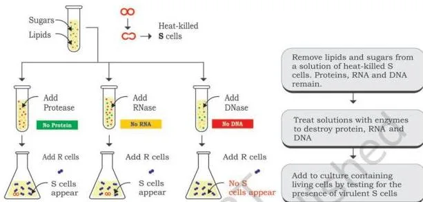
| Avery, MacLeod and McCarty, 1944 | An extract of heat-killed S bacteria, cleared of lipids and carbohydrates, was treated with RNase, DNase or protease, and each extract was tested for its power to transform R bacteria | Only the DNase-treated extract failed to transform. Destroying DNA destroys the transforming effect, so DNA is the transforming principle | p. 169 |
| Hershey and Chase, 1952 | T2 phages were grown in \(^{32}\text{P}\) and \(^{35}\text{S}\) media to label DNA and protein; infected E. coli was blended and centrifuged | \(^{32}\text{P}\) (DNA) was found in the bacterial pellet; \(^{35}\text{S}\) (protein) stayed in the supernatant. DNA enters the cell, protein does not | p. 170 |
All three share one logic: destroy the molecule, destroy the effect. Avery’s team destroyed DNA with DNase and transformation stopped; Hershey and Chase removed the protein coat and infection still produced new phages (NCERT, p. 170).
Gene organisation: prokaryotes compared with eukaryotes
Every cell faces the same packing problem: its DNA is far longer than the cell itself. Prokaryotes and eukaryotes solved it differently, and the difference shapes how their genes are built.
Prokaryotes keep one circular, double-stranded DNA in the nucleoid. Extra non-essential loops, called plasmids, may sit alongside it (NCERT, p. 171).
Packing happens by supercoiling — the DNA double helix is twisted on itself like a rubber band coiled into a ball. Twisting against the helix direction gives negative supercoiling; with it, positive supercoiling. Most bacterial genomes are negatively supercoiled during normal growth (NCERT, p. 171).
The most abundant protein in the bacterial nucleoid is HU, a histone-like protein that helps condense the DNA (NCERT, p. 172).
Eukaryotes pack linear DNA around histones — basic proteins. Eight histones (two each of H2A, H2B, H3, H4) form the core octamer; DNA wraps around it to make a nucleosome (NCERT, p. 172).
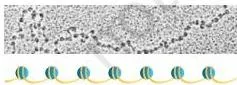
Nucleosomes are linked by short linker DNA, with one H1 histone per linker. Under a microscope they look like beads on a string — 10 nm beads packed into a 30 nm fibre, folded into 300 nm loops, and condensed to about 700 nm at metaphase (NCERT, pp. 172-173).
The book makes a neat historical point: when beads on a string were first seen in the 1940s, each bead was assumed to be a gene. Later work showed a bead is a nucleosome covering about 200 bp of DNA.
That is far too small to be a gene — a protein of 100 amino acids already needs a gene of about 300 nucleotides (NCERT, pp. 173-174).
Genome vocabulary: 1000 bp = 1 kb and 1000 kb = 1 Mb. The human genome is about 3,000 Mb, with more than 20,000 genes — only about 2% of the sequence codes for proteins, and most of a eukaryotic genome remains unexpressed (NCERT, pp. 174-175).
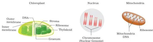
Eukaryotes also carry organellar genomes in mitochondria and chloroplasts: circular DNA in multiple copies, replicating semiconservatively and inherited mostly through the female gamete (NCERT, p. 176).
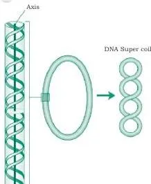
The supercoiling diagram (Fig 7.4) shows the idea behind the rubber band analogy: the helix’s own axis becomes coiled, which is how a circular chromosome several times the cell’s length fits inside it (NCERT, p. 171).

Fig 7.5 is the packaging ladder you should be able to reproduce: double helix → nucleosome beads on a string (10 nm) → 30 nm fibre → 300 nm loops → condensed chromosome (about 700 nm at metaphase). The caption carries an important fact: one chromosome is one DNA molecule, just tightly wrapped (NCERT, p. 172).
DNA replication: the evidence and the machinery
Before a cell divides, it must produce an exact second copy of its DNA. The chapter first proves how the copy is made, then names every enzyme that does the work.
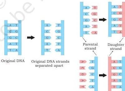
Watson and Crick’s structure immediately suggested the mechanism: because bases pair specifically (A with T, C with G), each strand can serve as a template for a new complementary strand. That would make replication semiconservative — each daughter duplex keeps one old strand and one new one (NCERT, p. 176).
Mathew Meselson and Franklin Stahl proved this in 1958 with the nitrogen isotopes \(^{14}\text{N}\) and \(^{15}\text{N}\). They grew E. coli in heavy \(^{15}\text{N}\) medium, switched the cells to light \(^{14}\text{N}\) medium, and spun the DNA in a cesium chloride density gradient — a centrifuge technique that separates DNA molecules by density (NCERT, pp. 177-178).
DNA sinks or floats in the gradient until its density matches the salt around it, so it collects as a sharp band. Heavy \(^{15}\text{N}\) DNA settles low in the tube, light \(^{14}\text{N}\) DNA stays near the top, and hybrid DNA — one heavy strand and one light strand — stops between them.
Step 1 — the bands you already know: Generation I (one round in \(^{14}\text{N}\) medium) gives a single intermediate band, because every molecule is hybrid.
Generation II gives two bands of equal intensity: half hybrid, half all-light.
Step 2 — think in molecules, not generations: each hybrid molecule replicates into one hybrid and one light molecule; each light molecule makes two light molecules.
Step 3 — the third generation: start from generation II’s 2 hybrid + 2 light DNA molecules.
The 2 hybrids produce 2 hybrid + 2 light; the 2 light molecules double to 4 light.
Total: 2 hybrid + 6 light — a ratio of 3 light molecules for every hybrid.
Predicted pattern: the intermediate band remains, and the light band is about three times as thick — which is exactly why the book says the light band becomes progressively thicker while the intermediate band stays unchanged (NCERT, pp. 178-179).
Taylor and colleagues reached the same conclusion independently in 1958, using radioactive thymidine on the chromosomes of Vicia faba (NCERT, p. 179).
The machinery table below collects the enzyme team. Replication needs more than polymerase — and Exercise 3 at the end of the chapter asks what happens when all these proteins disappear.
| Enzyme or protein | Its job in replication | NCERT pages |
|---|---|---|
| DNA polymerase III | The main replication enzyme; synthesises new DNA \(5′ \rightarrow 3’\) and proofreads with \(3′ \rightarrow 5’\) exonuclease activity | pp. 179-181 |
| DNA polymerase I | Removes RNA primers with \(5′ \rightarrow 3’\) exonuclease activity and replaces them with DNA | pp. 179-181 |
| DNA polymerase II | Involved in repair of DNA | pp. 179-181 |
| Primase | A DNA-dependent RNA polymerase that makes a short RNA primer (about 10-12 nucleotides) providing the \(3’\) OH group polymerase needs | pp. 179-181 |
| Helicase | Breaks the hydrogen bonds between strands, using ATP, to unwind the helix | pp. 179-181 |
| Topoisomerase | Relieves torsional strain ahead of the fork by nicking and religating the DNA | pp. 179-181 |
| SSB proteins | Bind to exposed single strands and stop them reannealing | pp. 179-181 |
| DNA ligase | Seals nicks between newly made fragments by forming phosphodiester bonds | pp. 179-181 |
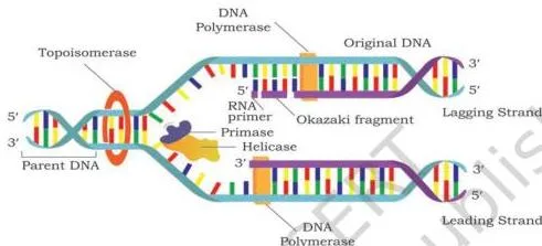
Replication begins at the origin of replication (oriC in E. coli) and moves in both directions, so the two forks are bidirectional. Synthesis is always \(5′ \rightarrow 3’\), which creates the leading and lagging strands (NCERT, pp. 180-182).
- Leading strand: synthesised continuously toward the moving fork.
- Lagging strand: synthesised discontinuously, away from the fork, in Okazaki fragments of about 1,000-2,000 nucleotides, each starting with its own RNA primer.
- Because one strand is continuous and the other is not, replication is called semi-discontinuous (NCERT, pp. 182-183).
After the fragments are made, DNA polymerase I removes the RNA primers and replaces them with DNA, and DNA ligase joins the fragments. Termination happens at a terminus roughly opposite the origin, where the two forks meet. Eukaryotes do the same job but with several origins per chromosome and several DNA polymerases (NCERT, pp. 183-184).
Gene expression: transcription and the genetic code
Information in DNA is written in four bases; proteins are built from twenty amino acids. Gene expression is the two-step bridge between them: DNA to RNA by transcription, then RNA to protein by translation.
The central dogma states that this flow is one-way: \( \text{DNA} \rightarrow \text{RNA} \rightarrow \text{protein} \) (Fig 7.17). The chapter gives two reasons an mRNA intermediate is useful: a single gene can be amplified into many mRNA copies, and in eukaryotes the mRNA must carry the message from the nucleus to the ribosomes in the cytoplasm (NCERT, p. 185).

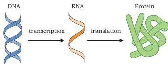
Read Fig 7.17 as two arrows and two processes: transcription copies DNA into mRNA, and translation decodes mRNA into a polypeptide chain. Retroviruses reverse the arrow — their RNA genome is copied back into DNA by reverse transcription (NCERT, p. 186).
Only one of the two DNA strands is normally copied. The template (non-coding) strand is the one RNA polymerase reads; the sense (coding) strand has the same sequence as the RNA, except that T in DNA is replaced by U in RNA (NCERT, p. 187).
RNA polymerase does not need a primer — a key difference from DNA polymerase. Prokaryotes have one enzyme (a core of \(\alpha_2 \beta \beta’\) plus a sigma factor forms the holoenzyme) that transcribes all RNAs. Eukaryotes have three: polymerase I makes rRNA, polymerase II makes hnRNA (the mRNA precursor), and polymerase III makes tRNA and 5S rRNA (NCERT, p. 187).
The transcription unit is the stretch of DNA transcribed into RNA. It has a start site, a terminator, and upstream a promoter where RNA polymerase binds. In bacteria the promoter contains the Pribnow box (TATAAT), where melting of the duplex begins (NCERT, p. 188).
- Initiation: the sigma subunit finds the promoter; about 17 bp of DNA unwind and RNA synthesis begins with no primer.
- Elongation: the polymerase moves along the template inside a transcription bubble, adding nucleotides to the \(3’\) end at about 40 nucleotides per second at 37°C.
- Termination: the enzyme reaches the terminator; some genes need a rho protein (rho dependent), others end without it (rho independent).
In prokaryotes a group of genes is often transcribed into one polycistronic mRNA, and because transcription and translation share the cytoplasm, translation can begin before transcription finishes (NCERT, pp. 188-190).
Eukaryotic genes are usually split — coding exons separated by non-coding introns. The primary transcript (hnRNA) must be processed before leaving the nucleus: capping adds a \(5’\) methyl G cap that protects against exonuclease, splicing removes introns and joins exons, and a poly-A tail is added to the \(3’\) end for stability (NCERT, pp. 190-191).
Put the two systems side by side and three differences stand out: prokaryotes use one RNA polymerase and no transcription factors, eukaryotes use three polymerases and several transcription factors; prokaryotic mRNA is used as soon as it is made, eukaryotic pre-mRNA must be capped, spliced and poly-adenylated first; and eukaryotic transcription and translation are separated by the nuclear membrane, so they cannot overlap the way they do in bacteria (NCERT, pp. 187-191).
The genetic code is the rulebook that connects codons to amino acids. Since there are 20 amino acids and only 4 bases, one base (4 codons) or two bases (16 codons) cannot be enough; three bases give \(4^3 = 64\) codons, more than enough — George Gamow’s argument (NCERT, p. 192).
The first codon was cracked by Nirenberg and Matthaei in 1961: an artificial poly-U mRNA made only the protein with radioactive phenylalanine, so UUU = phenylalanine. Poly-C gave proline (CCC) and poly-A gave lysine (AAA); Khorana, Ochoa and Leder finished the set of 64 (NCERT, pp. 192-193).
- 61 of the 64 codons code for the 20 amino acids; UAA, UAG and UGA are stop codons that code for nothing.
- AUG codes for methionine and also acts as the initiator codon.
- The code is unambiguous — one codon always picks one amino acid.
- The code is degenerate — most amino acids have more than one codon; only methionine and tryptophan have one each.
- The code is non-overlapping — each base belongs to only one codon.
- The code is universal, with a few exceptions in mitochondrial codons and some protozoans.
(NCERT, p. 193)
How to read the genetic code table
The codon table (Fig 7.25 on NCERT page 193) is a reference you must be able to use quickly. It works on one simple rule in three steps.
- Find the first base of the codon in the left column of the table.
- Find the second base along the top row.
- Find the third base in the right column; where the three meet is the amino acid.
Example: first base G, second base A, third base U gives GAU, which codes for aspartic acid. Trying C in the first position, U in the second and G in the third gives CUG, which codes for leucine.
Two details matter when you use the table: AUG is methionine plus the start signal, and the three stop codons (UAA, UAG, UGA) have no amino acid and no tRNA to read them (NCERT, p. 193).
Translation: from codons to polypeptide
Translation is the ribosome’s work: decoding the mRNA’s codon sequence into a chain of amino acids. The chapter splits it into four stages (NCERT, p. 194).
Charging prepares each amino acid for delivery. Aminoacyl-tRNA synthetase activates the amino acid with ATP in two steps and attaches it to the \(3’\) CCA end of its specific tRNA; there are 20 such synthetases, one per amino acid (NCERT, pp. 195-196).
Wobble pairing explains why the cell needs fewer than 61 tRNAs: base pairing between codon and anticodon is precise at the first two positions but flexible at the third, so one tRNA can read more than one codon (NCERT, p. 195).
Example with your own pair: the mRNA codon CCA, which codes for proline, is read by a tRNA whose anticodon pairs with it; the flexibility comes only at the third position of the codon, not at the first two.
- Prokaryotic initiation: the 30S ribosome subunit binds the Shine-Dalgarno sequence at the \(5’\) end of mRNA, placing AUG in the P site; the initiator tRNA carries formylated methionine (fMet); the 50S subunit joins to form the 70S initiation complex, helped by initiation factors and GTP (NCERT, pp. 196-198).
- Eukaryotic initiation: the 40S subunit recognises the \(5’\) cap and scans along the mRNA to the first AUG (NCERT, p. 198).
Elongation cycles in three beats: a new aminoacyl-tRNA enters the empty A site; peptidyl transferase forms the peptide bond between the P-site and A-site amino acids; translocation moves the ribosome three nucleotides along the mRNA in the \(5′ \rightarrow 3’\) direction, shifting the uncharged tRNA to the E site where it exits (NCERT, p. 198).
Termination happens when a stop codon (UAA, UAG or UGA) enters the A site. No tRNA binds these codons; release factors bind instead, and the polypeptide, tRNA, mRNA and ribosome subunits are all released. Newly made proteins then undergo post-translational modifications (NCERT, pp. 198-199).
One mRNA is usually translated by several ribosomes at once, forming a polyribosome — this is how a cell makes many copies of a protein from a single message (NCERT, p. 199).
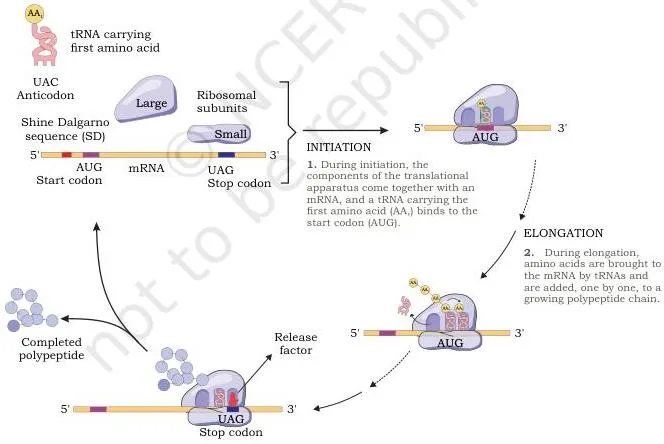
In Fig 7.29, locate the three tRNA-binding sites: A (where the new aminoacyl-tRNA arrives), P (where the growing chain is held), and E (where the emptied tRNA leaves). The diagram is the easiest way to remember the order of events in elongation (NCERT, pp. 196-197).
Mutation and DNA repair: how the code changes and how cells fix it
Replication, transcription and chromosome sorting are precise, but not perfect. Mutation is a sudden change in the genetic material, and repair systems are why changes stay rare.
- Chromosomal mutations: changes in chromosome structure (aberrations) or number (aneuploidy, polyploidy).
- Gene (point) mutations: molecular changes in DNA. Sickle cell anaemia is the chapter’s example — one nucleotide substitution produces abnormal haemoglobin.
(NCERT, pp. 200-201) At the molecular level, a gene mutation is an addition, a deletion or a substitution of one or a few nucleotides. Addition and deletion shift the reading frame, usually ruining the protein; substitution replaces a single nucleotide (NCERT, pp. 201-202).
A substitution is further classified by which base family is involved: transition swaps a purine for a purine or a pyrimidine for a pyrimidine, while transversion swaps a purine for a pyrimidine or the reverse (NCERT, p. 202).
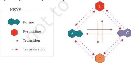
Fig 7.33 makes the distinction visual: look at whether the new base stays in the same chemical family. Same family in, that is a transition; family switch, that is a transversion (NCERT, p. 201).
Spontaneous mutations arise from the molecule’s own chemistry. A base normally exists in a keto form (C=O) or amino form (C–NH2); a hydrogen shift produces a rare enol or imino form — a tautomeric shift.
The rare imino form of guanine pairs with thymine instead of cytosine, so after two rounds of replication a \(G \equiv C\) pair becomes an \(A = T\) pair (NCERT, pp. 202-204).
Induced mutations are caused by external mutagens. Physical mutagens include X-rays, which can break phosphodiester bonds and delete nucleotides, and UV rays, which excite electrons and cause deletion or substitution. Chemical mutagens include alkylating agents (EMS, mustard gas), base analogs (5-bromouracil, 2-aminopurine) and deaminating agents (nitrous acid) (NCERT, pp. 204-205).
Follow 5-bromouracil as the chapter does: in its keto form it pairs with adenine and is incorporated into DNA; if it shifts to the enol form it pairs with guanine instead. The result is an \(A = T\) pair converted into a \(G \equiv C\) pair (NCERT, p. 205).
If mutations were really that common, heredity would be unstable. DNA repair systems keep errors low. The chapter presents excision repair and mismatch repair (NCERT, p. 206).
Base excision repair (BER) fixes a damaged single base. DNA glycosylase recognises and removes the damaged base, leaving an AP site (apurinic or apyrimidinic). An endonuclease cuts the sugar-phosphate backbone at the AP site, a polymerase adds the correct bases, and ligase seals the gap (NCERT, p. 206).
Nucleotide excision repair (NER) handles bigger damage, like the thymine dimers UV light creates. A protein complex called UVr finds the dimer (it distorts the helix), cuts the phosphodiester backbone four nucleotides downstream and eight upstream, unwinds and removes the damaged fragment, and then polymerase I and ligase restore the strand (NCERT, pp. 206-208).
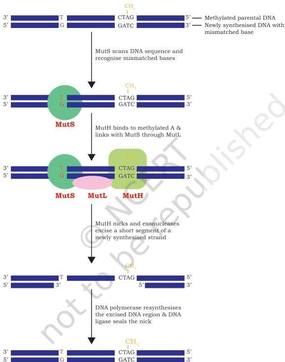
Fig 7.36(b) is the step-by-step for NER. It also connects directly to Exercise 4, which asks how UV changes DNA structure and how the cell corrects it: the distortion is the thymine dimer, and the correction is the UVr pathway you see here (NCERT, p. 207).
Mismatch repair (MMR) corrects nucleotides incorporated wrongly during replication. Proteins MutH, MutL, MutS and MutT recognise the mismatch, cut the new strand about 1,000 nucleotides from the error, and let polymerase and ligase refill and seal the gap (NCERT, p. 208).
Regulation of gene expression: the operon model
All the cells of one organism carry the same genes, yet a muscle cell and a nerve cell make different proteins. Regulation of gene expression is the switching on and off of genes so each cell makes only what it needs.
Some genes run at a constant level all the time — the housekeeping (constitutive) genes, such as those for citric acid cycle enzymes. Others are regulated, with their product levels rising and falling with the cell’s needs (NCERT, p. 209).
Regulation can act at several levels — chromatin, transcription, mRNA processing, mRNA transport, or translation. In both prokaryotes and eukaryotes, transcription initiation is a key control point, because it decides both which genes are expressed and how strongly (NCERT, p. 209).
In bacteria, genes with related jobs are clustered and transcribed together under one switch — an operon. It has structural genes (cistrons), a promoter where RNA polymerase binds, and an operator where a repressor can bind and block transcription.
The regulator gene sits upstream with its own promoter and is not part of the operon; it makes the repressor protein (NCERT, p. 211).
- Inducible operons — an inducer (usually a substrate) binds the active repressor, inactivates it, and switches the genes on.
- Repressible operons — a co-repressor (usually an end product) binds an inactive repressor, activates it, and switches the genes off.
- Negative control — the regulatory protein is a repressor that blocks transcription. Positive control — an activator stimulates transcription.
(NCERT, pp. 211-212) The lac operon is the chapter’s worked example of an inducible operon (NCERT, p. 212). Its three structural genes are lacZ (β-galactosidase, which splits lactose and also makes allolactose), lacY (permease, which transports lactose in) and lacA (transacetylase, whose lactose role is unknown). The regulator gene lacI sits upstream with its own promoter.
Why study this operon? Because of a striking observation: when E. coli growing without lactose is given lactose, its β-galactosidase level rises many-fold within 2 to 3 minutes (NCERT, p. 212). The operon model explains exactly that.
The mechanism is a clean cause-effect chain. No lactose: the active repressor from lacI binds the operator, physically blocks RNA polymerase, and no transcription happens — negative control.
Lactose present: β-galactosidase converts some lactose to allolactose; allolactose binds the repressor, changes its shape and makes it inactive. The operator is freed, RNA polymerase transcribes the three genes into one polycistronic mRNA, and the enzymes are made until most lactose is used up (NCERT, pp. 213-214).
Lac control also includes positive control: an activator is produced in an inactive form; when the inducer activates it, the activator binds DNA at a site other than the operator, allowing RNA polymerase to bind the promoter and initiate transcription (NCERT, p. 214).
Key figures in Basic Processes and how to read them




The eight diagrams below are the ones students actually get asked to interpret. For each, the short reading tells you what to look at first and what the figure is evidence for.
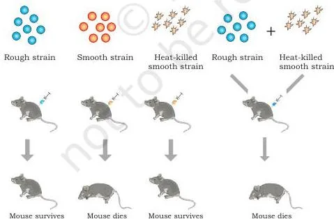
Fig 7.1 is the four-mouse panel. Read the fourth mouse — the one that dies after receiving heat-killed S plus live R — as the surprise: the live S bacteria recovered from it show that R was transformed, not that dead S bacteria were revived. Griffith called the unknown agent the transforming principle (NCERT, pp. 167-168).
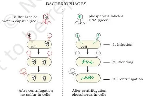
Fig 7.3 asks you to follow the radioactive label, not the virus. \(^{32}\text{P}\) (DNA) ends up in the pellet with the bacteria; \(^{35}\text{S}\) (protein) stays in the supernatant. Since the protein never entered the cell, protein cannot be the genetic material of the phage (NCERT, p. 170).
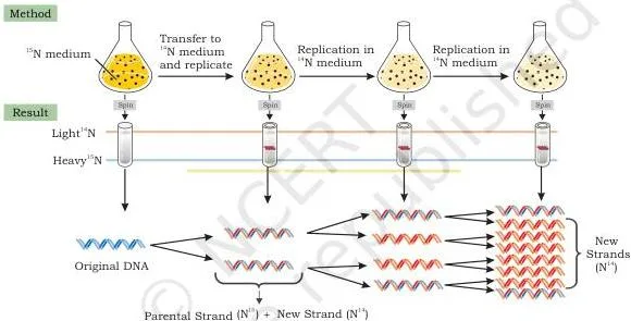
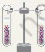
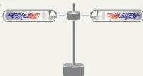
Fig 7.10 is three tubes you read as three sentences. Tube one: all-heavy DNA settles low. Tube two (generation I): one band between heavy and light — every molecule is hybrid. Tube three (generation II): two bands of equal intensity, hybrid and light. The heavy band never reappears, which is exactly what semiconservative replication predicts (NCERT, p. 178).
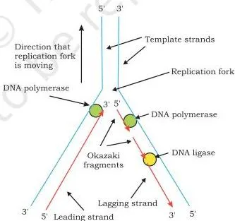
Fig 7.14 is about directions. Both new strands are built \(5′ \rightarrow 3’\), but the fork moves in only one direction, so the strand running away from the fork is assembled piecemeal as Okazaki fragments. The arrows in the diagram — one continuous arrow, one set of short arrows — are the whole answer (NCERT, p. 182).
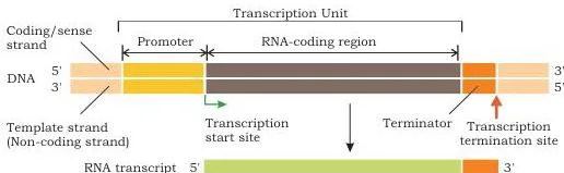
Fig 7.21 is the anatomy of a transcription unit. Find three landmarks: the promoter (where RNA polymerase binds, upstream of the start site), the start site itself, and the terminator where transcription ends. Then identify which strand is the template — RNA is copied from it, while the other strand matches the RNA sequence (NCERT, p. 188).
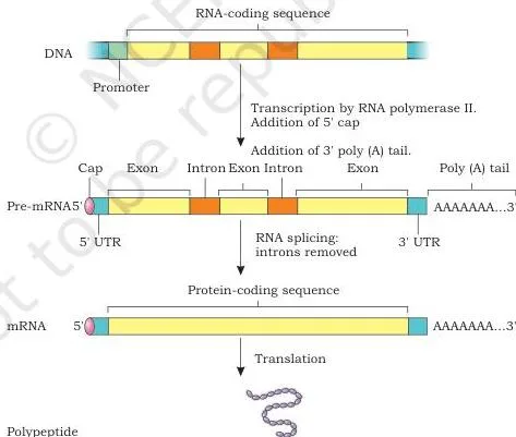
Fig 7.24 shows the three edits a pre-mRNA receives before leaving the nucleus. Read them in order: capping protects the \(5’\) end, splicing joins the exons, and the poly-A tail stabilises the \(3’\) end. The diagram’s point is that these modifications are part of gene expression, not an afterthought (NCERT, p. 190).
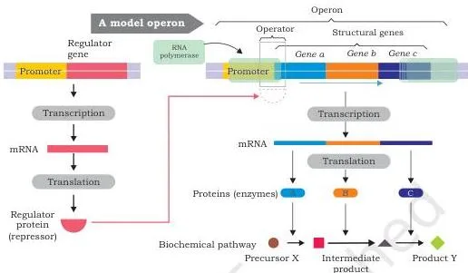
Fig 7.38 is the anatomy you must be able to label. Note the regulator gene upstream with its own promoter — the book is explicit that it is not part of the operon. The operator sits between the promoter and the structural genes, which is how a repressor can block polymerase (NCERT, p. 210).
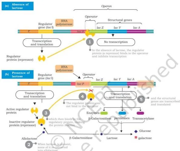
Fig 7.40 is the lac operon in its two states, drawn side by side. Compare the repressor: on the operator when lactose is absent, displaced when allolactose binds it. That single comparison between the two halves of the diagram is the complete answer to Exercise 2 (NCERT, p. 213).
Basic Processes definitions: the terms you must know
These are the chapter’s working terms, each defined in one line with the page where the book introduces it.
| Term | Meaning | NCERT page |
|---|---|---|
| Transformation | Transfer of genetic material from one cell to another that changes the recipient’s genetic makeup | p. 168 |
| Transforming principle | Griffith’s name for the unknown agent from dead S bacteria that transforms R bacteria; later shown to be DNA | p. 168 |
| Supercoiling | Further twisting of the DNA double helix on itself, so the helix axis coils into a superhelix | p. 171 |
| Nucleosome | Bead-like unit of eukaryotic packing: DNA wrapped around a histone octamer | p. 172 |
| Semiconservative replication | Replication that produces two duplexes, each with one old and one new strand | p. 176 |
| Origin of replication | The specific site where replication starts (oriC in E. coli) | p. 180 |
| Okazaki fragment | Short stretch of DNA (about 1,000-2,000 nucleotides) made discontinuously on the lagging strand | p. 183 |
| Leading strand | New DNA synthesised continuously in the \(5′ \rightarrow 3’\) direction toward the fork | p. 182 |
| Lagging strand | New DNA synthesised discontinuously as Okazaki fragments | p. 183 |
| Sense (coding) strand | DNA strand with the same sequence as the mRNA (T in place of U) | p. 187 |
| Template (non-coding) strand | DNA strand from which RNA is transcribed | p. 187 |
| Transcription unit | The stretch of DNA transcribed into RNA, with a start site, terminator and promoter | p. 188 |
| Promoter | Upstream DNA sequence where RNA polymerase binds to begin transcription | p. 188 |
| Genetic code | The set of rules by which triplet codons specify amino acids | p. 192 |
| Wobble pairing | Flexible base pairing at the third position of the codon, letting one tRNA read several codons | p. 195 |
| Point mutation | A change in the genetic material at the level of a single gene | p. 201 |
| Transition | Substitution of a purine by a purine, or a pyrimidine by a pyrimidine | p. 202 |
| Transversion | Substitution of a purine by a pyrimidine, or the reverse | p. 202 |
| Mutagen | An external agent, physical, chemical or biological, that induces mutation | p. 204 |
| Operon | A cluster of structural genes transcribed together with the promoter and operator controlling them | p. 211 |
| Operator | DNA segment where the repressor binds, blocking RNA polymerase | p. 211 |
| Repressor | Regulatory protein that binds the operator and prevents transcription | p. 211 |
| Inducer | Effector molecule (like allolactose) that inactivates a repressor and switches on an inducible operon | p. 212 |
| Housekeeping genes | Genes expressed at a constant level because their products are always needed | p. 209 |
Common mistakes in Basic Processes (and the correct version)
Each of these is a mistake the book’s own text warns against, in the order students usually make them.
| Mistake | Correct rule | Page and how to check |
|---|---|---|
| Griffith proved DNA is the transforming principle | Griffith proved a transforming principle exists; Avery, MacLeod and McCarty identified it as DNA | pp. 168-169 — check whose experiment used DNase |
| The sense strand is the one that is transcribed | RNA is copied from the template (non-coding) strand; the sense strand matches the RNA sequence with U instead of T | p. 187 — check which strand is actually read |
| The lagging strand is built \(3′ \rightarrow 5’\) | All DNA synthesis is \(5′ \rightarrow 3’\); the lagging strand is simply made discontinuously, in Okazaki fragments | pp. 182-183 — check the arrow directions in Fig 7.14 |
| RNA polymerase needs a primer | It does not — it can start a new RNA chain on a bare template | p. 187 — compare with DNA polymerase, which cannot |
| Lactose is the inducer of the lac operon | β-galactosidase converts some lactose into allolactose; allolactose inactivates the repressor | pp. 213-214 — check what actually binds the repressor |
| AUG is only a start signal | AUG also codes for methionine; the three stop codons code for nothing and no tRNA binds them | pp. 193, 198 — check the genetic code features |
| Wobble means pairing is loose everywhere | Pairing is precise at the first two positions and flexible only at the third | p. 195 — check where the flexibility sits |
| Transition means any base substitution | Transition is purine to purine or pyrimidine to pyrimidine; a family switch is a transversion | p. 202 — check the base families |
| The regulator gene is part of the operon | It sits upstream with its own promoter | p. 211 — check the operon diagram anatomy |
What the Basic Processes exercises ask you to do
The chapter ends with seven exercises. The table maps each to the concept it tests and where the chapter explains it, so you can revise the idea before attempting the question.
| Question | What it tests | Where the chapter explains it |
|---|---|---|
| Q1 — importance and steps of gene expression | Central dogma, transcription and translation | pp. 185-199 |
| Q2 — regulation of gene expression in prokaryotes with the lac operon | The operon model | pp. 209-214 |
| Q3 — effect of losing all proteins on DNA replication | The replication machinery, every part of which is a protein | pp. 179-183 |
| Q4 — UV damage, its molecular basis and its repair | Thymine dimers and nucleotide excision repair | pp. 204-208 |
| Q5 — four differentiations | Leading vs lagging strand; transcription vs translation; transition vs transversion; codon vs anticodon | pp. 182-183, 185-186, 194-195, 202 |
| Q6 — least harmful radiation | Radiations as mutagens, and what reaches DNA | pp. 204-205 |
| Q7 — where the AP site forms | Base excision repair | p. 206 |
Three questions need a little reading. Q3: every part of the replication machinery — helicase, primase, polymerases, SSB proteins, ligase — is a protein, so losing all proteins stops replication at the very first step (NCERT, pp. 179-183).
Q6: the chapter treats radiations as mutagens, explaining how X-rays break phosphodiester bonds and UV excites electrons (NCERT, pp. 204-205). Of the four options, alpha rays are the least penetrating, so they reach DNA the least — and are therefore the least likely to be harmful.
Q7: the AP site appears in base excision repair — DNA glycosylase removes a damaged base and leaves an apurinic or apyrimidinic site. The correct option is (a) excision repair (NCERT, p. 206).
One honest note: textbook contents and the examinable syllabus are not always identical. Check the current official CBSE syllabus for what is examinable in this academic session before you treat any chapter as fully testable.
Basic Processes in ten lines
The points below compress the chapter’s own SUMMARY (NCERT, pp. 215-216) — everything you should be able to say in one breath the night before an exam.
- Miescher isolated DNA (as ‘nuclein’) from pus cell nuclei in 1869 (p. 167).
- Griffith (1928) discovered transformation — a transforming principle changed R bacteria into S bacteria (p. 168).
- Avery, MacLeod and McCarty (1944) identified the transforming principle as DNA (p. 169).
- Hershey and Chase (1952) confirmed DNA is the genetic material — the phage’s DNA enters the cell, its protein does not (p. 170).
- The central dogma is the one-way flow \( \text{DNA} \rightarrow \text{RNA} \rightarrow \text{protein} \); retroviruses reverse it by reverse transcription (pp. 185-186).
- Meselson and Stahl proved semiconservative replication with \(^{15}\text{N}\) and \(^{14}\text{N}\) and density gradient centrifugation (p. 178).
- Replication runs on a team — polymerase III, primase, helicase, topoisomerase, SSB proteins, ligase — with the leading strand continuous and the lagging strand in Okazaki fragments (pp. 179-183).
- Transcription copies the template strand into RNA without a primer; eukaryotic transcripts are processed by capping, splicing and a poly-A tail (pp. 187-191).
- The genetic code has 64 triplets: 61 code for amino acids, 3 are stop signals, and AUG is both methionine and the start signal (p. 193).
- Translation has four stages — charging, initiation, elongation, termination — and several ribosomes can translate one mRNA at once as polyribosomes (pp. 194-199).
- Mutation is addition, deletion or substitution of nucleotides; excision repair and mismatch repair keep the rate low (pp. 200-208).
Related resources for Class 11 Biotechnology
This listing is maintained for the 2026-27 academic session using the NCERT textbook information available to us. NCERT remains the authority for confirming the latest edition.
Basic Processes sits inside the Class 11 Biotechnology book page; the Class 11 hub holds every NCERT book for the class. The chapter also builds on two earlier topics in this book: DNA structure, covered in Chapter 3, and genes on chromosomes from the previous chapter (NCERT, p. 167).
For revision, the Class 11 Biotechnology revision notes gather the subject’s key ideas, and the Class 11 study material plus CBSE revision notes cover the wider syllabus.
Two linked topics stay closely connected to this chapter: basic principles of inheritance, which the chapter assumes, and genetic disorders, where point mutations such as the sickle cell example surface in human genetics.
Sources and data verification
- The figure and page references on this page come from the official NCERT edition of the Class 11 Biotechnology textbook, Chapter 7 ‘Basic Processes’.
- This page covers that one chapter of that one book. It does not describe the wider CBSE subject scheme.
- It is maintained for the current academic session using the NCERT information available to us; the official PDF always remains the definitive text.
- NCERT settles textbook editions and official PDFs; CBSE settles curriculum, syllabus and examinations.
Reference: NCERT Class 11 Biotechnology textbook, chapter 7, official edition on ncert.nic.in.
Frequently asked questions about Basic Processes
Who proved DNA is the genetic material?
Hershey and Chase’s 1952 experiment with T2 bacteriophage gave the direct proof: only the phage’s DNA entered the E. coli cell, yet new phages were made. Before that, Avery, MacLeod and McCarty had already shown in 1944 that DNA is the transforming principle, and Griffith’s 1928 experiment had shown that a transforming principle exists at all (NCERT, pp. 168-170).
Why does the Meselson-Stahl experiment prove semiconservative replication?
Because the band pattern matches only the semiconservative prediction. After one generation in \(^{14}\text{N}\) medium all DNA is hybrid (one intermediate band); after two generations half is hybrid and half is all-light (two bands of equal intensity); and the heavy band never reappears.
Any scheme in which one old double helix stays intact would eventually show a heavy band — it never does (NCERT, pp. 177-178).
What is the difference between the leading and lagging strand?
Both are synthesised \(5′ \rightarrow 3’\); the difference is continuity. The leading strand is made continuously in the direction the replication fork moves, with a single RNA primer, while the lagging strand is made discontinuously, away from the fork, in Okazaki fragments each starting with its own primer (NCERT, pp. 182-183).
Why is the genetic code called degenerate?
Because most amino acids are specified by more than one codon — leucine, for example, has six codons. Only methionine and tryptophan have a single codon each, and the three stop codons code for nothing (NCERT, p. 193).
What is the inducer of the lac operon?
Allolactose. When lactose is present, β-galactosidase converts some of it into allolactose; allolactose binds the repressor, changes its shape and makes it inactive, so transcription of the operon’s three genes proceeds (NCERT, pp. 213-214).
How does a cell repair UV damage to DNA?
By nucleotide excision repair. UV light creates thymine dimers that distort the double helix; the UVr protein complex finds the dimer, cuts the backbone four nucleotides downstream and eight upstream, unwinds and removes the damaged fragment, and DNA polymerase I and ligase fill and seal the gap (NCERT, pp. 206-208).
Explore Class 11 Biotechnology Books
- Previous: Basic Principles of Inheritance
- Next: Genetic Disorder
Related chapters:
- An Introduction to Biotechnology
- G.N. Ramachandran (1922–2001)
- Biomolecules