
This page holds the official Body Fluids and Circulation Class 11 chapter — NCERT Biology Chapter 15 — which runs twelve printed pages of the textbook beginning at page 192. The chapter PDF as published by NCERT is right below, followed by a plain-language walkthrough of every section, figure and exercise in it.
Open the official Body Fluids and Circulation Class 11 NCERT Chapter 15 PDF to read the chapter exactly as printed — its tables, diagrams, summary and end-of-chapter exercise set in a single file.
| What the chapter holds | Count | Where it is used |
|---|---|---|
| Printed pages | 12 | |
| Sections in the chapter | 13 | |
| Figures with NCERT captions | 4 | |
| Tables | 1 | |
| Exercise questions | 14 | answered in our NCERT Solutions |
| Official NCERT PDF | Download the chapter PDF | the chapter exactly as NCERT publishes it |
Chapter 15 at a Glance
The table below lists what this chapter holds — its sections, figures, tables and exercises — counted directly from the NCERT textbook. The chapter spans about twelve pages of the book, starting at NCERT page 192.
What This Chapter Covers: From Blood to Double Circulation
The chapter does one job: it introduces the two fluids that carry materials around the body — blood and lymph — and the four-chambered heart and vessel network that keep both moving.
| Section | What it explains | NCERT pages |
|---|---|---|
| 15.1 Blood | Plasma, formed elements, blood groups and clotting | 192-197 |
| 15.2 Lymph (tissue fluid) | Where tissue fluid comes from and what lymph carries | 197 |
| 15.3 Circulatory pathways, the human heart, cardiac cycle and ECG | Open and closed systems, heart structure, the beating cycle and its electrical record | 197-201 |
| 15.4 Double circulation | The pulmonary and systemic routes of blood | 201-202 |
| 15.5 Regulation of cardiac activity | Neural and hormonal control of the heart | 202 |
| 15.6 Disorders of the circulatory system | Hypertension, coronary artery disease, angina and heart failure | 202-203 |
Key Concepts Explained
The Body Fluids and Circulation Class 11 chapter is about what flows through the body — blood and lymph — and the four-chambered pump that keeps both moving. Each concept below is explained in the order the book teaches it, with the textbook page beside it.
Blood: plasma and the formed elements
Blood is the transport fluid of the body, and it is classed as a connective tissue because it has a fluid matrix — plasma — with cells floating in it (NCERT page 192).
Plasma is the straw-coloured, viscous fluid that makes up about 55 per cent of blood; 90-92 per cent of it is water, and proteins contribute 6-8 per cent (NCERT page 192). The three major proteins have three different jobs: fibrinogen for clotting, globulins for defence and albumins for osmotic balance.
Plasma also carries minerals, glucose, amino acids and lipids in transit, plus clotting factors in an inactive form. Plasma without those clotting factors is called serum (NCERT page 193).
The formed elements — erythrocytes (RBCs), leucocytes (WBCs) and platelets — make up the other 45 per cent of blood (NCERT page 193).
RBCs form in red bone marrow, are biconcave and lack a nucleus in most mammals, and carry the iron-containing protein haemoglobin — 12-16 g per 100 mL of blood — which transports respiratory gases. An RBC lives about 120 days and is destroyed in the spleen (NCERT page 194).
WBCs are colourless, nucleated and fewer: 6000-8000 per mm³ of blood. They divide into granulocytes (neutrophils, eosinophils, basophils) and agranulocytes (lymphocytes, monocytes). Platelets, or thrombocytes, are fragments of bone marrow cells called megakaryocytes, present at about 1.5 to 3.5 lakh per mm³ of blood (NCERT pages 194-195).
The table below brings count, nucleus, life span and function of every formed element into one place, drawn from NCERT pages 194-195.
| Formed element | Count in blood | Nucleus | Life span | Main function |
|---|---|---|---|---|
| RBC (erythrocyte) | 5-5.5 million per mm³ | Absent in most mammals | About 120 days | Transports respiratory gases with haemoglobin |
| Neutrophil | 60-65 per cent of total WBCs | Present | Short-lived | Phagocytic — destroys foreign organisms |
| Eosinophil | 2-3 per cent of total WBCs | Present | Short-lived | Resists infections; linked to allergic reactions |
| Basophil | 0.5-1 per cent of total WBCs | Present | Short-lived | Secretes histamine, serotonin and heparin; inflammatory reactions |
| Lymphocyte | 20-25 per cent of total WBCs | Present | Short-lived | Immune responses; B and T forms |
| Monocyte | 6-8 per cent of total WBCs | Present | Short-lived | Phagocytic — destroys foreign organisms |
| Platelet (thrombocyte) | 1.5-3.5 lakh per mm³ | No — a cell fragment | Short-lived | Releases substances involved in clotting |
Blood groups: ABO and Rh
Blood cannot be transfused at random: if a donor’s red cells carry an antigen that the recipient’s plasma has antibodies against, the cells clump and are destroyed.
ABO grouping rests on two surface antigens, A and B, on the RBCs, and two natural antibodies, anti-A and anti-B, in the plasma (NCERT page 195).
| Blood group | Antigens on RBCs | Antibodies in plasma | Can receive from |
|---|---|---|---|
| A | A | anti-B | A, O |
| B | B | anti-A | B, O |
| AB | A and B | None | AB, A, B, O |
| O | None | anti-A and anti-B | O |
Read the compatibility from the antigen-antibody columns. Group O has no antigens, so no recipient’s antibodies can attack its cells — O is the universal donor. Group AB has no antibodies, so it has nothing with which to attack a donor’s cells — AB is the universal recipient (NCERT page 195).
There is a catch. O can donate to anyone but receive only O, because O plasma carries both anti-A and anti-B. AB can receive from anyone but donate only to AB, because AB cells carry both antigens.
Rh grouping uses a different antigen, first described in Rhesus monkeys and present in about 80 per cent of humans — the Rh positive majority. An Rh negative person exposed to Rh positive blood forms antibodies against it, so Rh must also be matched before transfusion (NCERT page 196).
The classic problem is an Rh negative mother with an Rh positive baby. The placenta keeps the two bloods separate in the first pregnancy, so nothing happens then. If the mother’s blood is exposed to the baby’s Rh positive cells during the first delivery, she starts making anti-Rh antibodies.
In later pregnancies these antibodies can leak into the foetus and destroy its red cells, causing erythroblastosis foetalis. Giving the mother anti-Rh antibodies immediately after the first delivery prevents it (NCERT page 196).
How blood clots
A cut stops bleeding because clotting factors that normally sit inactive in plasma switch on in a cascade when tissue is injured (NCERT page 196).
- Injury makes platelets release certain factors; tissues at the wound release their own factors too.
- A series of linked enzymic reactions forms the enzyme complex thrombokinase.
- Thrombokinase converts inactive prothrombin into active thrombin.
- Thrombin converts inactive fibrinogen into fibrin.
- Fibrin threads form a network that traps dead and damaged formed elements — the dark reddish-brown scum you see over a cut.
Calcium ions play a very important role in this clotting process (NCERT page 197).
Lymph: the tissue fluid
Not everything in blood stays inside the vessels — the fluid that leaks into the tissues returns through its own network.
As blood passes through capillaries, water and small water-soluble substances move out into the spaces between cells, forming the interstitial fluid or tissue fluid. Larger proteins and most formed elements stay behind. The lymphatic system collects this fluid and drains it back into the major veins; inside the system the fluid is called lymph (NCERT page 197).
Lymph is a colourless fluid that carries specialised lymphocytes for immune responses, as well as nutrients and hormones. Fats are absorbed through the lymph in the lacteals of the intestinal villi (NCERT page 197).
Circulatory pathways and the human heart
Circulation may be open or closed, and the vertebrate heart gained chambers as animals evolved — the table below shows the progression.
Open circulation occurs in arthropods and molluscs: the heart pumps blood through large vessels into open spaces called sinuses. Closed circulation, in annelids and chordates, keeps blood always within vessels, so the flow can be regulated more precisely (NCERT page 197).
| Group | Chambers | Circulation pattern |
|---|---|---|
| Fishes | 2 (one atrium, one ventricle) | Single circulation — heart → gills → body → heart |
| Amphibians | 3 (two atria, one ventricle) | Incomplete double circulation — the two bloods mix in the single ventricle |
| Reptiles (except crocodiles) | 3 (two atria, one ventricle) | Incomplete double circulation |
| Crocodiles, birds and mammals | 4 (two atria, two ventricles) | Complete double circulation — no mixing of the two bloods |
Fishes pump deoxygenated blood to the gills, where it is oxygenated and then sent to the body — a single circulation. Amphibians and most reptiles mix oxygenated and deoxygenated blood in the single ventricle. Crocodiles, birds and mammals keep the two bloods apart in separate ventricles (NCERT pages 197-198).
The human heart sits in the thoracic cavity between the two lungs, tilted slightly to the left, about the size of a clenched fist, and is wrapped in a double-walled bag, the pericardium (NCERT page 198). It has four chambers: two small upper atria and two larger lower ventricles, separated by the inter-atrial and inter-ventricular septa.
Valves keep blood moving one way. The opening between the right atrium and right ventricle is guarded by the tricuspid valve, with three flaps; the left opening by the bicuspid or mitral valve, with two. The exits of the ventricles into the pulmonary artery and aorta carry semilunar valves. All of them prevent backward flow (NCERT page 198).
Mnemonic: ‘tri’ means three flaps, ‘bi’ means two. Keep the sides straight by the alphabet — B comes before T, and Left before Right — so the bicuspid valve is on the left and the tricuspid on the right.
A patch of specialised cardiac muscle, the nodal tissue, runs the heart’s rhythm. The sino-atrial node (SAN), high in the right atrium, generates 70-75 action potentials per minute — more than any other part of the nodal system — so it sets the pace and is called the pacemaker (NCERT page 199).
The atrio-ventricular node (AVN) lies near the atrio-ventricular septum. From it, the AV bundle passes through the septum and divides into right and left bundles that end in fine purkinje fibres throughout the ventricular musculature (NCERT page 199).
Cardiac cycle, cardiac output and heart sounds
One heartbeat is a timed sequence of contraction (systole) and relaxation (diastole) that the heart repeats about 72 times a minute (NCERT page 199).
- All four chambers relax — joint diastole. The tricuspid and bicuspid valves are open, the semilunar valves closed, and blood flows from the veins into the atria and then the ventricles.
- The SAN fires. Both atria contract — atrial systole — pushing about 30 per cent more blood into the ventricles.
- The impulse travels through the AVN and AV bundle, and the ventricles contract — ventricular systole. The pressure shuts the tricuspid and bicuspid valves, then forces the semilunar valves open, driving blood into the pulmonary artery and aorta.
- The ventricles relax — ventricular diastole. Falling pressure closes the semilunar valves; when the atria refill, the tricuspid and bicuspid valves reopen and the cycle returns to joint diastole.
This whole sequence is the cardiac cycle. At 72 beats per minute, one cycle lasts 0.8 seconds (NCERT pages 199-200).
Each ventricle pumps about 70 mL per beat — the stroke volume. Stroke volume \( \times \) heart rate gives the cardiac output, the volume each ventricle pumps per minute — about 5000 mL, or 5 litres (NCERT page 200).
Two sounds mark the cycle: lub, the closure of the tricuspid and bicuspid valves, and dub, the closure of the semilunar valves (NCERT pages 199-200).
The electrocardiogram
The heart’s electrical activity can be recorded from the skin, and every wave on the trace has a name and a meaning (NCERT page 200).
An electrocardiogram (ECG) is a graphical record of the electrical activity of the heart during a cardiac cycle. A standard trace uses three leads — one on each wrist and one on the left ankle (NCERT page 201).
- P wave — depolarisation of the atria, which leads to their contraction.
- QRS complex — depolarisation of the ventricles, which begins ventricular systole.
- T wave — repolarisation of the ventricles; its end marks the end of systole.
Because one QRS complex appears per beat, counting them in a given time gives the heart rate. Healthy traces look similar, so any deviation from the normal shape suggests a possible abnormality (NCERT page 201).
Double circulation, regulation and disorders
Blood follows two fixed routes, and the pump driving both is tuned by nerves and hormones.
Pulmonary circulation runs from the right ventricle through the pulmonary artery to the lungs, where the blood is oxygenated, and back through the pulmonary veins to the left atrium. Systemic circulation runs from the left ventricle through the aorta to all tissues, and returns deoxygenated blood through the venae cavae to the right atrium.
The two together are the double circulation (NCERT page 201).
Artery and vein walls have three layers: the tunica intima (squamous endothelium), the tunica media (smooth muscle and elastic fibres — thinner in veins) and the tunica externa (fibrous connective tissue).
Two special routes complete the picture: the hepatic portal system, whose hepatic portal vein carries blood from the intestine to the liver, and the coronary system, which serves the heart muscle itself (NCERT pages 201-202).
The heart is myogenic: its own autoexcitable nodal tissue sets the pace. The medulla oblongata moderates this through the autonomic nervous system — sympathetic nerves raise heart rate and cardiac output, parasympathetic nerves lower them — and adrenal medullary hormones can also raise cardiac output (NCERT page 202).
The chapter closes with four disorders. Hypertension is blood pressure of 140/90 or higher, against a normal of 120/80. Coronary artery disease, or atherosclerosis, narrows the arteries of the heart muscle with deposits of calcium, fat, cholesterol and fibrous tissue. Angina is acute chest pain from insufficient oxygen reaching the heart muscle.
Heart failure is the heart not pumping effectively enough for the body’s needs — not the same as cardiac arrest, when the heart stops beating, or a heart attack, when the muscle is suddenly damaged by an inadequate blood supply (NCERT pages 202-203).
Figure Walkthrough: Reading the NCERT Diagrams
Every diagram in this chapter repays a careful look — each one is a compressed answer to an exam question. Here is each figure read stage by stage.
Figure 15.1: Formed elements in blood (NCERT page 194)

Start with the RBC — drawn biconcave and without a nucleus, exactly the features that let it carry haemoglobin and squeeze through capillaries. Then compare the five WBC types by their cytoplasm: neutrophils, eosinophils and basophils look granular (hence granulocytes), while lymphocytes and monocytes look clear (agranulocytes). Platelets appear as small fragments, because that is what they are — pieces of megakaryocytes.
Match each drawing to its job on NCERT pages 194-195: neutrophils and monocytes are phagocytic, eosinophils resist infections, basophils secrete histamine and heparin, lymphocytes run immune responses, and platelets drive clotting.
Figure 15.2: Section of a human heart (NCERT page 198)
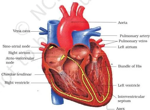
Find the four chambers first — the two thin-walled atria above, the thick-walled ventricles below, separated by the inter-atrial and inter-ventricular septa. The valves are the doors: tricuspid between the right atrium and right ventricle, bicuspid on the left, and semilunar valves at the exits into the pulmonary artery and aorta.
The nodal tissue is marked as small patches and strands — the SAN high in the right atrium, the AVN lower, with the AV bundle and purkinje fibres running into the ventricular walls. Read the arrows as the direction of flow: the valves ensure it never reverses (NCERT page 198).
Figure 15.3: A standard ECG (NCERT page 201)
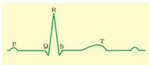
Read the trace left to right. The small P wave comes first — atrial depolarisation. The tall, sharp QRS complex is ventricular depolarisation; systole begins just after Q. The T wave is ventricular repolarisation, and its end marks the end of systole. The flat segments between waves are the rest periods.
Count the QRS complexes in a minute and you have the heart rate — which is exactly how doctors use a trace like this one (NCERT page 201). This figure is also the drawing asked for in exercise question 14.
Figure 15.4: Schematic plan of blood circulation (NCERT page 202)
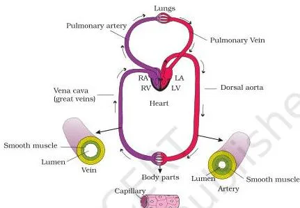
This figure answers ‘what is double circulation?’ visually. Follow the pulmonary loop first: right ventricle → pulmonary artery → lungs → pulmonary veins → left atrium. Then the systemic loop: left ventricle → aorta → body tissues → venae cavae → right atrium. Deoxygenated blood is drawn on one side, oxygenated on the other, and the two never mix.
Note the hepatic portal vein carrying blood from the intestine to the liver before it joins the systemic circulation (NCERT page 202).
Important Definitions from the Chapter
These are the terms the chapter defines and that its exercises ask you to use. Each is given in one or two plain sentences.
| Term | Meaning | NCERT page |
|---|---|---|
| Plasma | Straw-coloured fluid matrix of blood, about 55 per cent of blood volume, mostly water with proteins and dissolved substances | 192 |
| Serum | Plasma from which the clotting factors have been removed | 193 |
| Formed elements | The cellular part of blood — erythrocytes, leucocytes and platelets — about 45 per cent of blood | 193 |
| Haemoglobin | Red, iron-containing protein in RBCs that transports respiratory gases | 194 |
| Universal donor | Blood group O, whose RBCs carry no A or B antigens, so it can be donated to any group | 195 |
| Universal recipient | Blood group AB, whose plasma has no anti-A or anti-B antibodies, so it can accept any group | 195 |
| Erythroblastosis foetalis | Destruction of foetal RBCs by maternal anti-Rh antibodies in a pregnancy after the first | 196 |
| Lymph | Colourless fluid of the lymphatic system; collected tissue fluid carrying lymphocytes, nutrients and hormones | 197 |
| Systole | The contraction phase of a heart chamber | 200 |
| Diastole | The relaxation phase of a heart chamber | 200 |
| Cardiac cycle | One complete sequence of systole and diastole of all four chambers, repeated with each beat | 200 |
| Stroke volume | Blood pumped out by each ventricle in one beat, about 70 mL | 200 |
| Cardiac output | Blood pumped out by each ventricle in one minute; stroke volume \( \times \) heart rate, about 5 litres | 200 |
| Pacemaker | The SAN, which generates the most action potentials per minute and sets the heart’s rhythm | 199 |
| Myogenic heart | A heart whose rhythm originates in its own autoexcitable nodal tissue | 202 |
| Double circulation | Two separate circulatory pathways — pulmonary and systemic — with no mixing of the two bloods | 201 |
| Hypertension | Blood pressure of 140/90 or higher, against a normal of 120/80 | 202 |
| Angina | Acute chest pain from insufficient oxygen reaching the heart muscle | 203 |
Common Mistakes Students Make
Six confusion points in this chapter are so common they have names. Each row names the mistake, the rule that fixes it, and a way to check yourself.
| Mistake | Correct rule | How to check your answer |
|---|---|---|
| Assuming the universal donor O can receive blood from anyone | O has anti-A and anti-B antibodies in its plasma, so it can receive only O blood | Look at the antibody column: O’s own antibodies would attack A, B and AB cells |
| Thinking the first Rh-incompatible pregnancy is harmed | The placenta keeps the two bloods apart; the mother forms anti-Rh antibodies only after exposure during the first delivery, so later pregnancies are at risk | Trace the sequence: antibody formation happens after the first baby, harm happens in the next pregnancy |
| Swapping stroke volume and cardiac output | Cardiac output = stroke volume \( \times \) heart rate, about 5 L per minute | Check units: stroke volume is mL per beat, cardiac output is mL per minute |
| Treating heart failure, cardiac arrest and heart attack as the same | Heart failure is weak pumping; cardiac arrest is the heart stopping; a heart attack is sudden muscle damage from inadequate blood supply | Match each term to its own definition on NCERT page 203 |
| Assigning the wrong heart sound | Lub is the tricuspid and bicuspid valves closing; dub is the semilunar valves closing | Lub occurs as ventricular systole begins, dub as it ends |
| Muddling the ECG waves | P is atrial depolarisation, QRS is ventricular depolarisation, T is ventricular repolarisation | Order them by the heart’s action: atria first, then ventricles, then recovery |
Exam Preparation Notes
The chapter’s closing exercise set tests the content in four ways: direct definitions, one matching item, difference questions and one drawing question. The table below maps each exercise to the concept it tests and the textbook page where the answer is taught.
| Exercise | Concept tested | Where the answer is taught |
|---|---|---|
| 1 | Formed elements and one function of each | NCERT pages 194-195 |
| 2 | Importance of plasma proteins | NCERT pages 192-193 |
| 3 | Matching — eosinophils (resist infections), RBC (gas transport), AB group (universal recipient), platelets (coagulation), systole (contraction of heart) | NCERT pages 194-200 |
| 4 | Blood as a connective tissue | NCERT page 192 |
| 5 and 7(a) | Differences between lymph and blood | NCERT page 197 |
| 6 | Double circulation and its significance | NCERT pages 201-202 |
| 7(b) | Open and closed circulatory systems | NCERT page 197 |
| 7(c) | Systole and diastole | NCERT pages 199-200 |
| 7(d) | P-wave and T-wave | NCERT page 201 |
| 8 | Evolution of the vertebrate heart | NCERT pages 197-198 |
| 9, 10, 11 | Myogenic heart, SAN as pacemaker, role of AVN and AV bundle | NCERT pages 198-199 |
| 12 and 13 | Cardiac cycle, cardiac output and heart sounds | NCERT pages 199-200 |
| 14 | Drawing a standard ECG and explaining its segments | NCERT page 201 |
A numerical on cardiac output needs one formula and careful units. Here is one worked with fresh numbers.
Step 1: Write the formula.
Cardiac output = stroke volume \( \times \) heart rate.
Step 2: Substitute a heart rate of 80 beats per minute and a stroke volume of 70 mL per beat.
\[ \text{Cardiac output} = 70\ \text{mL/beat} \times 80\ \text{beats/min} = 5600\ \text{mL/min} \]
Step 3: Convert to litres, since 1000 mL = 1 L.
\[ 5600\ \text{mL} = 5.6\ \text{L} \]
Step 4: Find the duration of one cardiac cycle.
Time per cycle = 60 seconds divided by the heart rate.
\[ \frac{60\ \text{s}}{80} = 0.75\ \text{s} \]
Final answer: cardiac output is 5600 mL, or 5.6 litres, per minute, and each cardiac cycle lasts 0.75 seconds.
A complete numerical answer states the formula, substitutes with units at every step, and ends with the unit on the final value.
One honest caution: textbook contents and the examinable syllabus are not always identical, so check the current official CBSE syllabus for the exact scope of what is examinable.
Revision Summary
For a last-night recap: vertebrates move materials through the body in two fluids — blood, a fluid connective tissue, and lymph, the tissue fluid returned by the lymphatic system. Blood is plasma plus formed elements: RBCs carrying haemoglobin for gas transport, several classes of WBCs for defence, and platelets for clotting.
Transfusion depends on the ABO antigens and antibodies and on the Rh antigen, whose incompatibility threatens later pregnancies.
The heart is a four-chambered, myogenic pump whose SAN sets the pace, driving the cardiac cycle of systole and diastole; each ventricle ejects about 70 mL per beat, giving a cardiac output near 5 litres a minute. The ECG records the cycle’s electrical activity in the P wave, QRS complex and T wave.
Complete double circulation runs separate pulmonary and systemic pathways, and the medulla, autonomic nerves and adrenal hormones tune the whole system.
The numbers below are the ones worth memorising — each appears in the chapter and can anchor a short-answer question.
| Quantity | Value |
|---|---|
| Plasma share of blood | About 55 per cent |
| Formed elements share | About 45 per cent |
| Haemoglobin level | 12-16 g per 100 mL of blood |
| RBC life span | About 120 days |
| WBC count | 6000-8000 per mm³ of blood |
| SAN discharge | 70-75 action potentials per minute |
| Heart rate | About 72 beats per minute |
| Cardiac cycle duration | 0.8 seconds |
| Stroke volume | About 70 mL per ventricle |
| Cardiac output | About 5 litres per minute |
Related Resources
This listing is maintained for the 2026-27 academic session using the NCERT textbook information available to us. NCERT remains the authority for confirming the latest edition.
If you are revising Chapter 15 as part of the full book, the pages below carry the rest of Class 11 Biology on this site.
- Class 11 Biology notes — the chapter pages for this book on this site.
- Breathing and Exchange of Gases — Chapter 14, the chapter that precedes this one.
- Excretory Products and Their Elimination — Chapter 16, the chapter that follows.
- Class 11 hub — the Class 11 section of the notes on this site.
- CBSE notes index — the starting point for all notes on this site.
You can also browse this chapter on the official NCERT textbook page for Class 11 Biology, which lists every chapter of the book.
Sources and Data Verification
Four things are worth knowing about where this page’s facts come from.
- The figures, page references and chapter contents describe the current NCERT Class 11 Biology textbook, Chapter 15, Body Fluids and Circulation, which begins at page 192 of the prescribed English edition.
- This page covers that one chapter of the Class 11 Biology NCERT book — it does not cover other books, other subjects or other classes.
- The listing is maintained for the current academic session using the NCERT information available to us.
- NCERT settles textbook editions, chapter contents and the official PDFs; CBSE settles the curriculum and examinations. For what is examinable, check the current official CBSE syllabus.
Reference: NCERT Class 11 Biology textbook, chapter 15, official edition on ncert.nic.in.
Frequently Asked Questions
Why is O blood group called the universal donor and AB the universal recipient?
Group O red cells carry no A or B antigens, so no recipient’s plasma antibodies have a target to attack — O blood can be given to any group. Group AB plasma has no anti-A or anti-B antibodies, so an AB person can receive any group’s cells without clumping them (NCERT page 195).
Remember the catch: O can donate anywhere but receive only O, and AB can receive anywhere but donate only to AB.
Why does Rh incompatibility usually affect the second pregnancy and not the first?
In a first pregnancy, the placenta keeps the mother’s Rh negative blood and the baby’s Rh positive blood well separated, so no antibodies form. Exposure to the baby’s Rh positive cells can happen during the first delivery itself; only then does the mother begin making anti-Rh antibodies.
In subsequent pregnancies those antibodies can cross into the foetus and destroy its red cells, causing erythroblastosis foetalis (NCERT page 196).
What is the difference between cardiac output and stroke volume?
Stroke volume is the blood one ventricle pumps out in a single beat — about 70 mL. Cardiac output is the blood one ventricle pumps in a whole minute, and it equals stroke volume \( \times \) heart rate — about 5 litres (NCERT page 200). For a heart rate of 80 and a stroke volume of 70 mL, cardiac output = \( 70 \times 80 = 5600\ \text{mL} \) — that is 5.6 litres per minute.
How is heart failure different from cardiac arrest and a heart attack?
Heart failure means the heart is not pumping blood effectively enough for the body’s needs — it is a state, not a sudden event. Cardiac arrest is when the heart stops beating altogether. A heart attack is sudden damage to the heart muscle from an inadequate blood supply (NCERT page 203). The chapter is explicit that the three are not the same.
What do the P wave, QRS complex and T wave of an ECG represent?
The P wave is depolarisation of the atria, which leads to atrial contraction. The QRS complex is depolarisation of the ventricles, beginning ventricular systole. The T wave is ventricular repolarisation, the return of the ventricles to the resting state; its end marks the end of systole (NCERT page 201).
Where can I download the official Body Fluids and Circulation Class 11 PDF?
The official Body Fluids and Circulation Class 11 NCERT Chapter 15 PDF is hosted by NCERT itself, and the link near the top of this page opens it directly. You can also reach it through the official NCERT textbook page for Class 11 Biology.
Explore Class 11 Biology Books
- Previous: Breathing and Exchange of Gases
- Next: Excretory Products and Their Elimination
Class 11 Biology on LearnCBSE:
Related chapters:
- The Living World
- Biological Classification
- Plant Kingdom